From: Investigating Metastasis Using In Vitro Platforms

NCBI Bookshelf. A service of the National Library of Medicine, National Institutes of Health.

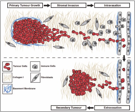
The metastatic cascade. (Top) Proteolytic disruption of the collagen and laminin-rich basement membrane enables the escape of tumor cells from the primary tumor. Facilitated by paracrine signals from stromal cells, migrating tumor cells invade the surrounding stroma rich in collagen-I and enter the vasculature (intravasation). (Bottom) Vascular arrest and extravasation of circulating tumor cells relies on their interaction with endothelial cells and the sub-endothelial basement membrane. Successful colonization of the secondary organ is dependent on the ability of metastatic cells to survive, proliferate and to promote vascularization, leading to the formation of a clinically relevant secondary tumor.
From: Investigating Metastasis Using In Vitro Platforms

NCBI Bookshelf. A service of the National Library of Medicine, National Institutes of Health.