From: Pharmacologic Approaches

NCBI Bookshelf. A service of the National Library of Medicine, National Institutes of Health.

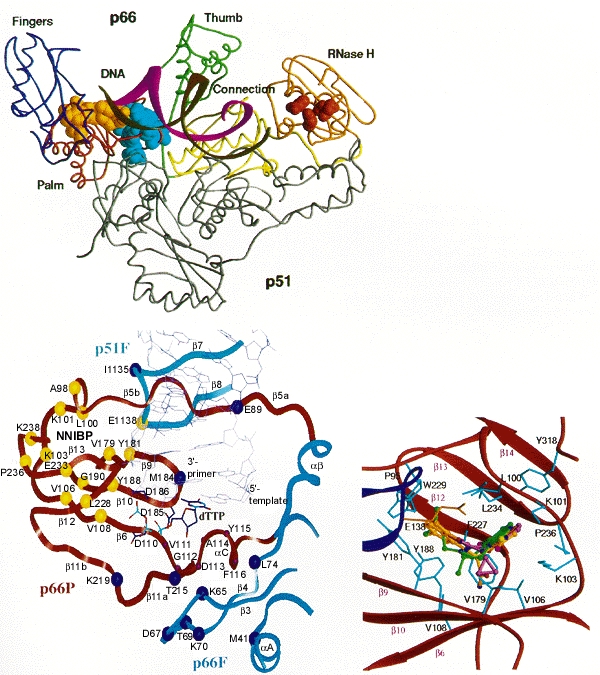
Binding sites for RT inhibitors. (Top) Structure of HIV-1 RT. The active sites for polymerase and RNase H are shown in gold and brown, respectively, and the nonnucleoside inhibitor binding pocket is shown in blue. (Reprinted from Arnold et al. 1996.) (Bottom, left) Active site and NNRTI-binding region bound to template-primer. The region shown comprises parts of the palm (P) and fingers (f) domains of p66 and the fingers domain of p51. Nucleoside analogs interact with the dNTP-binding site (shown with a docked dTTP substrate). Nonnucleoside inhibitors interact with the adjacent nonnucleoside inhibitor-binding pocket (NNIBP). (Blue spheres) Amino acid substitutions associated with nucleoside analog resistance; (yellow spheres) amino acid substitutions associated with nonnucleoside inhibitor resistance. (Reprinted from Tantillo et al. 1994.) (Bottom, right) The NNRTI-binding region shown with three bound inhibitors: TIBO (green); α-APA (red), and nevirapine (green). (Reprinted from Ding et al. 1995a.) Note that despite their very different structures (Fig. 14), the three inhibitors have a similar conformation when bound.
From: Pharmacologic Approaches

NCBI Bookshelf. A service of the National Library of Medicine, National Institutes of Health.