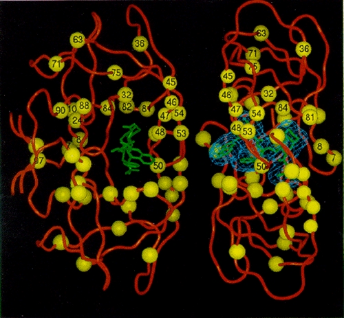
Figure 16. Location of mutations associated with resistance to inhibitors of HIV-1 protease.

Figure 16

Location of mutations associated with resistance to inhibitors of HIV-1 protease. Two views of the enzyme are shown, with a bound inhibitor shown in green, and mutations associated with resistance to at least one inhibitor shown as yellow spheres numbered on one subunit. (Courtesy of J. Alexandratos and A. Wlodawer.)

From: Pharmacologic Approaches

Cover of Retroviruses
Retroviruses.
Coffin JM, Hughes SH, Varmus HE, editors.
Cold Spring Harbor (NY): Cold Spring Harbor Laboratory Press; 1997.
Copyright © 1997, Cold Spring Harbor Laboratory Press.

NCBI Bookshelf. A service of the National Library of Medicine, National Institutes of Health.