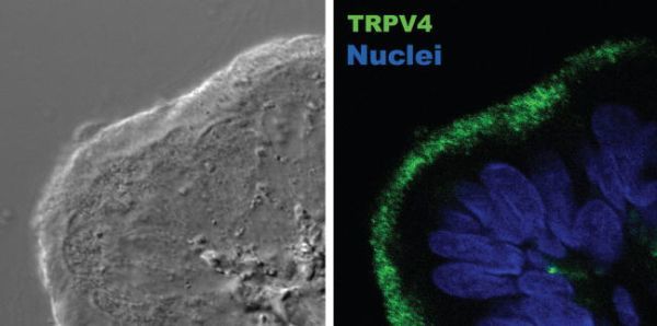
FIGURE 30.2. Confocal microscopy localization of TRPV4 to motile cilia in a tissue section of the female hamster oviduct.

FIGURE 30.2

Confocal microscopy localization of TRPV4 to motile cilia in a tissue section of the female hamster oviduct. (Left) Nomarsky image. (Right) Detection of TRPV4 (green) and TOPRO-3 nuclear staining (blue). The antibody used to detect TRPV4 is described in references [6, 8], and [22].

From: Chapter 30, The TRPV4 Channel in Ciliated Epithelia
Cover of TRP Ion Channel Function in Sensory Transduction and Cellular Signaling Cascades
TRP Ion Channel Function in Sensory Transduction and Cellular Signaling Cascades.
Liedtke WB, Heller S, editors.
Boca Raton (FL): CRC Press/Taylor & Francis; 2007.
Copyright © 2007, Taylor & Francis Group, LLC.

NCBI Bookshelf. A service of the National Library of Medicine, National Institutes of Health.