From: Chapter 29, The Role of TRPV4 in the Kidney

NCBI Bookshelf. A service of the National Library of Medicine, National Institutes of Health.

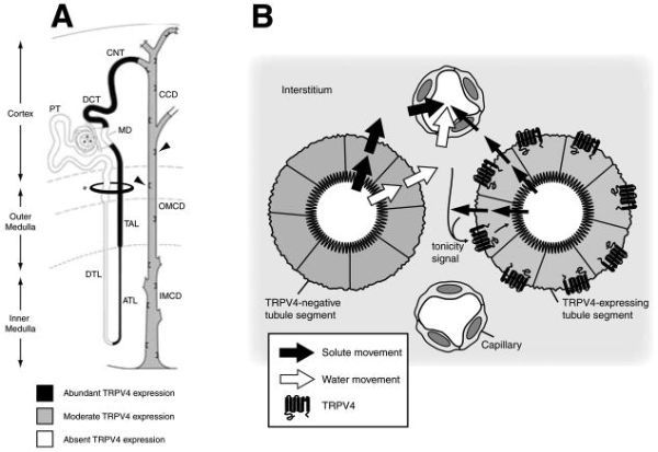
TRPV4 in the mammalian kidney. (A) TRPV4 expression along the mouse and rat nephron. Black shading indicates strong TRPV4 expression; gray shading indicates less intense TRPV4 expression. Throughout the course of the collecting duct, expression was stronger in the intercalated cells (e.g., arrowheads). The kidney is divided into the cortex and medulla; the latter consists of outer and inner medulla. The outer medulla is further divided into outer stripe and inner stripe (not labeled). PT, proximal tubule; DTL, descending thin limb of the Loop of Henle; ATL, ascending thin limb of the Loop of Henle; MD, macula densa; DCT, distal convoluted tubule; CNT, connecting tubule; CCD, cortical collecting duct; OMCD, outer medullary collecting duct; IMCD, inner medullary collecting duct. The ellipse (marked with *) denotes the section depicted in panel B. (B) In all kidney tubule segments where TRPV4 is present, it is predominantly expressed on the basolateral membrane. TRPV4 expression is also confined to cell types lacking constitutive apical water permeability (see text). Nonetheless, TRPV4 is well positioned to respond to local cues of tonicity in the interstitium rather than in the tubular lumen. As shown in panel A, TRPV4-expressing tubule segments coexist with TRPV4-negative segments at all levels of the kidney from the cortex through the inner medulla. The TRPV4-negative segments transport solute (filled arrow) and water (open arrow) from the tubule lumen to the interstitium; from the interstitium, solute and water are reabsorbed via capillaries. TRPV4-expressing segments transport primarily solute (although the TRPV4-expressing collecting duct cells abundantly transport water in a nonconstitutive fashion, i.e., only in the presence of the water-retentive hormone AVP). Therefore, although TRPV4 is not in contact with lumenal fluid, it is exposed to the dynamic environment of the interstitium; hence, changes in solute or water absorption from TRPV4-negative segments can generate a “tonicity signal” (curved arrows) that, through basolaterally expressed TRPV4, can influence solute transport in the downsteam TRPV4-positive tubule segments. It is in these distal tubule segments where “fine-tuning” of tubular lumenal fluid composition is achieved. This cross-section is approximately at the level of the ellipse (marked with an *) in panel A. Modified from references 30, 32, and 54.
From: Chapter 29, The Role of TRPV4 in the Kidney

NCBI Bookshelf. A service of the National Library of Medicine, National Institutes of Health.