Patient population: Adult patients with suspected or confirmed small bowel obstruction. It does not apply to medical problems that mimic bowel obstruction (eg, scleroderma, Hirschsprung-type disease, opioid induced ileus, etc.).
Objectives: Develop a standard for the diagnosis, triage, and management of small bowel obstruction to improve patient outcomes at Michigan Medicine.
Key points
Clinical Presentation
Patients presenting with abdominal pain, nausea, abdominal distention, vomiting, and/or obstipation/constipation, should be evaluated for a small bowel obstruction (SBO). Clinical signs and symptoms of SBO are in .
Findings Consistent with SBO Without Bowel Compromise.
Definitions
Small bowel obstruction (SBO): An intrinsic, extrinsic, or endoluminal process which narrows the bowel lumen and delays the passage of luminal contents.
Complete small bowel obstruction: Obstruction with no passage of luminal contents beyond the point of obstruction. Clinically recognized when patients exhibit the signs, symptoms, and radiographic findings consistent with SBO (). Bowel movements and flatus are absent with complete obstruction.
Partial small bowel obstruction (pSBO): Incomplete obstruction with luminal narrowing but some contents continue to pass through the intestine. This is distinguished from complete obstruction by the continued passage of bowel movements and flatus, and a benign abdominal exam.
Ileus: Functional motility disorder characterized by adynamic paralytic bowel, leading to many of the same symptoms of mechanical obstruction, but without a single site of obstruction demonstrable on imaging. Common causes include recent abdominal surgery, medications such as opiates, and electrolyte disturbances.
Small bowel dilatation: Abnormally dilated when it reaches a diameter of >3 cm.
Transition zone: Short segment area between dilated proximal bowel and decompressed distal bowel. Sometimes called a transition point and is radiographic evidence of SBO.
Bowel compromise: Compromised when there is ischemia or injury that has led, or may lead, to necrosis and/or perforation of the bowel wall. There is a high risk of morbidity and mortality if not treated in an expedient manner. Signs and symptoms of bowel compromise are shown in .
Findings Consistent With Small Bowel Compromise.
Diagnosis
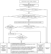
Evaluation for SBO is summarized in . The diagnosis of SBO is suspected in patients with abdominal pain, distention, nausea and vomiting.
General Approach to Evaluating SBO.
Definitions: UA = urinalysis, AKI = acute kidney injury, IBD = Irritable bowel disease, CKD = chronic kidney disease
Diagnosis is further supported by a history, including a complete discussion of prior intra-abdominal surgery.
Majority of stable patients presenting with symptoms consistent with SBO require CT imaging of the abdomen and pelvis with IV but not oral contrast to confirm the diagnosis and determine if urgent surgical intervention is warranted. Risks of intravenous contrast in patients with kidney disease or history of contrast allergy must be considered.
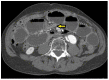
Diagnosis of SBO is highly suspected in patients with appropriate history and physical findings, AND with CT imaging demonstrating a transition zone (). Without a transition zone, SBO is unlikely.
CT Image of Transition Zone. Axial CT of the abdomen demonstrate small bowel dilatation with single abrupt transition zone in the mid abdomen (arrow) consistent with mechanical SBO secondary to adhesions.
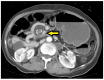
CT Image of Swirl/ Whirl Sign. Axial CT of the abdomen demonstrate swirling of the mesenteric vessels in in the mid abdomen (arrow) consistent with volvulus.
Triage ()
Patients with signs of bowel compromise (), including imaging evidence of free air in the abdomen, pneumatosis, portal venous gas, or clinical evidence of peritoneal signs, and/or signs of sepsis in the context of suspected SBO, require urgent Acute Care Surgery consultation and consideration of urgent surgery.
Patients with complete SBO, as supported by clinical findings consistent with SBO and a transition zone on CT imaging (), should be admitted to Acute Care Surgery.
Patients with pSBO as defined by a transition zone on imaging, accompanied by only mild abdominal pain, minimal distention, and the continuing passage of flatus and/or stool can be admitted to a medicine service, as pSBOs are likely to resolve without surgical intervention.
Patients presenting with partial or complete SBO within 30 days following an intra-abdominal surgery should be evaluated by and admitted to a surgery service (ideally, the original operative surgical service).
SBO patients with Inflammatory Bowel Disease (IBD) require gastroenterology and surgery consultation to guide evaluation and treatment. These patients are usually admitted to the gastroenterology service, with the surgery service providing consultation (unless emergent surgery is required).
SBO patients with advanced malignancy require consultation with surgery, and possibly oncology and/or palliative care to guide evaluation and treatment. These patients are usually admitted to a general medicine service, with the surgery service providing consultation (unless emergent surgery is required).
SBO or Partial SBO in patients with a history of Roux-en-Y gastric bypass (RYGB) or Duodenal Switch (DS) always requires emergent surgical evaluation and treatment. A small bowel obstruction in this patient population is almost always a surgical emergency that is treated with urgent or emergent operative intervention.
Treatment
SBO with bowel compromise
Usually require emergent surgical treatment.
SBO with signs of complete obstruction
Most patients are treated with non-operative management initially (), as most episodes of SBO resolve spontaneously.
Non-Operative Medical Management for SBO.
A Gastrografin challenge may be recommended by surgery (usually after 24 - 48 hours of non-operative management) to predict if the obstruction will spontaneously resolve.
Surgical management is reserved for those who fail to resolve their obstruction after Gastrografin challenge, or in those whose exam worsens or, those who develop signs of compromised bowel.
Partial SBO
Most patients are treated with non-operative management ().
Surgical consultation is warranted for any pSBO patient that does not improve after 24 - 48 hours of non-operative management.
A Gastrografin challenge may be recommended by surgery (usually after 24 - 48 hours of non-operative management) to predict if the obstruction will spontaneously resolve.
Surgical management is reserved for those that fail to resolve the obstruction after 3 - 5 days or after a Gastrografin challenge, those with a worsening exam, or those that develop signs of compromised bowel.
- *
Strength of Recommendation: I = generally should be performed; II = may be reasonable to perform; III = generally should not be performed.
Level of evidence supporting a diagnostic method or an intervention: A = Systematic review of randomized controlled trials; B = Randomized controlled trials, C = Systematic review of non-randomized controlled trials; group observation studies, D = Individual observation descriptive studies, E = Expert opinion.
Guideline Creation Process and Considerations
Related National Guidelines and Performance Measures
The University of Michigan Health System (UMHS) Clinical Guideline on Small Bowel Obstruction is generally consistent with the Bologna Guidelines for Diagnosis and Management of Adhesive Small Bowel Obstruction (ASBO).
There are no national performance measures associated with small bowel obstruction.
Funding
The development of this guideline was funded by the University of Michigan Health System.
Guideline Development Team and Disclosures
The multidisciplinary guideline development team consisted of:
The medical team: Gary Vercruysse MD, FACS, Mahmoud Al-Hawary, MD; Jill Cherry-Bukowiec, MD; Derek Dimcheff, MD, PhD; Richard J. Saad, MD, MS, FACG; David Somand, MD; Lauren Wanacata MD; Rebecca Busch MD; Luke Pumiglia, BA
Guideline development methodologists: Dave Megan Mack, MD; Dave Wesorick, MD; April Proudlock, BBA, RN
Literature search services were provided by informationists at the Taubman Health Sciences Library, University of Michigan Medical School.
The University of Michigan Health System endorses the Guidelines of the Association of American Medical Colleges and the Standards of the Accreditation Council for Continuing Medical Education that the individuals who present educational activities disclose significant relationships with commercial companies whose products or services are discussed. Disclosure of a relationship is not intended to suggest bias in the information presented, but is made to provide readers with information that might be of potential importance to their evaluation of the information.
No relevant personal financial relationships with commercial entities:
Relevant personal financial relationships with commercial entities: None.
Strategy for Literature Search
Three different Main searches were used to search this topic. The comprehensive main search on all intestinal obstruction search retrieved 5,193 references. When the search hedges for Guidelines, Clinical Trials, and Cohort Studies were added, the base results are as follow:
Bowel Obstruction -Guidelines, total results were 48
Bowel Obstruction -Clinical Trials, total results were 316
Bowel Obstruction -Cohort Studies, total results were 1,041
The retrieval was much smaller for the small bowel and large bowel searches.
Small Bowel Obstruction -Guidelines, total results were 12
Small Bowel Obstruction -Clinical Trials, total results were 62
Small Bowel Obstruction -Cohort Studies, total results were 289
Large Bowel Obstruction -Guidelines, total results were 11
Large Bowel Obstruction -Clinical Trials, total results were 115
Large Bowel Obstruction –Cohort Studies, total results were 394
The MEDLINE In-Process database was also searched using the strategy in the search strategies document. The search retrieved 100 documents. The results with the hedges applied are:
Guidelines, total results were 6
Clinical Trials, total results were 12
Cohort Studies, total results were 40
Within the Cochrane Database of Systematic Reviews, 11 reviews were found using the strategy in the search strategies document.
Level of evidence supporting a diagnostic method or an intervention:
A = systematic reviews of randomized controlled trials with or without meta-analysis,
B = randomized controlled trials,
C = systematic review of non-randomized controlled trials or observational studies, non-randomized controlled trials, group observation studies (cohort, cross-sectional, case-control),
D = individual observation studies (case study/case series),
E = expert opinion regarding benefits and harm
Search details and evidence tables available at http://www.uofmhealth.org/provider/clinical-care-guidelines.
Recommendations
Guideline recommendations were based on prospective randomized controlled trials if available, to the exclusion of other data; if RCTs were not available, observational studies were admitted to consideration. If no such data were available for a given link in the problem formulation, expert opinion was used to estimate effect size. The “strength of recommendation” for key aspects of care was determined by expert opinion.
The strength of recommendations regarding care were categorized as:
I = Generally should be performed
II = May be reasonable to perform
III = Generally should not be performed
Review and Endorsement
Drafts of this guideline were reviewed in clinical conferences and by distribution for comment within departments and divisions of the University of Michigan Medical School to which the content is most relevant: General Medicine; Department of Emergency Medicine; Department of Radiology; Department of Surgery; Gastroenterology Division. The Executive Committee for Clinical Affairs of the University of Michigan Hospitals and Health Centers endorsed the final version.
Created: April 2021.