To achieve the objectives of STI case management, high-quality care and treatment for STIs must be available to people who need such services at their first point of contact with the health-care system. Regardless of the choice an individual makes for obtaining advice and treatment, whether in the public or private sector, STI programmes should ensure that appropriate and effective comprehensive case management is available. Integrated care for STIs must be offered at as many primary health-care facilities as possible to ensure readily accessible services, reduction of stigma and promotion of the use of such facilities. This will be achieved more effectively if appropriate training in providing STI care is given to all health-care providers posted to work at primary health care facilities. Further, primary health care facilities should be appropriately equipped with relevant commodities and equipment to enable the staff to deliver high-quality care for people who need such services.
The rest of this section briefly describes the elements of case management.
3.2.1. Consultation with the person with an STI to establish the problem
To establish a correct diagnosis, the health-care provider needs to ensure that there is a conducive environment to enable people with STI symptoms to discuss them freely. This requires the following as basic minimum items:
adequate privacy for people to feel comfortable to discuss personal sexual matters;
provision of private facilities for a good clinical examination, ideally with good lighting;
an examination couch and a modesty blanket or draw sheet to cover the person in preparation for a physical examination; and
examination gloves for the health-care provider.
3.2.1.1. History-taking and risk assessment
History-taking, with emphasis on sexual history, is important in establishing an understanding of the person’s likelihood of being infected with an STI. During history-taking, the patient should be asked about the last unprotected sexual contact and whether with a regular or casual sex partner.
The importance of taking a sexual history cannot be emphasized enough in preventing STIs and managing a person suspected of having an STI, including HIV. Health-care providers should be non-judgemental in their approach to history-taking and make their patients feel comfortable to discuss personal and intimate issues about their sex life. Health-care providers need to integrate history-taking of common health risk factors together with sexual history risk factors. For example, as health-care providers ask about alcohol consumption and smoking, they should proceed at the same time to questions about sexual behaviour. Once this is done, it will all become part of general history-taking and will reduce the stigma and embarrassment associated with “talking about sex”.
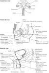
A sexual history involves talking about the genitalia. Discussing using illustrated diagrams can be helpful to health-care providers and patients alike in talking about risks of STIs in the genital area or the anorectal area ().
Common sites of infections in the female and male genital tract.
History-taking, especially personal sexual history, is important in understanding the likelihood that the person has an STI. During history-taking, the person should be asked about the last sexual contact and sexual contacts before that and their sexual practices, including penile-vaginal, penile-anal, oral sex, use of sex toys and others and whether any protection, such as a condom, was used consistently. Documenting the type of sex partner(s), whether regular, casual or sex in exchange of money or favours, is also important.
One area in which risk assessment can be useful for a man is when he presents with dysuria without urethral discharge. The risk assessment may be considered to be positive for an STI if he has had unprotected sex within the last 7–21 days, to allow for the incubation period of both N. gonorrhoeae and C. trachomatis.
Verifying whether there has been any recent self-treatment is also important as well as when he last passed urine since urination within the past hour or two may temporarily wash away the discharge.
Usually, women who present to a health-care facility with a vaginal discharge do so when they perceive it as being unusual for them (such as the quantity, thickness or smell being abnormal for them), and usually their perception is correct (30–33). The majority will have either bacterial vaginosis or T. vaginalis infections as well as candidiasis.
Thus, during history-taking, risk assessment of a woman with abnormal vaginal discharge requires a good sexual history to estimate her risk of cervical infection with N. gonorrhoeae and/or C. trachomatis. In the published literature, clinical observations that have consistently been found to be associated with cervical infection are:
the presence of cervical muco-purulent discharge;
cervical erosions or cervical friability; and
bleeding between menses or during sexual intercourse.
The risk assessment needs to consider these parameters together with some demographic and behavioural risk factors frequently associated with cervical infection, and the risk may be considered positive for STIs if the following criteria are met:
Positive responses to the risk assessment increase the likelihood that the client has an STI. In that situation, encouraging and discussing about partner treatment with the same regimen as the index client, even if it is not certain that the client has an STI, is therefore prudent, while recognizing that the commonest cause of vaginal discharge, bacterial vaginosis, is not considered to be sexually transmitted and neither is Candida albicans.
Thus, all women presenting with abnormal vaginal discharge should have a thorough medical and sexual history taken and be physically examined, ideally with a speculum, to view the cervix. However, external examination of the genitalia is better than no examination at all.
3.2.1.2. Clinical examination of people with STI-related symptoms
Once the medical and sexual history has been taken and assessment of risk of STIs duly noted, the person must be physically examined.
The person should be informed what the examination will entail and consent obtained. Any examination of the anogenital area should preferably be conducted in the presence of a chaperone. A male health-care provider must have a female chaperone in attendance, and vice versa, at all times, unless this is not feasible because of staff capacity. In that case, the person’s consent to be examined without a chaperone should be obtained.
The examination must particularly focus on the anogenital area, but a general examination must also look for other manifestations of STIs, such as lymphadenopathy, cutaneous manifestations of some STIs, such as syphilis and HIV, and abdominal abnormalities, especially for women with pelvic inflammatory disease.
Steps to follow when examining men
Wash hands before the examination and put on clean gloves with each patient.
Inform the patient what is going to take place at each step of the examination.
Ask the patient to lie down on a couch and expose the genital area from umbilicus to knee level. Where a couch is not available, the patient may be examined in a standing position, but this should be avoided as much possible. To avoid embarrassment and to show respect, the patient must be covered with a modesty blanket or a draw sheet and expose the part of the body to be examined when ready.
The examination of a man must include inspection in good light to look for rashes, sores, swellings, warts and urethral or anal discharge and general inspection including the following:
looking inside the mouth for signs of oral thrush, oral sores or other lesions;
looking at the skin over the abdomen for rashes and obvious swelling;
checking the pubic area for evidence of other STIs, such as pubic lice and nits, scabies, sores and inguinal lymph nodes;
checking for any skin rashes on the palms of the hands, soles of the feet, thighs and buttocks;
checking the external genitals – penis and scrotum – and noting any discharge and other lesions, such as ulcers and warts;
checking the area around the anus for a discharge, rashes (such as condylomata lata) and warts; and
checking the groin for swellings and sores.
Palpation must be done gently to ensure that, if there are any tender areas, they are not pressed in a way that hurts unintentionally. This will enable the health-care provider to identify the following:
palpating the inguinal region (groin), axillae, submandibular areas and neck looking for enlarged lymph nodes and buboes;
palpating the scrotum, feeling for the testis, epididymis and spermatic cord on each side, and note any signs of discomfort suggestive of tenderness;
examining the penis, noting any rashes, warts or sores;
asking the person to pull back the foreskin, if present, and looking at the glans penis and urethral meatus for discharge or any other lesions;
palpating any genital ulcers for tenderness and induration and looking for phimosis and paraphimosis; and
examining the glans penis and urethral meatus for discharge or any other lesions.
If no obvious discharge is present, the patient may be asked to milk the urethra gently from the base towards the urethral meatus to determine any discharge. The patient may then be asked to bend the knees towards the chest to expose the perineum, buttocks and anal region. If the patient is examined in the standing position, he may be asked to turn his back to you and bend over, spreading his buttocks slightly, and the anus is then examined for ulcers, warts, rashes or discharge.
At the end of the examination, the gloves are removed, and both the health-care provider and the patient must wash their hands.
All the findings must be recorded, to complement the history, including the risk assessment and the clinical findings, such as the presence or absence of ulcers, buboes, genital warts and urethral discharge. Once the syndrome is determined, the appropriate flow chart should be followed for managing the patient.
Steps to follow when examining women
A woman must be examined in good light and in privacy. It is important to inform the patient what the examination will entail. A male health-care provider must have a female chaperone in attendance at all times, unless this is not feasible because of staff capacity. In that case, the patient’s consent to be examined without a chaperone should be obtained.
Examination of a woman during menstruation is not contraindicated, and testing for STIs, such as N. gonorrhoeae and C. trachomatis can be performed if the woman gives consent. Urine samples, vaginal swabs and blood tests can all be collected for STI tests during menstruation.
Before proceeding with examination, ensure the following:
washing hands before the examination and putting on clean gloves with each patient;
asking the patient to undress to enable examination from the chest down; and
getting the patient to lie down on an examination couch in good light; a woman should not be examined standing up and should be covered with a draw sheet or a modesty blanket, exposing only the part of the body to be examined when ready.
The inspection must be general to ensure that other conditions are captured. The examination must include the following steps:
looking inside the mouth for signs of oral thrush, oral sores or other lesions;
looking at the skin over the abdomen for rashes and any obvious swellings;
checking the pubic area for evidence of other STIs, such as pubic lice and nits, scabies, sores and inguinal lymph nodes;
checking for any skin rashes on the palms of the hands and soles of the feet;
checking the thighs and buttocks for rashes;
checking the area around the anus for rashes and warts;
checking the groin for swellings and sores; and
checking the external genitalia and taking note of any discharge, or other lesions, such as warts, condylomata lata and excoriations on the vulva.
Palpation must be done gently to avoid hurting unintentionally. The abdomen must be palpated gently, watching the face for any indication of areas of tenderness and feeling for any masses and swellings, including pregnancy. In women with lower abdominal pain or vaginal discharge, the examination must focus on the pelvis to assess for signs of pelvic inflammatory disease (see section on pelvic inflammatory disease).
The general palpation by the health-care provider should include the groin, axillae, submandibular areas and neck for enlarged lymph nodes, noting whether they are painful.
Then the patient should be asked to bend her knees towards the chest and then separate them and the following should continue to be observed:
inspection of the vulva, perineum (between the vagina in front, the buttocks behind and the medial sides of the thighs on both sides), and the perianal skin for rashes, sores, warts and swelling;
inspect between the labia of the vagina and the urethral opening for any obvious lesions or discharge and any vaginal discharge;
note: the colour of the discharge, whether it is yellow, white and/or blood stained; the smell, whether a “fishy smell” can be discerned; and the type of vaginal discharge: whether it is frothy, thick or sticky;
two fingers should be inserted into the vagina and a bimanual examination carried out with one hand on the pelvic area of the abdomen and the other inside the vagina, feeling for masses and tenderness and checking for cervical motion tenderness by moving the cervix gently from side to side to elicit uterine and/or adnexal tenderness (see pelvic inflammatory disease); and
a speculum examination should be performed next to visualize the cervix and vaginal mucosa.
3.2.1.3. How to perform a speculum examination
A speculum is a medical device used to examine inside the vagina. A speculum examination is often performed alongside a bimanual examination as part of a good practice gynaecological workup, especially for women with anogenital symptoms. Metal specula must be sterilized before use, and plastic specula must not be reused.
Before a speculum examination, the patient should be informed about the device, what the health-care provider is going to do and the patient reassured that the procedure should not be painful but if the patient is uncomfortable or experiences pain, the procedure will be discontinued.
Explain that the patient needs to remove the underwear and lie on the examination couch, covering herself with the sheet provided. The patient must be provided with privacy to undress.
In preparation for performing a speculum examination, the following steps should be taken.
The patient should have an empty bladder to make the examination more comfortable;
The speculum should be properly sterilized before use.
All the secondary equipment needed should be laid out ready on a trolley, such as warm water, gloves, swabs and a waste disposal bin.
The light source should be prepared and tested before beginning the procedure;
The privacy screen, curtain or door should be closed for the examination.
The procedure should be done as follows.
Wet the speculum with clean warm water before inserting it;
Insert the first finger of the gloved hand in the opening of the woman’s vagina (some clinicians use the tip of the speculum instead of a finger for this step). As the finger is put in, it is gently pushed downward on the muscle surrounding the vagina, and then the speculum is inserted slowly while asking the woman to relax her muscles;
With the other hand, the speculum is held with the speculum blades together between the index and middle fingers and turned sideways as the speculum is slipped into the vagina, while taking care not to press on the urethra or clitoris because these areas are very sensitive.
When the speculum is halfway in, it is turned so the handle is facing downward. (Note: some examination couches do not have enough room to insert the speculum with the handle down – in this case, it is turned up)
The blades of the speculum are then gently opened a little while searching for the cervix.
The speculum is then moved around slowly and gently until the cervix can be seen between the blades – at this point the screw (or otherwise lock on the speculum) can be tightened so it will stay in place.
Now the cervix can be examined, in good light, and it should look pink, round and smooth. There may be small yellowish cysts, areas of redness around the opening of the cervix (cervical os) or a clear mucoid discharge – these are normal findings.
Look for signs of cervical infection by checking for yellowish discharge or easy bleeding when the cervix is touched with a swab and any abnormal growths or sores.
Note whether the cervical os is open or closed and whether there is any discharge or bleeding.
If there was blood in the vagina, the clinician should look for any biological tissue coming from the cervix, which could be signs of induced abortion or miscarriage.
If any specimens are to be taken, this would be the stage to perform endocervical swabs, swabs from the posterior fourchette of the vagina – as well as biopsy, if applicable.
To remove the speculum, it should first be gently pulled out until the blades are clear of the cervix. Then the blades are brought together but not completely closed to avoid pinching the vaginal wall and gently pulled out, turning the speculum gently to look at the walls of the vagina.
The patient can then be thanked and informed that the procedure has been completed and to get dressed while the patient’s privacy is observed. After that, the patient can wash her hands and be asked to sit down to receive feedback on the findings of the examination.
The health-care provider should remove the gloves before touching anything, wash hands and sit with the patient to give feedback on the examination findings.
As noted above, the next step is either to establish the diagnosis at this stage after the history-taking and examination and manage the patient syndromically using the appropriate flow chart(s) based on the examination findings or proceed to perform any additional diagnostic tests.
3.2.1.4. How to perform an anoscopy examination
An anoscope is an instrument used for visualizing the anus and lowest portion of the rectum. It is tubular and can be inserted with a lubricant into the anal canal. Once inserted, the examiner visualizes the walls of the anus and lower rectum using an appropriate light source. It can be used to identify abnormalities, such as haemorrhoids, inflammation and tumours in this part of the gastrointestinal tract.
Anoscopy can be performed within a health-care facility if sufficient training has been undertaken and equipment such as an examination couch, gloves, a light source and lubricants are available. No special preparations are needed, such as emptying the bowels or topical anaesthetic. However, some health facilities apply a topical anaesthetic 30 minutes before the procedure. Caution is needed among patients who have undergone recent anal surgery or are known to have anal fissures.
Before performing the procedure, the patient should be informed about the device, what the health-care provider is going to do and the patient informed that the procedure is painless, but pressure similar to that of a bowel movement may be felt.
In preparation for performing anoscopy, the following steps should be taken.
The anoscope should have been properly sterilized before use.
All the secondary equipment needed should be laid out ready on a trolley, such as lubricant, gloves, light source and cotton swabs, preferably with large tips.
Privacy should be assured – a privacy screen, curtain or door that can be closed for the examination.
The procedure should be carried out as follows.
Lie the patient down in the left lateral position.
Separate the buttocks or ask the patient or an assistant to help and examine the perianal area for warts, haemorrhoids or polyp prolapses.
Perform a digital rectal examination with a lubricated, gloved index finger, taking note of sphincter tone and any prostate abnormalities.
Remove the finger and change glove to a new one.
Lubricate the anoscope and insert it into the anus gently and, pointing the anoscope towards the umbilicus, advance it completely into the anus or as far as the patient can tolerate;
Remove the obturator of the anoscope to examine the anal mucosa, removing any faecal matter with a swab.
Check for blood, mucus, pus or haemorrhoidal tissue.
Gently remove the anoscope, when done, and observe the sides of the anal canal in the process.
The health-care provider should remove the gloves before touching anything, and both the provider and the patient must wash their hands before the provider sits with the patient to give feedback on the examination findings.
Any observation of suspicious growths or bleeding lesions should be referred for gynaecological assessment.
3.2.1.5. Establishing a diagnosis
Traditionally, laboratory tests have been used to address STI prevention and control to achieve the following.
to provide a definitive diagnosis, thus, allowing for cause-guided treatment;
to provide screening services for asymptomatic individuals at risk of infection;
to provide statistical information on the prevalence of various infections;
to determine the antimicrobial susceptibility of causative organisms; and
to assist in managing sex partners.
Thus, ideally, everyone presenting with a condition assumed to be an STI should be diagnosed through a process of obtaining the medical and sexual history, physical examination and laboratory testing of relevant specimens from either the lesion, blood or urine. The diagnosis could then be made through a combination of direct microscopy in syndromes with genital discharges, culture of the organisms, such as N. gonorrhoeae, serological testing as in syphilis and HIV infection and molecular detection. The health-care level at which these tests can be done varies with availability of resources, both financial and human, as well as the skills required to conduct the tests. Regardless of which system is set up for diagnosing STIs, there should be a mechanism to refer to a level at which more tests can be done, especially for patients with recurrent or persistent infections and those with unusual clinical presentations.
However, in many parts of the world, such a process is constrained by a lack of inexpensive diagnostic tests, and especially in the regions where the burden of STIs is highest and laboratories and laboratory technicians have insufficient capacity. In many instances, the appropriate reagents necessary to detect STI pathogens are not locally available and would be expensive to procure. Further, even if laboratory-based tests were available, substantial financial resources would be required for such an approach and probably unaffordable for the programme and for the patients. A further disadvantage of laboratory-based etiological diagnosis is the delay in access to treatment if treatment is withheld until the results are available.
Affordable, rapid point-of-care diagnostic tests for STIs provide a means to strengthen the diagnosis of STIs more readily. This would be such a welcome advance in the diagnosis of STIs, especially for women with vaginal discharge, a syndrome commonly labelled as an STI syndrome but that neither indicates nor predicts gonococcal or chlamydial cervical infections among women. Genital ulcers would also benefit enormously from a rapid point-of-care test, since recent studies indicate that most genital ulcers are caused by viral infections, especially HSV-2. The rapid diagnostic tests for gonococcal and chlamydial infections currently commercially in circulation are of poor sensitivity and specificity and expensive. Their use would negatively affect the reliability of laboratory testing for STI diagnosis. However, rapid diagnostic tests for syphilis (treponemal test) are available and cheap and allow for a same-day “screen and treat” approach. Dual HIV and syphilis rapid tests are also available and provide an opportunity for increasing access to HIV and syphilis testing.
In the absence of diagnostic tests, a syndrome-based approach to managing people with STIs has been developed and adopted in many countries. The approach is more rational and scientific than a clinical approach in which a health-care provider reaches a diagnosis based on the clinical appearance of the lesion or the nature of a genital discharge. Several studies have shown that clinical judgement based on the experience and appearance of an ulcer, for example, has poor sensitivity in the diagnosis (34–36).
The syndromic management approach is based on identifying consistent groups of symptoms and easily recognized signs (syndromes) and providing treatment that will take care of most of or the most serious organisms responsible for producing the syndrome. By giving treatment for the most common causative pathogens for a particular syndrome, the syndromic approach, generally, has high sensitivity at the expense of specificity, thus resulting in overtreatment. WHO developed simplified flow charts to guide health-care workers in implementing the syndromic management of STIs. These WHO flow charts have been designed to be adapted at the local level, using locally available data and information.
3.2.1.6. Health education and counselling
People seeking care for STIs are especially worried about the condition and are more receptive to education messages than at other times. This is probably because they are aware of their vulnerability when they face an infection. Health-care providers should take advantage of this time to educate their patients about STIs, including HIV, and how they are transmitted and acquired. Further, the counselling can help them to assess their own risk and take responsibility to reduce the risk, if feasible, or change sexual behaviour and start using preventive interventions, such as the male and female condoms.
Health education is the provision of accurate and evidence-informed information about STIs so that a person becomes knowledgeable about the subject and can make informed choice.
Counselling is a two-way interaction between patients and provider intended to help the patients to understand themselves better in their feelings, attitudes, values and beliefs and to empower them to execute changes for healthy living in their life.
The key messages to give during an encounter with a person seeking care for STIs is how the infection may have been contracted, how to prevent future infections and the importance of completing a course of treatment and abstaining from further sexual intercourse until treatment has been completed and the infection has been controlled or cured. This should be emphasized to patients. However, patients should also be strongly advised to use condoms if abstinence from sex is not possible.
During the encounter with the patients, screening for other infections should be offered, especially for HIV infection and syphilis, both of which have rapid diagnostic tests currently available.
Health education and counselling are covered in other relevant WHO publications, such as Brief sexuality-related communication: recommendations for a public health approach (37).
3.2.1.7. Partner notification and treatment
A person with STIs has contracted the infection from a sex partner who also had the infection. Equally, from the time that the attending (index) patient was infected, he or she has also been infectious – able to transmit the STI to other sex partners or the same partner (the source of the infection) who, in the meantime, may have been treated. Thus, the chain of transmission of the STI can be broken only if all the mutual sex partners are treated for the infections before they have further sex with each other.
Many STIs, such as gonococcal infection, chlamydial infections, syphilis and HIV, are asymptomatic, and people may not be aware that they are infected. Thus, partner notification can be one way to detect and treat asymptomatic individuals.
Some reproductive tract infections are not sexually transmitted, such as the bacteria responsible for bacterial vaginosis among women with vaginal discharge. Although C. albicans can be sexually transmitted, it is not classified as an STI. The sex partners of people with candidiasis do not need treatment unless they exhibit symptoms. Partner notification therefore needs to be approached with caution for women with vaginal discharge since they may not have a sexually transmitted pathogen. This is one reason affordable, rapid diagnostic tests to screen for STIs in such situations and guide appropriate partner notification and treatment are so urgently needed.
There are several approaches to partner notification for STIs. The patient can be issued with a contact-tracing card to give to the sex partner(s) to invite them to attend for an assessment for STIs and be treated accordingly (patient referral partner notification). The other method is for the health-care provider to obtain contact details from the index patient and then to attempt to contact the sex partners (provider referral partner notification).
Other methods of partner notification and treatment are variations of these two in which the index case may be given a prescription or medicines to give to their sex partners without the health-care provider having the opportunity to examine the sex partner (expedited partner therapy). Another method, sometimes referred to as contractual partner referral, is agreement between the service provider and the index patient that the latter will reach the sex partner(s) within an agreed time frame, after which the health-care provider will then try to contact the sex partner if the agreement period has elapsed without the sex partners presenting for examination and treatment.
Regardless of which method of partner notification and treatment is followed, confidentiality, non-judgemental attitudes and absence of coercion must be observed. Health education and counselling are important to equip people with STIs to embark on informing their sex partners about their STI.
3.2.1.8. Follow-up and referral for people with STIs
WHO encourages that people diagnosed with STIs be provided immediate treatment and, if any diagnostic tests are to be carried out, that they do not delay the provision of treatment. This would ensure an immediate break in the chain of transmission and prevent STI-related complications and long-term sequelae of STIs.
Giving treatment during the same visit reduces infectiousness and onward transmission, even more so if single-dose therapies are available.
If effective medicines are given and any test results are available on the same visit, then follow-up may be restricted only to those with persistent symptoms after a stipulated period. This will reduce costs for both the patient and the health-care system. Treatment given on the same visit is especially relevant in settings in which patient return rates are inconsistent for several reasons, such as distances to the clinic, user fees, transport fees, user-friendliness of health services and attitudes of health-care providers.
Follow-up may be specifically requested in certain conditions, such as a woman being treated for acute pelvic inflammatory disease as an outpatient or a neonate with ophthalmia neonatorum to ensure that the treatment has been effective, since delays in cure may result in severe consequences such as loss of eyesight.
The patient may return for further assessment either because the condition has not resolved when treatment ends or it has recurred. The health-care provider will need to determine whether this resulted from poor compliance, which is unlikely if single-dose therapy was given and taken on the same day as the patient attended the clinic or the patient has a persistent infection because of antimicrobial resistance or has been reinfected.
Depending on the assessment, health-care providers have the following options for treatment:
poor compliance – such as a patient taking a 7-day or 21-day course of doxycycline for chlamydial infections, including for lymphogranuloma venereum;
reinfection – perhaps because sex took place, without a condom, with an untreated sex partner or a new partner;
antimicrobial resistance – this is of particular importance in gonococcal and M. genitalium infections since antimicrobial resistance in N. gonorrhoeae and M. genitalium are being experienced with recommended treatments for these infections; and
the presence of an untreated infection – such as T. vaginalis and/or M. genitalium among men with urethral discharge treated only for N. gonorrhoeae and C. trachomatis at the first visit.
The health-care provider should assess the most likely scenario for the individual and treat appropriately.
Sometimes the patient needs to be referred to another level of care. The health-care provider at the first point of care should then determine whether the referral should be made to clinicians who have extensive specialized training or experience in diagnosing, treating and following up complex STI cases or to a facility with laboratory-based tests to exclude antimicrobial resistance or to any other specialist centre. For example, someone with an abnormality in the anorectal area may need to be referred to a colorectal surgeon or oncologist and someone with a testicular problem to a urologist.
In any specific country or setting, providers of care for people with STIs need to have information on referral channels at their disposal for complex genitourinary symptoms they feel unable to handle.


