From: JAK/STAT Pathway Signalling in Drosophila Melanogaster

NCBI Bookshelf. A service of the National Library of Medicine, National Institutes of Health.

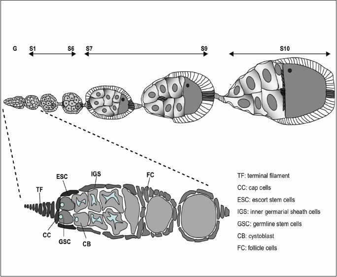
A) Diagram of adult ovariole showing the stages of oogenesis starting from the anterior germarium to stage 10 with the developing egg at the posterior end of the egg chamber. From stage 7 the anterior polar cells (dark grey) recruit a small cohort of surrounding follicle cells and begin to migrate between the nurse cells towards the egg. By stage 10 these migrating border cells have arrived at the anterior face of the oocyte where they spread out to encapsulate the future egg. B) A more detailed view of the germarium showing the terminal filament (TF) and the cap cells (CC) which form the stem cell niche maintaining the germline stem cells (GSC). GSC can be recognized by rounded spectrosomes. Division of the GSC produced another stem cell plus a daughter which is committed to form the cystoblasts (CB) that move posteriorly and is surrounded by the inner germarial sheath cells (IGS). At the anterior of the IGS lie the escort stem cells. As the CB divides forming a cyst, it is encompassed by follicle cells (FC).
From: JAK/STAT Pathway Signalling in Drosophila Melanogaster

NCBI Bookshelf. A service of the National Library of Medicine, National Institutes of Health.