From: Vascular Endothelial Growth Factor

NCBI Bookshelf. A service of the National Library of Medicine, National Institutes of Health.

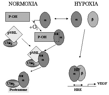
Model of oxygen sensing, signaling and gene regulation. Under hypoxic conditions, HIF-1α (α) is stable and can translocate to the nucleus where it heterodimerizes with HIF-1β/ARNT (β) and after binding to hypoxia-response elements (HRE), activates gene transcription, of e.g., VEGF. When, however, the intracellular oxygen tension is high, a prolyl hydroxylase enzyme (P-OH) modifies the HIF-1α subunits resulting in the hydroxylation of a specific proline residue (OH). This modification is sufficient for binding of HIF-1α to the von Hippel-Lindau tumor suppressor protein (pVHL) which then targets the α subunit for proteolytic degradation in the proteasome via ubiquitination (ubiq.). (Adapted from refs. 53,82).
From: Vascular Endothelial Growth Factor

NCBI Bookshelf. A service of the National Library of Medicine, National Institutes of Health.