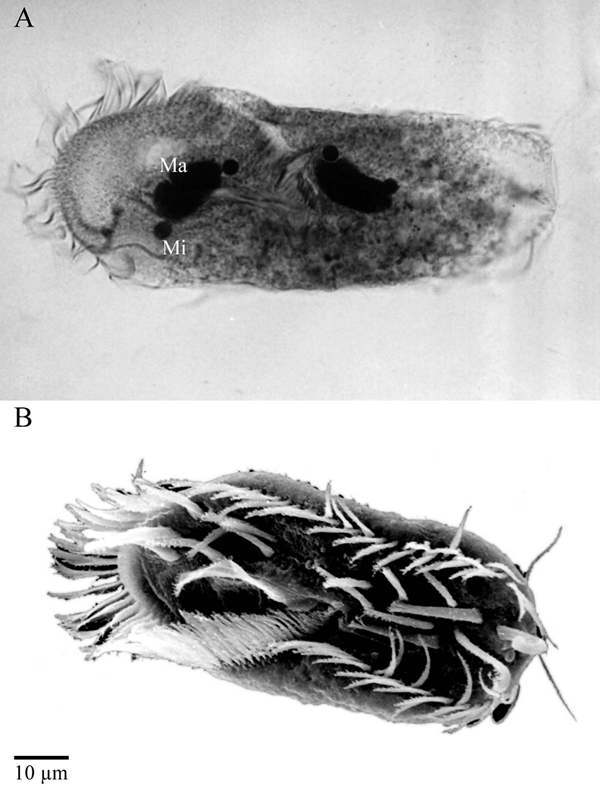
Figure 1. Morphology of the hypotrichous ciliate Stylonychia lemnae.

Figure 1

Morphology of the hypotrichous ciliate Stylonychia lemnae.(A) Light microscopic photograph of S. lemnae showing the large macronuclei and the diploid micronuclei (kindly provided by D. Ammermann). (B) Scanning electron micrograph of S. lemnae (kindly provided by M. Schlegel)

From: The Biology of Telomeres in Hypotrichous Ciliates

Cover of Madame Curie Bioscience Database
Madame Curie Bioscience Database [Internet].
Austin (TX): Landes Bioscience; 2000-2013.
Copyright © 2000-2013, Landes Bioscience.

NCBI Bookshelf. A service of the National Library of Medicine, National Institutes of Health.