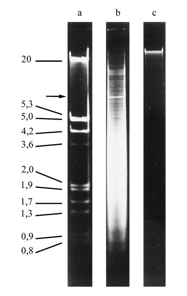
Figure 3. Agarose gel electrophoresis of chromosomal DNA.

Figure 3

Agarose gel electrophoresis of chromosomal DNA. (a) Size markers. (b) The typical low molecular weight DNA banding pattern of macronuclear DNA. The arrow points to the gene-sized DNA molecule encoding ribosomal RNAs. (c) high molecular weight micronuclear DNA.

From: The Biology of Telomeres in Hypotrichous Ciliates

Cover of Madame Curie Bioscience Database
Madame Curie Bioscience Database [Internet].
Austin (TX): Landes Bioscience; 2000-2013.
Copyright © 2000-2013, Landes Bioscience.

NCBI Bookshelf. A service of the National Library of Medicine, National Institutes of Health.