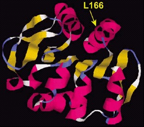
Figure 3. Structural model of the DJ-1 protein.

Figure 3

Structural model of the DJ-1 protein. The dark and light ribbons indicate regions with α-helix and β-sheet structure, respectively. The position of the amino acid mutated in one PD family (L166) is shown.

From: Chromosome 1 and Other Hotspots for Parkinson's Disease Genes

Cover of Madame Curie Bioscience Database
Madame Curie Bioscience Database [Internet].
Austin (TX): Landes Bioscience; 2000-2013.
Copyright © 2000-2013, Landes Bioscience.

NCBI Bookshelf. A service of the National Library of Medicine, National Institutes of Health.