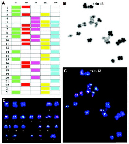
Figure 6. A) Centromere-specific multicolor-FISH probes used in cenM-FISH.

Figure 6

A) Centromere-specific multicolor-FISH probes used in cenM-FISH. B) MII oocyte chromosome complement seen by phase-contrast. C) The same complement after cenM-FISH treatment seen by fluorescent microscopy. D) Karyotype of the MII oocyte chromosome complement by cenM-FISH showing a gain of chromatid 13.

From: Preimplantation Genetic Diagnosis (PGD): Screening for Aneuploidy in Human Oocytes and Polar Bodies

Cover of Madame Curie Bioscience Database
Madame Curie Bioscience Database [Internet].
Austin (TX): Landes Bioscience; 2000-2013.
Copyright © 2000-2013, Landes Bioscience.

NCBI Bookshelf. A service of the National Library of Medicine, National Institutes of Health.