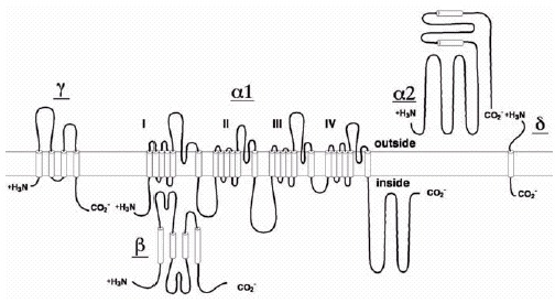
Figure 2. Structures of the Subunits of CaV Channels.

Figure 2

Structures of the Subunits of CaV Channels. Transmembrane folding models and site of regulation of the Ca2+ channel subunits. Predicted alpha helices are depicted as cylinders. The lengths of lines correspond approximately to the lengths of the polypeptide segments represented.

From: Biochemical Studies of Voltage-Gated Ca2+ Channels

Cover of Madame Curie Bioscience Database
Madame Curie Bioscience Database [Internet].
Austin (TX): Landes Bioscience; 2000-2013.
Copyright © 2000-2013, Landes Bioscience.

NCBI Bookshelf. A service of the National Library of Medicine, National Institutes of Health.