From: Centrins, a Novel Group of Ca2+-Binding Proteins in Vertebrate Photoreceptor Cells

NCBI Bookshelf. A service of the National Library of Medicine, National Institutes of Health.

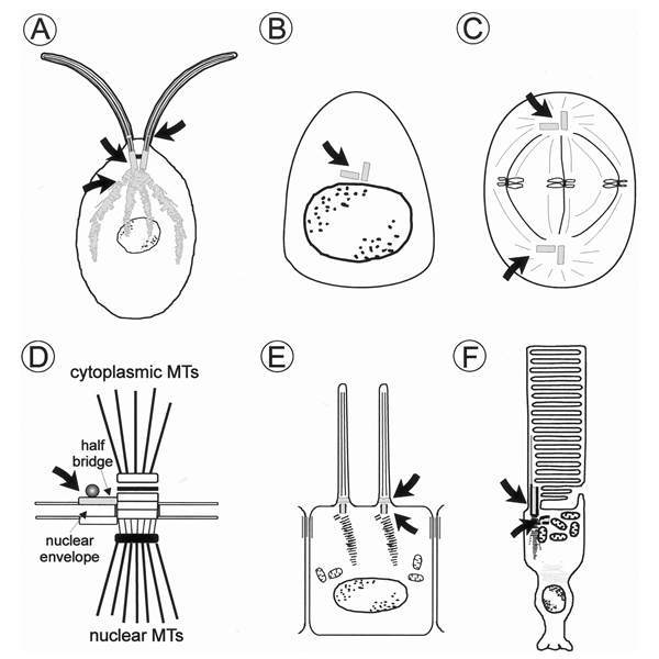
Localization of centrin in diverse cell types. Schematic diagrams of A unicellular green algae (e.g., Chlamydomonas reinhardtii); B animal cell in G1 or G0 phase (e.g., retinal nonphotoreceptor cells, cells of the retinal pigment epithelium) and C in metaphase; D spindle pole body of the yeast Saccharomyces cerevisiae, MTs = microtubules; E ciliated epidermal cell; F vertebrate photoreceptor cell. Centrin cellular localization is coloured and indicated by arrows. In the yeast, cdc31p (yeast centrin) is associated with the half bridge of the spindle pole body which accts as the major microtubule organizing centre (MTOC). Centrin is also commonly found at the MTOC, the centrosome of animal cells and at the centrosome-related basal bodies of ciliated cells. In cilia, centrin is also a component of the transition zone which links the basal body region with the axoneme.
From: Centrins, a Novel Group of Ca2+-Binding Proteins in Vertebrate Photoreceptor Cells

NCBI Bookshelf. A service of the National Library of Medicine, National Institutes of Health.