From: Centrins, a Novel Group of Ca2+-Binding Proteins in Vertebrate Photoreceptor Cells

NCBI Bookshelf. A service of the National Library of Medicine, National Institutes of Health.

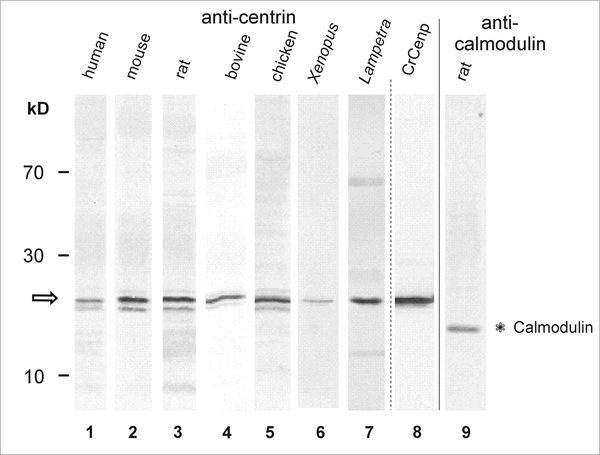
Western blot analysis reveals centrin expression in retina of various vertebrate species. Lane 1-Lane 8: Anti-centrin (mAb clone 20H5) Western blots. Lane 9: Anti-calmodulin Western blot of rat retina. Lane 1: human retina. Lane 2: mouse retina. Lane 3: rat retina. Lane 4: bovine retina. Lane 5: chicken retina. Lane 6: Xenopus retina. Lane 7: Lampetra retina. Lane 8: Bacterially expressed Chlamydomonas centrin. Anti-centrin antibodies detect bands at about the predicted molecular weight of 20 kDa (arrow) and do not crossreact with the calmodulin migrating at 17 kDa. Note: in some lanes (e.g., lane 5) several bands around 20 kDa are anti-centrin positive. These bands do neither represent different centrin isoforms nor different Ca2+-binding status of centrin. The higher bands most probably resemble phosphorylated centrin,43,44 and some lower bands may result from proteolytic digestion.
From: Centrins, a Novel Group of Ca2+-Binding Proteins in Vertebrate Photoreceptor Cells

NCBI Bookshelf. A service of the National Library of Medicine, National Institutes of Health.