From: Centrins, a Novel Group of Ca2+-Binding Proteins in Vertebrate Photoreceptor Cells

NCBI Bookshelf. A service of the National Library of Medicine, National Institutes of Health.

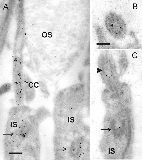
Immunoelectron microscopic localization of centrin in the connecting cilium of rod photoreceptor cells. A Silver-enhanced immunogold labeling of centrin in a longitudinal section of parts of rat rod photoreceptor cell. Centrin labeling is exclusively localized in the connecting cilium (CC) and the basal body complex (arrow) in the inner segment (IS) of photoreceptors. B Transversal section through the connecting cilium reveals that centrin is localized in the subciliary domain of the ciliary lumen encircled by axonemal microtubule doublets. C Slightly tangential section through the apical part of rat rod photoreceptor cell inner segment. Centrin antibodies react in the connecting cilium at the inner surface of the axonemal microtubule doublets (arrowhead). The arrow indicates basal body labeling.
Bars: A: 265 nm, B, C: 175 nm
From: Centrins, a Novel Group of Ca2+-Binding Proteins in Vertebrate Photoreceptor Cells

NCBI Bookshelf. A service of the National Library of Medicine, National Institutes of Health.