NCBI Bookshelf. A service of the National Library of Medicine, National Institutes of Health.
Chapell R, Turkelson CM, Coates VH, et al. Diagnosis and Treatment of Worker-Related Musculoskeletal Disorders of the Upper Extremity. Rockville (MD): Agency for Healthcare Research and Quality (US); 2002 Dec. (Evidence Reports/Technology Assessments, No. 62.)
This publication is provided for historical reference only and the information may be out of date.

Diagnosis and Treatment of Worker-Related Musculoskeletal Disorders of the Upper Extremity.
Show detailsScope and Objectives
Worker-related upper-extremity disorders (WRUEDs) result in pain, disability, and loss of productivity. This report is a systematic analysis of the evidence pertaining to thirteen key questions and four specific disorders. These disorders are considered worker-related not because they are necessarily caused by working, but because they effect workers.
Conditions of Interest
Although a wide variety of WRUEDs have been described in the medical literature, this report is limited to four. They are:
- Carpal tunnel syndrome
- Cubital tunnel syndrome
- Epicondylitis
- De Quervain's disease
Key Questions
This report addresses 13 questions regarding worker-related disorders of the upper extremity. Eleven of these are condition specific. Therefore, we individually address them for each of the disorders we consider. Questions 12 and 13 are not condition-specific. Therefore, they are answered only once. The questions we address are:
Condition-Specific Questions:
Question #1: What are the most effective methods and approaches for the early identification and diagnosis of worker-related musculoskeletal disorders of the upper extremity?
Question #2: What are the specific indications for surgery for worker-related musculoskeletal disorders of the upper extremity?
Question #3: What are the relative benefits and harms of various surgical and nonsurgical interventions for persons with worker-related musculoskeletal disorders of the upper extremity?
Question #4: Is there a relationship between specific clinical findings and specific treatment outcomes among patients with worker-related musculoskeletal disorders of the upper extremity?
Question #5: Is there a relationship between duration of symptoms and specific treatment outcomes among patients with worker-related musculoskeletal disorders of the upper extremity?
Question #6: Is there a relationship between factors such as patients' age, gender, socioeconomic status and/or racial or ethnic grouping and specific treatment outcomes among patients with worker-related musculoskeletal disorders of the upper extremity?
Question #7: What are the surgical and nonsurgical costs or charges for treatment of worker-related musculoskeletal disorders of the upper extremity?
Question #8: For persons who have had surgery for worker-related musculoskeletal disorders of the upper extremity, what are the most effective methods for preventing the recurrence of symptoms, and how does this vary depending on subject characteristics or other underlying health problems?
Question #9: What instruments, if any, can accurately assess functional limitations in an individual with a worker-related disorder of the upper extremity?
Question #10: What are the functional limitations for an individual with a worker-related musculoskeletal disorder of the upper extremity before treatment?
Question #11: What are the functional limitations of an individual with a worker-related musculoskeletal disorder of the upper extremity after treatment?
Non-Condition-Specific Questions:
Question #12: What are the cumulative effects on functional abilities among individuals with more than one worker-related musculoskeletal disorder of the upper extremity in the same limb?
Question #13: What level of function can patients achieved in what period of time when they are required to change hand dominance as a result of injury to their dominant hand?
Worker-Related Upper-Extremity Disorders
Carpal Tunnel Syndrome
Carpal tunnel syndrome (CTS) results from compression of the median nerve as it passes through the carpal tunnel from the wrist to the hand. This leads to progressive sensory and motor disturbances.
Signs and Symptoms
Symptoms of CTS include paresthesia (tingling), anesthesia (numbness), diminished or altered sensation (hypoesthesia or dysesthesia) in the affected area of the hand; pain in the hand and arm, and/or the impairment of motor function, particularly of the abilities to grip and grasp.2 Usually the symptoms appear first (and worst) at nighttime.3 In about 1% of cases, permanent nerve damage results, resulting in impaired use of the hands.4 Continued denervation can lead to atrophy of the innervated muscle.5
Anatomy
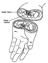
The median nerve is a mixed sensory and motor nerve that supplies the thumb, all of the index and middle fingers, and part of the ring finger. It enters the hand on the palmar side of the wrist, through a narrow, rigid, osteoligamentous passageway (the carpal tunnel, see Figure 1) that is bordered on three sides by the carpal bones and on the other by the flexor retinaculum (or transverse carpal ligament). The median nerve shares the carpal tunnel with nine flexor tendons that displace the nerve to the superficial (palm-most) side of the tunnel, directly against the transverse carpal ligament (See figure 2). The nerve is the softest and most sensitive element in the tunnel. Anything that decreases the size of the tunnel or increases the size of its contents can cause CTS. This may include space-occupying lesions, arthritis, trauma, edema, and dislocation of the lunate bone.

Figure
Figure 1. Location of the carpal tunnel.

Figure
Figure 2. Structures associated with carpal tunnel syndrome.
Etiology
Carpal tunnel syndrome is often idiopathic. The most common attributed cause of CTS is tenosynovitis or hypertrophy of the tendon sheaths of the finger flexor tendons due to overuse, often from the repetitive hand motions associated with certain occupations.6, 7 Assemblers, cashiers, and secretaries are among those most prone to the disease, with data-entry keyers, typists, and office clerks also at high risk.4 It is not clear, however, whether occupational activities cause or merely contribute to development of CTS.8 Female sex, middle age, diabetes, alcoholism, hypothyroidism, obesity, pregnancy, menopause, and the use of birth control pills are all associated with CTS.9
CTS is associated with several conditions. Rheumatoid involvement in the wrist joint may lead to carpal tunnel compression.3 Bone growth due to acromegaly may lead to shrinking of the carpal tunnel and median nerve compression.10 Patients receiving hemodialysis may develop CTS because of edema or amyloid deposits in the carpal tunnel.7, 11 Tissue deposits due to gout may also cause or exacerbate CTS.12
Carpal tunnel syndrome may be exacerbated by other nerve injuries, such as at the neck, shoulder, elbow, or by generalized peripheral neuropathies. This phenomenon, known as double-crush syndrome,13 has not been definitively established to exist, and remains controversial.14 Comorbidities causing peripheral neuropathy such as diabetes or thyroid disturbances may both exacerbate CTS and interfere with its diagnosis.15–18 CTS associated with pregnancy, childbirth and lactation may resolve spontaneously.19
Epidemiology
The overall prevalence of CTS in the United States may be as high as 1.9 million people, and each year there are 300,000–500,000 operations for the condition, at a total cost of more than $2 billion.20 There are no widely accepted figures for the fraction of cases requiring surgery. Estimates range from nearly half of all CTS patients with occupational disease to a “small percentage” of all patients.20
The incidence of CTS is higher in women than in men, and differences in carpal tunnel volume between men and women may contribute to these differences.21 Idiopathic CTS occurs in women three to five times more frequently than in men.22 Many of the occupations associated with CTS are held disproportionately by women, and several of the causal medical conditions are found more often in women than in men.20 In addition, the prevalence for men generally increases steadily with increasing age while, for women, the prevalence peaks dramatically during middle age (45–55 years of age) and then levels off.23, 24
About 60% of cases are seen in patients between 40 and 60 years of age.25 Whites have been reported to have a 1.8 times higher prevalence of carpal tunnel syndrome than do non-whites.26
The U.S. Bureau of Labor Statistics reported 29,937 cases of CTS that resulted in work days lost in 1996, and the National Institute of Occupational Safety and Health (NIOSH) reported that, in 1993, CTS occurred at a rate of 5.2 per 10,000 full-time workers. This syndrome required the longest recuperation period of all conditions that result in lost work days, with a median of 30 work days lost.4 A study of all surgeries performed to treat carpal tunnel syndrome in Wisconsin from July 1990 to March 1993 found that 75% of the individuals had only one surgery, 24.7% had two surgeries, and 0.3% had three or more surgeries. Workers' Compensation paid for 26.1% of these surgeries.23
Diagnosis
Diagnosis of carpal tunnel syndrome is complicated by the fact that there is no “gold standard” method for verifying its presence or absence.27 A variety of diagnostic instruments have been used by investigators including clinical signs, sensory tests, nerve conduction studies, and imaging tests. It is not known which modality or combination of modalities are optimal for the diagnosis of carpal tunnel syndrome.
Most clinical tests to diagnose carpal tunnel syndrome involve specific maneuvers that elicit pain, numbness, or tingling in the median-nerve portion of the wrist. For example, in Phalen's test, the patient places both elbows on a horizontal surface with the forearms vertical, and allows the wrists to flex by gravity. If the patient feels numbness or tingling within one minute, the test is positive.28 In Tinel's test, the examiner taps lightly on the palmar aspect of the wrist, over the carpal tunnel. If the patient feels tingling, the test is positive.29
Sensory tests for carpal tunnel syndrome typically involve measurement of a patient's threshold for detection of a sensory stimulus. For example, in the Semmes-Weinstein test, the examiner touches the patient with monofilaments, and the test is positive if the patient's sensitivity to the monofilaments falls outside normal limits.30 Another example is the two-point discrimination test in which the examiner touches two closely-spaced prongs to the patient's fingers. The test is positive if the patient cannot discriminate the prongs when they are 5 millimeters apart.31
Nerve conduction tests are also used to diagnose CTS. In such tests, electrodes are placed in two locations along a nerve; the nerve is stimulated from one electrode, and the impulse is recorded from the other electrode. Tests can be performed on either the median nerve, ulnar nerve, or radial nerve, and can assess either motor or sensory function. The placement of electrodes in sensory nerve conduction tests can be either orthodromic (in which stimulating electrodes are placed distal to recording electrodes) or antidromic (in which stimulating electrodes are placed proximal to recording electrodes). Other aspects of the nerve impulse can also be measured such as latency, amplitude, and velocity. Some investigators compare two or more nerve conduction tests in an attempt to assist the diagnosis of carpal tunnel syndrome (e.g., compute a difference between two latencies). We refer to these comparisons as composite nerve conduction tests.
Imaging tests for carpal tunnel syndrome include magnetic resonance imaging (MRI), computed tomography (CT), scan x-ray film, and ultrasound. Using these methods, investigators attempt to measure the size of anatomical areas within the carpal tunnel or other areas that may be affected by carpal tunnel syndrome.
Treatment
Conservative treatment
Nonsurgical interventions that have been used to treat CTS include wrist splints, avoidance of precipitating activities, anti-inflammatory drugs, vitamin B6, diuretics, ultrasound, injection of anti-inflammatory steroids and physical therapy.17, 32–36 Treatment of comorbid conditions contributing to CTS may also be effective.37, 38
Surgical treatment
The standard surgery for CTS is the transection of the transverse carpal ligament.39 This transection may be accomplished by endoscopic or open surgery. For virtually all patients it is an outpatient procedure performed in an ambulatory surgical center under regional anesthesia, but a few patients request general anesthesia. A variety of endoscopic techniques have been reported.40–46 Variations in technique include the specific types of equipment used and whether the technique requires one or two incisions. No published evidence is available quantifying the relative advantages and disadvantages of the various methods.
Additional procedures, such as ligament repair or neural surgery may also be used. Ligament reconstruction involves the reattachment of the transected ends of the transverse carpal ligament in such a way that the overall ligament is lengthened. This results in an enlargement of the carpal tunnel and relief of the pressure on the median nerve.47–49
Neural surgery for CTS (external or internal neurolysis or epineurotomy) is generally performed immediately following the division of the transverse carpal ligament. The term “neurolysis” is used to encompass several different procedures.50 These include removal of adhesions from the connective tissue surrounding the nerve (the epineurium), relieving pressure within the epineurium by means of a longditudinal incision, or removal of a segment of epineurium. There is confusion due to the nonstandard usage of terms, compounded by the different subspecialties and nationalities of surgeons. The common goal in all techniques is to remove adhesions and scar tissue to decompress the nerve and allow it to glide freely.
Cubital Tunnel Syndrome
Patients with cubital tunnel syndrome are affected by a weak grip, lack of hand coordination, hand clumsiness, and numbness, paresthesia, and pain in the hand, particularly in the fourth and fifth digits. These symptoms are thought to be caused by compression of the ulnar nerve at multiple sites in the area of the elbow, where the nerve passes through an anatomically restricted area called the cubital tunnel.
Signs and symptoms
Patients presenting with cubital tunnel syndrome usually complain of a weak grip, hand clumsiness and lack of coordination, and dropping of objects. Numbness and paresthesia in the fourth and fifth digits may also be present, in particular after prolonged flexion of the elbow.51 Pain in the hand may be present, but is neither as severe or as common as in carpal tunnel syndrome.52 The medial aspect of the elbow may be painful.53 Severe cases may present with atrophy of the intrinsic muscles and clawing of the fourth and fifth fingers.51
Diagnosis
Upon examination, patients with cubital tunnel syndrome are positive for Tinel's sign (tingling in the fingers after tapping over the ulnar nerve at the elbow), and the ulnar nerve may feel swollen and hard upon palpation.52 In addition, patients have diminished sensation in the fourth and fifth digits (pin-prick or Semmes-Weinstein monofilament testing), weak intrinsic hand muscles, a progressive inability to separate the fingers, and a loss of power grip and dexterity.53 Patients with more advanced cases may exhibit a positive Wartenberg's sign (upon extension of the fingers abduction of the fifth digit occurs) and/or a positive Froment's sign (patient cannot pinch between the index finger and thumb without flexion of the distal phalanx of the thumb).53
Electrodiagnostic tests can be used to confirm a lesion of the ulnar nerve, and to help locate the exact site of compression. Two examples of such tests are motor and sensory conduction velocities across the elbow.54, 55 For motor conduction velocity, stimulating electrodes are placed above and below the elbow, and a recording electrode is placed on the abductor digit minimi (a muscle in the hand that is innervated by the ulnar nerve).54 The measured latencies, along with the measured distances between stimulating and recording electrodes, are used to compute the motor conduction velocity in the across-elbow portion.54 For sensory conduction velocity, the ulnar nerve can be stimulated below the elbow and recorded above the elbow (this placement of electrodes is termed orthodromic because the stimulating electrode is distal to the recording electrode).54 Alternatively, the electrodes can be reversed to yield an antidromic sensory measurement.55 Regardless of whether orthodromic or antidromic placement is employed, the latencies and distances are used to calculate the sensory conduction velocity across the elbow.54, 55
Cubital tunnel syndrome can be confused with compression of nerves at other points. Cervical root lesions, such as compression of the eighth cervical root by a bulging disc, may produce symptoms similar to that of cubital tunnel syndrome.56 Other nerve compression disorders that may produce symptoms similar to that of cubital tunnel syndrome included compression of the medial components of the brachial plexus (thoracic outlet syndrome), compression of the ulnar nerve at the wrist in Guyon's canal (ulnar tunnel syndrome), and compression of the ulnar nerve at more than one point.56
Anatomy
The ulnar nerve carries nerve fibers from the eighth cervical and first thoracic nerves. It passes down the upper arm medial to the brachial artery, then passes through the intermuscular septum and travels towards the elbow near the medial head of the triceps. At the elbow, the ulnar nerve passes behind the medial epicondyle of the humerus in a groove between it and the heads of the flexor carpi ulnaris, the cubital tunnel. The ulnar nerve then enters the forearm between the two heads of the flexor carpi ulnaris muscle and enters the hand.57–59 It is not until the ulnar nerve passes between the two heads of the flexor carpi ulnaris muscle that it begins supplying motor and sensory innervation. It supplies motor innervation to the muscles of the forearm and hand, and sensory innervation to the medial half of the hand, the palm, and the fourth and fifth digits.57
The groove that the ulnar nerve passes through at the elbow is referred to as the cubital tunnel. This tunnel is bounded by the medial epicondyle of the humerus anteriorly (See Figure 3), the ulnohumeral ligament laterally, and posteromedially, a fibrous arcade of fascial strands that extends from the olecranon to the medial epicondyle, bridging the two heads of the flexor carpi ulnaris muscle.57, 58 Under normal conditions, the capacity of the ulnar tunnel is greatest during elbow extension. Flexion of the elbow decreases the volume of the cubital tunnel by tightening the arcuate ligament, bulging of the medial elbow ligament, and contraction of the flexor carpi ulnaris muscle.58

Figure
Figure 3. The cubital tunnel and associated structures.
Inside the cubital tunnel, the motor fibers to the flexor carpi ulnaris and flexor digitorum profundus are located deep inside the ulnar nerve, while the motor fibers to the hand muscles and sensory fibers to the fingers are located more superficially. This peripheral location places these fibers to the hand at increased risk of damage from compression, and accounts for their early involvement in the development of cubital tunnel syndrome.56
Etiology
Cubital tunnel syndrome is caused by compression of the ulnar nerve within or near the cubital tunnel. The site of entrapment of the ulnar nerve in the region of the elbow can occasionally occur in locations other than the cubital tunnel, including proximal to the elbow by the medial head of the triceps (the arcade of Struthers), at the elbow by the arcuate ligament, or in the mid-forearm by the flexor carpi ulnaris muscle.53 Chronic reduction in volume of the cubital tunnel results in compression damage and focal ischemia of the nerve. Compression of the ulnar nerve within the cubital tunnel is most often due to constriction of the nerve by the overlying fibrous arcade. Compression can be caused by repetitive trauma, inflammation, idiopathic thickening of Osborne's band, arthritis, hematomas, tumors, bone fragments, and idiopathic persistent epitrochleoanconeus muscle.57, 59 Fractures, dislocations, and direct blunt trauma near the elbow can cause acute compression of the ulnar nerve.59 Cubital tunnel syndrome can be precipitated by general anesthesia, and is thought to be related to compression of the ulnar nerve caused by poor limb positioning, tourniquets, and/or blood pressure cuffs.58, 59 Systemic diseases such as diabetes, kidney disease, amyloidosis, acromegaly, alcoholism, hemophilia, and leprosy can contribute to the development of cubital tunnel syndrome.58
In many patients, no precipitating event can be identified. Compression of the ulnar nerve can be the end result of a pathological cycle of chronic irritation of the nerve. Mild irritation of the nerve can causeinflammation and swelling. These processes restrict movement of the nerve through the cubital tunnel. Failure of the ulnar nerve to slide smoothly during elbow flexion and extension causes the nerve to be stretched, and to rub against surrounding surfaces, damaging the nerve and surrounding tissues, leading to more inflammation, swelling, and the formation of adhesions between the nerve and surrounding tissues, which further restricts nerve movement. Eventually this process leads to chronic compression of the nerve.59 Activities thought to result in repetitive trauma to the ulnar nerve include habitual leaning on the elbow, sleeping with the arms flexed, or performing repetitive elbow flexion-extension motions.
Epidemiology
The incidence and prevalence of this disorder has not been established. In Connecticut, 3% of claims for Workers' Compensation for occupational disorders of the upper extremity were reported to be for cubital tunnel syndrome.60 Cubital tunnel syndrome affects men 1.3 to 3 times more often than women.61, 62 Thin women (BMI<22) are reported to have a greater prevalence of cubital tunnel syndrome than heavier women. No association between BMI and cubital tunnel syndrome has been reported for men.61
Treatment
Conservative treatment
The choice of how to treat cubital tunnel syndrome is based upon the severity of symptoms upon presentation. Mild cases are usually treated by minimizing elbow flexion through behavioral changes and splinting, minimizing direct pressure on the elbow using pads and pillows, and reducing inflammation with non-steroidal anti-inflammatory drugs (NSAIDs). If symptoms are severe, or do not respond to conservative treatment, then surgery may be performed.63
Surgical treatment
Surgical techniques used to relieve the compression of the ulnar nerve can be divided into three categories: decompression, epicondylectomy, and transposition of the ulnar nerve.
Decompression is the simplest of the procedures and usually involves cutting the tissues that form the roof of the cubital tunnel.64 The tissues commonly cut during decompression are the medial intermuscular septum, the arcade of Struthers, the superficial fascia, and the deep flexor pronator aponeurosis. Decompression can be performed through an open incision or by endoscopic techniques.65 Cutting the tissues in this fashion is thought to relieve the compression on the nerve that is causing the problem.
Medial epicondylectomy consists of removal of the medial epicondyle, and reattachment of the flexor-pronator muscle groups to the site of removal.66 Decompression is usually performed at the same time. Removal of the epicondyle is thought to allow greater anterior migration of the ulnar nerve upon elbow flexion.63
Transposition of the ulnar nerve describes several different procedures, all of which reposition the ulnar nerve outside of the cubital tunnel, anterior to the medial epicondyle.67 Moving the nerve in this fashion is thought to decrease or eliminate nerve tension and avoid further irritation and compression of the nerve.67 Subcutaneous transposition refers to shifting the ulnar nerve and forming a sling of fascia to hold it in place.68 The nerve can also be placed in a trough inside the flexor-pronator muscle mass (intramuscular transposition). Submuscular transposition (the Learmonth procedure) involves detaching the flexor-pronator muscle mass from the medial epicondyle, moving the ulnar nerve anteriorly and underneath the flexor-pronator muscle to lie on the brachialis fascia near the median nerve, and then re-attaching the flexor-pronator muscles to the epicondyle. Sometimes when using this technique the flexor-pronator muscle is elongated to prevent tension from being placed on the underlying ulnar nerve.69
Epicondylitis
Patients with epicondylitis experience pain at the elbow. The pain is localized over the affected epicondyle, and becomes severe upon use of the affected muscles when grasping objects.
Signs and symptoms
The chief complaint of patients affected by epicondylitis is an insidious onset of elbow pain. The pain is described as dull and aching when at rest, but becomes sharp and severe upon use of the affected muscles when grasping objects.70 There is tenderness upon palpation over the affected epicondyle. In severe cases, the afflicted person may complain of grip weakness. Upon resisting wrist extension (flexion, for medial epicondylitis), severe pain occurs at the affected epicondyle.53
Diagnosis
Diagnosis of epicondylitis is reached by clinical exam and history. In addition to pain upon resisted wrist extension, other clinical signs of epicondylitis include pain upon resisted supination of the forearm, reduced grip strength, and pain upon resisted extension of the middle finger.71–73 In clinically diagnosed cases that do not improve with conservative management, MRI of the elbow has been used to clarify the diagnosis and assess the degree of tendon disease.74
Anatomy
Epicondylitis refers to pain in the area where the muscles of the forearm attach to the epicondyle of the elbow, pain that is worsened by use of these muscles. Epicondylitis is divided into two distinct syndromes: lateral and medial epicondylitis. Lateral epicondylitis, also referred to as tennis elbow, refers to pain in the attachment of the extensor muscles, most commonly the insertion of the extensor carpi radialis brevis tendon, into the lateral epicondyle. Medial epicondylitis, also referred to as golfer's elbow, refers to pain in the attachment of the flexor muscles of the forearm to the medial epicondyle. Lateral epicondylitis is more common than medial epicondylitis.75
A tendon attaches muscle to bone or fascia. The power of the muscle contraction is transmitted down the tendon and causes the attached bone to move. The site of attachment of the tendon to the bone is thus subject to considerable force with each contraction of the muscle.76 Tendonitis and tenosynovitis refer to disorders of the tendon and the synovial membrane of the tendon sheath, respectively. Although historically inflammation was thought to be the pathology underlying tendonitis, chronic degenerative changes in the tendon and synovial tissue appear to be the predominant pathological processes.53, 77
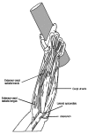
The exact pathology that underlies epicondylitis is not known.70 The problem appears to be confined to the tendinous and fascial attachments to the bone (See Figure 4). The tendons become dull, gray, friable, and edematous. The normal tendon fibers become disrupted by invading fibroblasts and granulation tissue.78 Adhesions may form between the tendon and surrounding tissues. The extensor carpi radialis brevis tendon appears to be most often affected because it is intimately attached to the joint capsule, and because of this proximity adhesions readily form between it and the joint.

Figure
Figure 4. Structures associated with lateral epicondylitis.
Etiology
Lateral epicondylitis is thought to be a degenerative process caused by overuse of the wrist extensors. Repetitive strong synergic and fixator action of the wrist extensors during gripping are believed to result in minor trauma to the muscle attachment to the epicondyle.75 Continued muscle use prevents healing. Medial epicondylitis is thought to be a similar process affecting the flexor, rather than the extensor, muscles. Forceful, repetitive motions of the forearm are thought to be the initial precipitating factor.79
Epidemiology
Epicondylitis has been reported to affect 4.23 individuals per 1000 adults per year in the U.S.80 The mean age of diagnosis is 45 years, and men and women appear to be equally affected.80 Lateral epicondylitis is six times more common than medial epicondylitis.80 Individuals who have been diagnosed with carpal tunnel syndrome have a greater prevalence of lateral epicondylitis than do those without carpal tunnel syndrome.81 Persons who engage in forceful, repetitive forearm work such as mechanics, butchers, and construction workers have a higher prevalence of the condition than the general population.82
Treatment
Conservative treatment
Initial treatment of epicondylitis usually involves rest and massage. In addition, a number of conservative therapies are used to treat epicondylitis. These are briefly described below.
Pharmacologic treatments for epicondylitis include NSAIDs, either taken orally or applied topically, topical dimethyl sulfoxide (DMSO), injections of glucocorticoid steroids, injections of anesthetics, and oral glucosamines.
Rest, ice, massage, physiotherapy, manipulations, splints, braces, and exercise programs are commonly used when treating epicondylitis.
Other treatments for epicondylitis include acupuncture, low level red or infrared lasers, ultrasound, phonophoresis, transcutaneous electrical nerve stimulation (TENS), extracorporal shock-wave therapy (ESWT), and pulsed electromagnetic fields (PEMF).
Surgical treatment
Surgery is not generally a first-line treatment for epicondylitis. However, in cases that are resistant to more conservative treatments, a variety of surgical techniques have been used. Some of the techniques are listed in Table 1. They can be broken down into four broad categories: denervation, nerve decompression, excision of various tissues, and lengthening of the extensor tendon (ERCB).83
Table
Table 1. Surgical procedures used to treat epicondylitisa.
De Quervain's Disease
Signs and Symptoms
De Quervain's disease is characterized by pain localized on the radial border of the wrist that may also radiate into the thumb and forearm.85 The pain is usually worsened by abduction and/or extension of the thumb.53 Other symptoms may include weakness of the thumb and loss of grip. Range of motion of the wrist and thumb is usually unaffected or only slightly limited.85
Anatomy
De Quervain's disease is a stenosis (thickening) of the fibrous sheath of the first extensor compartment of the extensor retinaculum.86 This compartment surrounds two tendons, the extensor pollicis brevis and the abductor pollicis longus (See Figure 5). In the past, de Quervain's disease has been described as a type of stenosing tenosynovitis of the hand and wrist. Because recent studies have shown that there is no inflammatory process associated with de Quervain's disease, some experts believe that the term tenosynovitis is not accurate for describing this condition.53, 86

Figure
Figure 5. Structures associated with De Quervain's disease.
Etiology
Possible etiologic factors include acute trauma, recurrent trauma, or an underlying collagen disease.87
Epidemiology
De Quervain's disease appears most frequently in the 30 to 50 year age group and has been reported to be 10 times more common among women than men.85 Work occupations commonly associated with this condition include musicians, weavers, typists, nurses, knitters, golfers, switchboard operators, and manual workers.53, 85 However, there is disagreement among experts as to whether these types of work cause de Quervain's disease or merely exacerbate the symptoms.53, 86 Anatomic variations of the first extensor compartment have also been reported to be associated with de Quervain's disease.86
Diagnosis
Diagnosis of de Quervain's disease is usually accomplished by the Finkelstein test. While the patient flexes the thumb within the palm while holding it tightly with the other fingers, the examiner performs an ulnar deviation of the patient's wrist. Intense pain on the styloid process of the radius indicates a positive test. The pain disappears after the thumb is released and extended.85 Additional diagnostic criteria include patient-reported pain at the radial wrist and tenderness to palpation at the radial wrist.53
Treatment
Conservative treatment
A number of conservative therapies have been used to treat de Quervain's disease. These include workplace modification, hand rest, neutral wrist splinting with a thumb spica, anti-inflammatory medication, and iontophoresis.53 If these therapies fail, injection of cortisone may be used to supplement splinting and anti-inflammatory medication.
Surgical treatment
Persistent pain after four to six weeks of conservative therapy is usually considered an indication for surgery.85, 87 This procedure consists of unroofing the retinaculum to release the abductor pollicis longus and extensor pollicis brevis tendon sheaths.87 As noted earlier, anatomic variation exists in that these tendon sheaths may be contained in one or two compartments. Reported complications of surgery include radial sensory nerve injury and painful surgical scarring.88
- Introduction - Diagnosis and Treatment of Worker-Related Musculoskeletal Disorde...Introduction - Diagnosis and Treatment of Worker-Related Musculoskeletal Disorders of the Upper Extremity
- Results - Garlic: Effects on Cardiovascular Risks and Disease, Protective Effect...Results - Garlic: Effects on Cardiovascular Risks and Disease, Protective Effects Against Cancer, and Clinical Adverse Effects
- Mus musculus tubulin, alpha-like 3 (Tubal3), transcript variant 1, mRNAMus musculus tubulin, alpha-like 3 (Tubal3), transcript variant 1, mRNAgi|224809299|ref|NM_001033879.3|Nucleotide
Your browsing activity is empty.
Activity recording is turned off.
See more...