U.S. flag

An official website of the United States government

NCBI Bookshelf. A service of the National Library of Medicine, National Institutes of Health.

Adam MP, Feldman J, Mirzaa GM, et al., editors. GeneReviews® [Internet]. Seattle (WA): University of Washington, Seattle; 1993-2024.

Cover of GeneReviews®

GeneReviews® [Internet].

Show details

Familial Cerebral Cavernous Malformations

Synonyms: Familial Cavernous Hemangioma, Familial Cerebral Cavernous Angioma, Familial Cerebral Cavernous Malformation Syndrome

, MD, , MD, , PhD, and , PhD.

Author Information and Affiliations

Initial Posting: ; Last Update: July 27, 2023.

Estimated reading time: 55 minutes

Summary

Clinical characteristics.

Familial cerebral cavernous malformations (FCCM) is a disorder characterized by multiple vascular lesions in the brain and spinal cord that consist of clustered, endothelial-lined caverns ranging in diameter from a few millimeters to several centimeters. Cerebral and/or spinal cavernous malformations may increase in number over time, and individual lesions may increase or decrease in size. The number of cerebral cavernous malformations (CCMs) identified in an individual ranges from one or two to hundreds of lesions (typical number 6-20 CCMs) depending on the individual's age and the quality and type of brain imaging used. Although CCMs have been reported in infants and children, the majority become evident between the second and fifth decades of life either incidentally or associated with seizures, focal neurologic deficits, headaches, and/or cerebral hemorrhage. Cutaneous vascular lesions are found in 9% and retinal vascular lesions in almost 5% of affected individuals. Up to 50% of individuals with FCCM remain symptom free throughout their lives.

Diagnosis/testing.

The diagnosis of familial cerebral cavernous malformations (FCCM) is established in a proband with multiple CCMs, one CCM and at least one other family member with one or more CCMs, or a heterozygous germline pathogenic variant in KRIT1, CCM2, or PDCD10 identified by molecular genetic testing.

Management.

Treatment of manifestations: Surgical removal of symptomatic lesions may be considered in individuals with acute hemorrhage and/or a mass effect presenting with focal neurologic deficit, headache, or seizure or in those with intractable seizures (with or without associated hemorrhage). Treatment of epilepsy is symptomatic. Headaches are managed symptomatically and prophylactically. Rehabilitation may aid in management of acute and chronic neurologic deficits.

Surveillance: Brain MR imaging with susceptibility-weighted imaging (SWI) is indicated in individuals experiencing new neurologic manifestations.

Agents/circumstances to avoid: Caution is recommended with medications such as analgesics such as NSAIDs, antithrombotic medications such as heparin and warfarin (Coumadin®), thrombolytic agents, and oral female hormones. Note: When antithrombotic and thrombolytic medications are necessary for treatment of life-threatening thrombosis, careful consideration of appropriate dosage and close monitoring are warranted. Radiation to the central nervous system may lead to new lesion formation.

Evaluation of relatives at risk: Asymptomatic at-risk relatives of all ages may be evaluated by molecular genetic testing if the family-specific pathogenic variant is known to allow early diagnosis and monitoring of individuals at risk of developing CCMs. Symptomatic relatives may undergo brain MRI with SWI sequences to determine presence, size, and location of lesions.

Pregnancy management: Pregnant women with FCCM who have had recent brain or spinal cord hemorrhage, epilepsy, or headaches require close monitoring during pregnancy. Seizures are the most common manifestations of CCM hemorrhage during pregnancy; exposure to anti-seizure medication during pregnancy may increase the risk for adverse fetal outcomes but is generally recommended, as the fetal risk is typically less than that associated with an untreated maternal seizure disorder. Any focal neurologic deficits or severe headaches during pregnancy should be evaluated and other neurologic causes (e.g., ischemic stroke, cerebral venous thrombosis) ruled out.

Genetic counseling.

FCCM is inherited in an autosomal dominant manner. Many individuals diagnosed with FCCM have a symptomatic parent. The proportion of individuals with FCCM caused by a de novo pathogenic variant is unknown. Each child of an individual with FCCM has a 50% chance of inheriting an FCCM-related pathogenic variant. If a pathogenic variant has been identified in an affected family member, prenatal testing of an at-risk pregnancy and preimplantation genetic testing are possible.

Diagnosis

No consensus clinical diagnostic criteria for familial cerebral cavernous malformations (FCCM) have been published.

Suggestive Findings

FCCM should be suspected in individuals with the following clinical findings, brain and/or spinal cord imaging findings, and family history.

Clinical findings

  • Seizures
  • Focal neurologic deficits
  • Headaches
  • Systemic vascular lesions
    • Vascular skin lesions. Three main types of cutaneous vascular malformations (CVMs) have been associated with FCCM: hyperkeratotic cutaneous capillary-venous malformations (HCCVMs), punctate capillary malformations (PCMs), and deep blue nodules (DBNs).
    • Retinal cavernomas and rare choroidal hemangiomas
    • Liver cavernous hemangiomas
    • Renal angiomas
    • Atypical vertebral hemangiomas (capillary-venous malformations within the bone that can rarely result in pathologic fractures)
  • Other
    • Brain tumors (e.g., meningioma, acoustic neuroma, cerebellar astrocytoma)
    • Adrenal gland calcifications

Brain and spinal cord imaging. FCCM is characterized by multiple cerebral and/or spinal cavernous malformations without developmental venous anomalies. There are several types of cavernous malformation based on imaging findings and in correlation with pathology (see Figure 1).

Figure 1.

Figure 1.

Zabramski classification of cavernous malformations A type 1 cavernous malformation is one with acute or subacute hemorrhage characterized radiographically by hyperintense T1 signal and hyper- or hypointense T2 core (1st panel, axial T2 brain MRI). Pathologically, (more...)

  • The characteristic, Zabramski type 2 lesion is comprised of mixed signal intensity with a central reticulated core surrounded by a dark hemosiderin ring. A Zabramski type 3 lesion is hypointense on T2 without the reticulated central portion.
  • A hemorrhagic cerebral cavernous malformation (CCM) may demonstrate acute or subacute blood (high T1 signal) with or without edema [Nikoubashman et al 2015, Flemming et al 2019].
  • Zabramski type 4 lesions appear as hypointense lesions on gradient echo or susceptibility-weighted imaging (SWI) only [Zabramski et al 1994].

Note: Brain MRI with SWI on a 3 Tesla or higher-magnet MRI is preferred [Akers et al 2017]. Intravenous gadolinium contrast administration is not needed for the identification of cavernous malformations but is useful in initially distinguishing CCMs from other entities such as telangiectasias, arteriovenous malformations, and hemorrhagic metastases. Spinal cord imaging should include both the cervical and thoracic spine and preferably include gradient-based sequences [Mabray et al 2020].

Family history. Family history may suggest autosomal dominant inheritance with affected males and females in multiple generations. Absence of a known family history does not preclude the diagnosis.

Note: A family member with a pathogenic variant in one of the FCCM-associated genes may or may not be clinically symptomatic (asymptomatic vascular lesions are fairly common in families segregating an FCCM-related pathogenic variant); therefore, the presence of a single CCM in an individual with no known family history of CCM does not exclude the diagnosis of FCCM.

Establishing the Diagnosis

The clinical diagnosis of FCCM can be established in a proband with multiple CCMs or one CCM and at least one other family member with one or more CCMs [Santos et al 2022]. The molecular diagnosis can be established in a proband with suggestive findings and a heterozygous germline pathogenic (or likely pathogenic) variant in KRIT1, CCM2, or PDCD10 identified by molecular genetic testing (see Table 1).

Note: (1) Per ACMG/AMP variant interpretation guidelines, the terms "pathogenic variant" and "likely pathogenic variant" are synonymous in a clinical setting, meaning that both are considered diagnostic and can be used for clinical decision making [Richards et al 2015]. Reference to "pathogenic variants" in this section is understood to include likely pathogenic variants. (2) Identification of a heterozygous variant of uncertain significance in KRIT1, CCM2, or PDCD10 does not establish or rule out the diagnosis.

Molecular genetic testing approaches can include a combination of gene-targeted testing (serial single-gene or concurrent gene testing, multigene panel) and comprehensive genomic testing (exome sequencing, genome sequencing). Gene-targeted testing requires that the clinician determine which gene(s) are likely involved (see Option 1), whereas comprehensive genomic testing does not (see Option 2).

Option 1

Serial single-gene or concurrent gene testing including sequence analysis of KRIT1, CCM2, and PDCD10 can be performed first followed by gene-targeted deletion/duplication analysis if no pathogenic variant is found on sequence analysis.

A multigene panel that includes KRIT1, CCM2, and PDCD10 and other genes of interest (see Differential Diagnosis) may be considered to identify the genetic cause of the condition while limiting identification of variants of uncertain significance and pathogenic variants in genes that do not explain the underlying phenotype. Note: (1) The genes included in the panel and the diagnostic sensitivity of the testing used for each gene vary by laboratory and are likely to change over time. (2) Some multigene panels may include genes not associated with the condition discussed in this GeneReview. (3) In some laboratories, panel options may include a custom laboratory-designed panel and/or custom phenotype-focused exome analysis that includes genes specified by the clinician. (4) Methods used in a panel may include sequence analysis, deletion/duplication analysis, and/or other non-sequencing-based tests.

Note: Targeted analysis can be considered first in individuals of Ashkenazi Jewish and Hispanic ancestry due to recurrent and/or founder variants in CCM2 and KRTI1 (see Table 7).

For an introduction to multigene panels click here. More detailed information for clinicians ordering genetic tests can be found here.

Option 2

When the diagnosis of FCCM has not been considered because an individual has atypical phenotypic features, comprehensive genomic testing does not require the clinician to determine which gene is likely involved. Exome sequencing is most commonly used; genome sequencing is also possible. To date, the majority of pathogenic variants reported in KRIT1, CCM2, and PDCD10 are within the coding region and are likely to be identified on exome sequencing.

For an introduction to comprehensive genomic testing click here. More detailed information for clinicians ordering genomic testing can be found here.

Table 1.

Molecular Genetic Testing Used in Familial Cerebral Cavernous Malformations

Gene 1, 2Proportion of FCCM Attributed to Pathogenic Variants in GeneProportion of Probands with a Pathogenic Variant 3 Detectable by Method
Sequence analysis 4Gene-targeted deletion/duplication analysis 5
CCM2 20%40%-70% 630%-60% 6, 7
KRIT1 53%-65%85%-95% 65%-15% 6, 7
PDCD10 10%-16%80%-90% 60%-10% 6
Unknown 85%-15%NA

FCCM = familial cerebral cavernous malformations; NA = not applicable

1.

Genes are listed in alphabetic order.

2.
3.

See Molecular Genetics for information on variants detected in these genes.

4.

Sequence analysis detects variants that are benign, likely benign, of uncertain significance, likely pathogenic, or pathogenic. Variants may include small intragenic deletions/insertions and missense, nonsense, and splice site variants; typically, exon or whole-gene deletions/duplications are not detected. For issues to consider in interpretation of sequence analysis results, click here.

5.

Gene-targeted deletion/duplication analysis detects intragenic deletions or duplications. Methods used may include a range of techniques such as quantitative PCR, long-range PCR, multiplex ligation-dependent probe amplification (MLPA), and a gene-targeted microarray designed to detect single-exon deletions or duplications. Gene-targeted deletion/duplication testing will detect deletions ranging from a single exon to the whole gene; however, breakpoints of large deletions and/or deletion of adjacent genes may not be detected by these methods. Exome and genome sequencing may be able to detect deletions/duplications using breakpoint detection or read depth; however, sensitivity can be lower than gene-targeted deletion/duplication analysis.

6.

Riant et al [2013] and data derived from the subscription-based professional view of Human Gene Mutation Database [Stenson et al 2020]

7.

Variability in the detection rate of deletion/duplication testing results from the high prevalence of a founder CCM2 deletion (exons 2-10) in the United States. This deletion was rare in an Italian population [Liquori et al 2007, Liquori et al 2008]. To date, very few large intragenic deletions/duplications have been reported in individuals with FCCM. Cohorts who had no detectable KRIT1, CCM2, or PDCD10 pathogenic variant by sequence analysis had (multi)exon deletions of KRIT1 at a frequency of 5% in the US, 4% in France, and 50% in Italy [Liquori et al 2008, Riant et al 2013].

8.

Following stringent inclusion criteria for FCCM (multiple lesions and/or family history consistent with FCCM), a heterozygous pathogenic variant in either KRIT1, CCM2, or PDCD10 is detected in at least 75% of affected families [Denier et al 2006, Liquori et al 2007, D'Angelo et al 2011, Riant et al 2013, Spiegler et al 2014], with some studies reporting ~97% detection rates [Cigoli et al 2014].

Clinical Characteristics

Clinical Description

Familial cerebral cavernous malformations (FCCM) is a disorder characterized by multiple cerebral cavernous malformations (CCMs) without a developmental venous anomaly. Individuals with FCCM may present with seizure, headaches, or focal neurologic deficits with or without associated cerebral hemorrhage. In some instances, individuals present for screening or evaluation of unrelated concerns and the cavernous malformation(s) are asymptomatic [Riant et al 2010].

To date, it is estimated that more than 1,000 families have been reported with FCCM and a pathogenic variant in one of the FCCM-associated genes. The following is a description of the phenotypic features associated with this disorder.

Table 2.

Familial Cerebral Cavernous Malformations: Frequency of Select Features

Feature 1% of Persons w/Feature 2Comment
Cavernous malformation of the central nervous system presenting with:
Seizures20%-40%The cumulative incidence of childhood seizures is ~20% (~60% by age 80 yrs). 3
Focal neurologic deficits25%-30%Focal neurologic deficits most commonly occur w/intracranial hemorrhage but may occur w/o hemorrhage as well. Focal neurologic deficits should localize to the CCM to be considered symptomatic.
Nonspecific headaches10%-30%Primary headache disorders are common in persons w/CCMs; however, headaches may also occur due to hemorrhage &/or hydrocephalus.
Symptomatic intracerebral hemorrhage30%-40%30%-40% of persons initially present w/symptomatic hemorrhage. 4
Spinal cord cavernous malformations~16%-70%Up to 70% of persons w/pathogenic variants in KRIT1 may have at least 1 spinal cavernous malformation, although many persons remain asymptomatic.
Extraneuronal manifestations
Cutaneous vascular malformations (CVMs)9%-20%Three types of CVMs have been reported:
  • Hyperkeratotic cutaneous capillary-venous malformations (HCCVMs)
  • Punctate capillary malformations (PCMs)
  • Deep blue nodules (DBNs) 5
Retinal vascular lesions~5%May incl retinal cavernomas & choroidal hemangiomas 6

CCM = cerebral cavernous malformation; CVM = cutaneous vascular malformation; FCCM = familial cerebral cavernous malformations

1.

Up to 40%-50% of individuals with FCMM are asymptomatic.

2.

Data reflect percentage of individuals with the feature at the time of initial presentation.

3.
4.
5.
6.

Neurologic findings. Up to 50% of individuals with a heterozygous germline pathogenic variant in either KRIT1, CCM2, or PDCD10 may be initially asymptomatic, although at least half of these individuals have identifiable CCM lesions on brain imaging [Battistini et al 2007, Fischer et al 2013]. CCMs have been reported in infants and children, but most individuals with FCCM present with symptoms between the second and fifth decades. In a study of individuals who have undergone genetic testing, 20% of symptomatic individuals presented under the age of 15 years [Denier et al 2006].

Clinically affected individuals may present with seizures (20%-40%), focal neurologic deficits (35%-50%), and nonspecific headaches (10%-30%). These symptoms may or may not be associated with symptomatic hemorrhage in the brain or the spinal cord [Zafar et al 2019]. Brain or spinal cord hemorrhage are common and occur in up to 30%-40% of individuals initially presenting with cavernous malformation(s) [Zafar et al 2019, Alalfi et al 2023, Weinsheimer et al 2023].

In large natural history studies, the risk of prospective, symptomatic hemorrhage does not seem to differ between familial and sporadic CCMs [Horne et al 2016, Taslimi et al 2016, Gross & Du 2017]. However, these studies had limited follow-up data and did not discern the effect of the number or type of lesions on hemorrhage risk. Prior studies had estimated the risk of prospective, symptomatic hemorrhage in FCCM to be between 2.8% and 16.5% per year depending on prior history of hemorrhage [Zabramski et al 1994, Labauge et al 2000, Carrión-Penagos et al 2020, Santos et al 2022, Weinsheimer et al 2023]. Most recently, the largest study of individuals with FCCM (n=321) estimated an overall hemorrhage risk of 2.8 per 100 person years [Weinsheimer et al 2023]. However, the risk was higher in individuals with prior hemorrhage, at 4.5 per 100 person years compared to 2.0 per 100 person years in those without prior hemorrhage. Furthermore, this study noted a higher risk of future hemorrhage based on total lesion count. Another study of children with FCCM (n=41) reported a five-year risk of hemorrhage of 5% per person year, with higher rates in those with pathogenic variants in CCM2, positive family history, and prior symptomatic hemorrhage [Geraldo et al 2023].

Seizures are common in FCCM during an individual's lifetime. One large study of individuals with FCCM (n=479) reported that by age 80 years, 60.4% of individuals had at least one seizure [Fox et al 2021]. The prospective risk of seizures has been estimated at approximately 1.2%-1.5% per person year in those not initially presenting with seizures [Taslimi et al 2019, Alalfi et al 2023]. Overall, seizure risk is estimated to be increased with higher lesion counts, larger CCMs, and possibly a pathogenic variant in PDCD10 [Fox et al 2021].

Brain imaging. Recommended standard imaging in individuals with cavernous malformations includes standard MRI sequences with either susceptibility-weighted imaging (SWI) on a 3 Tesla or higher-magnet machine [Akers et al 2017]. SWI allows the visualization of microscopic lesions, which are common in FCCM. Gradient echo (GRE) is acceptable but is less sensitive than SWI at assessing lesion count.

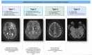
Four characteristic types of lesions have been described [Zabramski et al 1994] by MRI and histology (see Figure 1). Dividing CCMs into these radiologic and histologic types can be clinically useful in predicting hemorrhage risk [Nikoubashman et al 2015].

The clinical significance of small lesions (classified as Zabramski type 4) seen on MRI (sometimes referred to as cerebral dot-like cavernous malformations) is unclear. For these lesions, a mean hemorrhage rate of 1.3% per year was found over a period of 5.5 years in 18 children with either an inherited or a de novo heterozygous pathogenic variant in KRIT1 or PDCD10. However, these individuals were all asymptomatic [Nikoubashman et al 2015].

FCCM is a dynamic disease based on neuroimaging. Several studies suggest that new lesions appear at a rate between 0.2 to one lesion per person year [Brunereau et al 2000, Labauge et al 2001, Taslimi et al 2019, Santos et al 2022]. Studies reporting on de novo lesions can be limited if sequences (SWI vs GRE), slice thickness, and magnet strength differ. In both FCCM and sporadic CCM, lesions may change in size and signal characteristics over time.

In FCCM, 70%-86% of lesions are supratentorial and 16%-24% infratentorial in location [Brunereau et al 2000, Labauge et al 2001]. Of the infratentorial lesions, almost half occur in the brain stem. In addition, one study of individuals with FCCM found a higher prevalence of white matter intensities in younger individuals with FCCM compared to those with sporadic CCM [Golden et al 2015a], the clinical consequences of which are not yet known.

Spinal cord cavernous malformations (SCCMs). SCCMs are common in FCCM. In one study of individuals with known KRIT1 pathogenic variants, at least one lesion was found in up to 70% of individuals using standard and GRE imaging of the cervical and thoracic spine [Mabray et al 2020]. While spinal lesions may be common, symptomatic spinal cord lesions in FCCM are rare. In one small study (n=75) of individuals with FCCM, seven (9.3%) initially presented with symptomatic hemorrhage in the spinal cord and only one had a prospective spinal cord hemorrhage [Alalfi et al 2023].

Systemic vascular abnormalities. Vascular lesions outside the central nervous system have been reported in association with multiple intracranial cavernous malformations.

  • Vascular skin lesions have been reported in 9%-20% of individuals with FCCM [Manole et al 2020, Hart et al 2021].
    • Cutaneous vascular malformations were found in 38 out of 417 individuals with FCCM [Sirvente et al 2009]. Of the 38, 13 had skin lesions classified as capillary malformations; 15 individuals had hyperkeratotic cutaneous capillary-venous malformations (HCCVMs); eight had venous malformations; and two had unclassified lesions.
    • Bluish nodules and other subcutaneous nodules have been described as subtypes of venous malformations.
    • Some affected individuals have skin lesions removed secondary to bleeding, pain, protrusion, cosmetic concerns, or concern for malignancy.
  • Retinal vascular lesions, reported in 5% of affected individuals, may include retinal cavernous malformations and (rarely) choroidal hemangiomas [Wood et al 1957, Sarraf et al 2000, Labauge et al 2006, Hart et al 2021].
  • Liver hemangiomas have been reported in two Italian families with a heterozygous pathogenic variant in CCM2 but have not been confirmed to occur more frequently in individuals with FCCM compared to the general population [Drigo et al 1994, Toldo et al 2009].
  • Renal angioma has been reported in one affected individual of Italian descent [Battistini et al 2007].
  • Atypical vertebral hemangioma. Osseous vascular malformations of the vertebral bodies of the spine may also be present in individuals with FCCM. Rarely these lesions can be associated with pathologic fractures. Histopathology demonstrated capillary-venous malformation. These lesions are visible on spine MRI and are more atypical appearing than most vertebral hemangiomas, as they are hyperintense on T2-weighted images but hypointense on T1-weighted images [Tandberg et al 2020].

Other systemic manifestations

  • Scoliosis. In a series of 18 affected individuals with pathogenic variants in PDCD10, scoliosis was identified in 39% of individuals [Shenkar et al 2015].
  • Brain tumors. In a series of 18 affected individuals with pathogenic variants in PDCD10, 5 of 18 individuals had a brain tumor, including meningioma (n=2), acoustic neuroma (n=2), and cerebellar astrocytoma (n=1) [Shenkar et al 2015].
  • Adrenal gland calcifications have been rarely reported and are suspected to represent small vascular lesions, with limited histologic confirmation [Strickland et al 2017].

Histopathology. Histopathologic findings on resected CCMs show:

  • Closely clustered enlarged capillary channels (caverns) ranging from two to 55 mm (mean: 8 mm) with a single layer of endothelium without normal mature vessel wall elements or intervening brain parenchyma;
  • Thrombosis and intra- and extralesional hemorrhage. Edema may surround lesions with recent hemorrhage.

Phenotype Correlations by Gene

Several FCCM manifestations have been reported in association with specific genes.

  • One study of children with FCCM found a higher risk of hemorrhage in individuals with pathogenic CCM2 variants and positive family history [Geraldo et al 2023].
  • One small study suggested that individuals with pathogenic variants in KRIT1 may have a higher risk of de novo CCM appearance [Lanfranconi et al 2021]. Up to 70% of individuals with pathogenic variants in KRIT1 may have at least one spinal cavernous malformation, although many individuals remain asymptomatic.
  • Individuals with a heterozygous pathogenic variant in KRIT1 may also be more likely to have skin lesions than those with a heterozygous pathogenic variant in CCM2 or PDCD10 [Eerola et al 2000, Zlotoff et al 2007, Sirvente et al 2009, Toldo et al 2009, Kurlemann 2012, Campione et al 2013, Brownlee & Roxburgh 2014, Cigoli et al 2014, Bilo et al 2016, Hart et al 2021].
  • Individuals with pathogenic variants in PDCD10 are more likely to present early (<15 years) with hemorrhage [Denier et al 2006]. There was a trend toward earlier age at onset in symptomatic individuals with pathogenic variants in PDCD10 and a significant difference in age of symptomatic cerebral hemorrhage (average age 12.9 ± 11.6 years vs 22.9 ± 13.9 years in individuals with pathogenic variants in KRIT1 and 38.5 ± 21.9 years in individuals with pathogenic variants in CCM2).

Genotype-Phenotype Correlations

KRIT1 (c.1363C>T, p.Gln455Ter). A higher prevalence (estimated between 9% and 20%) and a different predominant morphologic subtype of cutaneous vascular malformations were observed in individuals from northern Mexico and the southwestern United States with this founder variant compared to other FCCM cohorts [Manole et al 2020] (see Table 7).

Penetrance

KRIT1. Among 64 families with 202 individuals who were heterozygous for a KRIT1 pathogenic variant [Denier et al 2004]:

  • Sixty-two percent were symptomatic;
  • Fifty-eight percent of those who were at least age 50 years had symptoms related to CCM;
  • Forty-five of 53 symptom-free individuals had lesions on MRI (three had indications of a type 4 lesion; see Figure 1) and five had no clinical or MRI findings of CCM.
  • Note: SWI MRI, the most sensitive imaging technique for identifying CCMs, was not performed in this study.

PDCD10. Penetrance may be decreased in families with a heterozygous pathogenic variant in PDCD10 compared to KRIT1 [Denier et al 2006].

Penetrance in individuals with pathogenic variants in CCM2 is not known.

Prevalence

It is estimated that 0.4%-0.9% of the general population has a CCM. Population-based studies estimate the incidence of CCMs to be 0.56 in 100,000 individuals older than age 16 years [Al-Shahi et al 2003]. In one study, the prevalence of CCMs was estimated between 0.16% and 0.5% based on clinical and nonclinical MRI series that mirror autopsy studies [Flemming et al 2017].

The estimated population-based prevalence of FCCM is 0.07% [Flemming et al 2017]. In a population-based study in individuals aged 50-89 years undergoing brain MRI for nonclinical study purposes, the prevalence of CCMs was 0.44%. In those with presumed FCCM, prevalence was 0.07% [Flemming et al 2017]. The common occurrence of asymptomatic vascular lesions in individuals with FCCM suggests that the population prevalence may be underestimated [Verlaan et al 2002, Johnson et al 2004].

The KRIT1 founder variant (c.1363C>T, p.Gln455Ter) is common among individuals of Hispanic descent with FCCM [Johnson et al 1995, Sahoo et al 1999] (see Table 7).

Differential Diagnosis

Sporadic cerebral cavernous malformations (CCMs). Histologically, familial CCMs (FCCM) and sporadic CCMs are similar. However, sporadic CCM is typically characterized by a single cavernous malformation and often associated with a developmental venous anomaly (DVA); multiple cavernous malformations can occur in the sporadic form, but they are always situated around or near a DVA. Limited evidence suggests that a DVA is an anatomic and genetic primer of sporadic CCM [Snellings et al 2022]. Some series indicate that up to 30% of sporadic CCMs are associated with a DVA. In a study using 7 Tesla MRI imaging, 100% of individuals with sporadic CCMs were found to have abnormal venous output from the cavernous malformation. Of note, DVAs are rarely seen in individuals with FCCM.

  • Somatic activating variants in PIK3CA have been identified in CCMs [Peyre et al 2021, Ren et al 2021, Weng et al 2021]. Somatic PIK3CA pathogenic variants have been identified in sporadic lesions as well as CCM lesions in individuals with germline pathogenic variants in any of the three known FCCM-related genes, suggesting that – in combination with an FCCM-related pathogenic variant – PI3 kinase activation accelerates growth of CCM lesions [Ren et al 2021].
  • Somatic pathogenic variants in MAP3K3 have been identified in sporadic CCMs [Weng et al 2021, Snellings et al 2022]. Overt hemorrhage appears to be less frequent in CCMs with somatic MAP3K3 pathogenic variants compared with those associated with a pathogenic variant in one of the FCCM-associated genes [Huo et al 2022].
  • Putting all recent somatic variant data together, it appears that (1) CCMs can develop as the result of biallelic loss of any of the three FCCM-related genes or as a result of somatic pathogenic variants in MAP3K3 or PIK3CA and (2) a PIK3CA pathogenic variant can confer an additional growth advantage for the lesion. Importantly, somatic pathogenic variants are only acquired within the developing CCM; they are not inherited through the germline and do not appear in peripheral blood leukocytes. Thus, while the identification of somatic variants is significant in terms of potential therapies, they are not useful as diagnostic tools, as each CCM lesion can have a novel combination of somatic pathogenic variants.

"Large lesion" cavernous malformation mimics. CCMs represent 5%-15% of all cerebral vascular malformations [Al-Shahi et al 2003]. Other vascular malformations occurring in the brain should be distinguishable from CCMs by neuroimaging and clinical manifestations (see Figure 2). Typical "large lesion" cavernous malformations are often very distinctive. However, in an acute hemorrhage from a CCM, the distinctive characteristics are often lacking [Flemming & Lanzino 2020, Kumar et al 2020]. In these situations, repeat imaging can be useful [Flemming et al 2019, Flemming & Lanzino 2020, Kumar et al 2020]. "Large lesion" cavernous malformation mimics include:

Figure 2.

Figure 2.

Differential diagnosis of cavernous malformations The left panel lists the differential diagnosis for "large lesion" cavernous malformations. The right panel lists the differential diagnosis for cavernous malformations not visible on standard MRI sequences (more...)

  • Hemorrhagic metastases. Renal cell, melanoma, papillary thyroid, lung, breast, and choriocarcinoma can create multiple hemorrhagic lesions of the brain that can be mistaken for FCCM. In general, metastases are more likely to enhance with contrast and more likely to have significant edema as compared to CCMs.
  • Primary brain tumors, often with calcification (ganglioglioma)
  • CAPNON (calcified pseudoneoplasm or the neuroaxis). The mixed density and susceptibility-weighted imaging (SWI) changes associated with this lesion are due to calcium rather than hemosiderin. These are usually singular lesions; thus, they are more likely to mimic sporadic CCM than FCCM.
  • Infectious or inflammatory nodules
  • Myxomatous emboli. An atrial myxoma embolizing to the brain can be confused with FCCM. However, these lesions typically have significant edema and a more complex appearance than CCMs.
  • Arteriovenous malformations (AVMs). Most AVMs are readily distinguishable from cavernous malformations by the multiple T2 flow voids, enhancement with contrast, and presence on angiography. However, small, thrombosed AVMs may have a similar appearance.

Zabramski type 4 cavernous malformation mimics. It is not uncommon to encounter situations in which an individual has multiple SWI lesions and no typical "large lesion" cavernous malformations. In this context, findings suggestive of FCCM include a family history (e.g., history of seizures, focal neurologic deficits, or brain bleeds) and lesions in the brain stem, cerebellum, deep and superficial regions (rather than just superficial regions), and at a younger age (i.e., age 45 years or younger). Genetic testing and ruling out Zabramski type 4 cavernous malformation mimics can be helpful in establishing a diagnosis of FCCM (see Figure 2). Zabramski type 4 cavernous malformation mimics include:

  • Hemorrhagic metastases (suggestive features include an older individual with a known primary malignancy and areas of enhancement using gadolinium as well as persistent edema)
  • Cerebral microbleeds due to amyloid angiopathy, hypertension, vasculitis
    • Amyloid angiopathy typically occurs in individuals older than age 60 years, and lesions are predominantly in the superficial cortex and sometimes associated with sulcal subarachnoid hemorrhage. If in doubt, a spinal fluid evaluation including tau and amyloid levels can be helpful in addition to apolipoprotein E testing.
    • Hypertensive angiopathy has a pattern of SWI abnormalities in the deep subcortical structures as well as the brain stem and typically occurs in older individuals with a history of uncontrolled or poorly controlled hypertension.
    • Vasculitis can be associated with superficial SWI changes, but is usually associated with leptomeningeal enhancement and areas of cerebral ischemia as well.
  • Head trauma. Classically, the microhemorrhages associated with trauma are located at the gray-white matter junction where microvasculature is susceptible to the effects of shearing trauma.
  • Calcium deposits. Calcium may appear in the brain for many reasons and will appear dark on SWI. A pattern in the bilateral basal ganglia may suggest benign calcification of the basal ganglia. If uncertain, a head CT scan can be helpful along with an MRI to diagnose calcium deposits.
  • Neurocysticercosis. The SWI signal abnormality in these individuals arises due to calcification rather than hemosiderin deposition. Neurocysticercosis is more likely than FCCM to have areas of enhancement and edema.
  • Brain irradiation. Two to 20 years after brain irradiation (whole brain or focused), exposed individuals may develop multiple cavernous malformations and/or capillary telangiectasias that may mimic the appearance of FCCM. History of radiation helps make the distinction between this entity and FCCM.
  • Capillary telangiectasias. Capillary telangiectasias can generally be distinguished from cavernous malformations by their small size and enhancement. These are commonly singular, are often located in the pons, appear dark on SWI and uniformly enhanced on contrasted studies, and do not typically appear on the standard T1/T2 images. Rarely, these can be large (>1cm) and mimic tumors and inflammatory conditions.

Management

The management of cerebral cavernous malformations (CCMs) is largely based on epidemiologic, non-randomized surgical and radiosurgical studies, as well as expert opinion. In 2017, The Alliance to Cure Cavernous Malformation (Formerly Angioma Alliance) Scientific Committee published guidelines for the management of CCMs including familial CCM (FCCM) [Akers et al 2017].

Evaluations Following Initial Diagnosis

To establish the extent of disease and needs in an individual diagnosed with FCCM, the evaluations summarized in Table 3 (if not performed as part of the evaluation that led to the diagnosis) are recommended.

Table 3.

Familial Cerebral Cavernous Malformations: Recommended Evaluations Following Initial Diagnosis

System/ConcernEvaluationComment
CCMs 1 MRI imaging of brain & spinal cord
  • MRI w/standard sequences (T1, T2, FLAIR) in addition to SWI
  • For initial diagnosis, contrast is helpful to distinguish from other entities.
  • Afterward, contrast is not necessary to detect hemorrhage but may be useful for preoperative planning.
Epilepsy EEG &/or video EEGTo help establish diagnosis of epilepsy or aid in seizure localization (CCM vs alternate pathologies) that may facilitate surgical epilepsy planning
Neuropsychological evalTo determine which hemisphere is language dominant & overall eligibility for surgical resection
Brain imagingTo confirm localization of epilepsy & exclude other epileptogenic foci
SCCMs Spinal imagingGradient-based sequences are most sensitive & should be used when SCCMs are suspected.
Retinal cavernous malformation Eye examWhile retinal cavernous malformations do not typically hemorrhage, baseline exam is recommended. 2
Genetic counseling By genetics professionals 3To inform affected persons & their families re nature, MOI, & implications of FCCM to facilitate medical & personal decision making
Family support
& resources
Assess need for:

CCM = cerebral cavernous malformation; FCCM = familial cerebral cavernous malformations; MOI = mode of inheritance; SCCM = spinal cord cavernous malformations; SWI = susceptibility-weighted imaging

1.

CCMs were previously called "angiographically occult vascular lesions" due to their relatively poor visualization on catheter angiograms. Cerebral arteriography is rarely necessary unless there is concern for an underlying arteriovenous malformation.

2.

Guidelines on the frequency of surveillance have not been established to date.

3.

Medical geneticist, certified genetic counselor, certified advanced genetic nurse

Treatment of Manifestations

Supportive care to improve quality of life, maximize function, and reduce complications is recommended. This ideally involves multidisciplinary care by specialists in relevant fields (see Table 4).

Table 4.

Familial Cerebral Cavernous Malformations: Treatment of Manifestations

Manifestation/ConcernTreatmentConsiderations/Other
Recurrent hemorrhage or mass effect Surgical resection 1
  • May be assoc w/↑ risk of short-term neurologic disability, new focal deficits, &/or seizures
  • Complete resection can cure a single lesion; postoperative serial imaging can be helpful to assure there was complete resection.
  • Persons w/hydrocephalus may require shunting or ETV.
Vitamin D3 (cholecalciferol) supplementation (adequate vitamin D intake recommended; 600 IU daily for adults age ≤70 yrs, 800 IU daily for those age >70 yrs)A repurposing drug screen identified vitamin D3 as a potential therapeutic option for CCMs. Vitamin D3 restored endothelial integrity in endothelial-deficient CCM2 cells in vitro. 2 Vitamin D3 is proposed to inhibit activation of RHOA involved in the leakiness of endothelium. Two cohort studies suggest more aggressive behavior of CCMs in persons w/low 25-hydroxy-vitamin D levels. 3 While no clinical trial has been performed to assess the benefits or risks to date, adequate vitamin D intake is recommended for persons w/FCCM.
Blood pressure controlSeveral studies show no ↑ risk of symptomatic hemorrhage in persons w/hypertension. However, 1 study found a relationship between systolic blood pressure & lesion burden. 4 Given other health benefits of lowering blood pressure, standard recommendations for blood pressure control are recommended for persons w/FCCM.
Epilepsy Standard ASMsEarly eval for possible surgical resection is recommended (see Hemorrhage, recurrent hemorrhage, mass effect, and intractable seizures).
Headaches Standard treatment & mgmtStandard treatment is recommended for primary headache disorders. If severe, prolonged, or progressive, or assoc w/new or worsening neurologic deficits, urgent brain imaging could lead to prompt mgmt.
Neurologic deficits Rehab servicesFor persons w/temporary or permanent neurologic deficits

ASM = anti-seizure medication; ETV = endoscopic third ventriculocisternostomy

1.

Of note, stereotactic radiosurgery maybe associated with risk of radiation injury or, rarely, radiation-induced cavernous malformation; it is not generally recommended in FCCM [Akers et al 2017].

2.
3.
4.

Hemorrhage, recurrent hemorrhage, mass effect, and intractable seizures. The natural history of an individual CCM must be compared to the risk of surgery to aid in treatment decisions. Common indications for surgical removal include: first hemorrhage in a surgically accessible CCM, recurrent hemorrhage with increasing disability, high risk of further deterioration in less accessible lesions, intractable seizures, or mass effect with declining neurologic function [Heros & Heros 2000, Selman et al 2000, Akers et al 2017]. An observational study reported that surgical excision increased the overall risk of short-term neurologic disability, symptomatic intracranial hemorrhage, and new focal neurologic deficits [Moultrie et al 2014]. This study did not take into consideration CCM location, presence of declining functional status preoperatively, or advances in surgical techniques and scoring systems that aid in that process. Due to remaining uncertainty on the exact timing and indications for surgery, this led to the first clinical trial assessing surgery and radiosurgery versus observation (see Therapies Under Investigation). Importantly, children often experience better outcomes with surgical resection, including better seizure control and high rates of neurologic recovery, possibly secondary to increased neuroplasticity in this cohort [Gao et al 2022, Rauschenbach et al 2023].

Individuals with hydrocephalus may require treatment with shunting or endoscopic third ventriculocisternostomy in those with unresectable midbrain lesions compressing the cerebral aqueduct or those with ventricular compression.

Laser interstitial thermal therapy is an evolving tool for the treatment of CCMs. Small, single-institution series suggest potential benefit in individuals with epilepsy, but further larger studies are needed.

Surveillance

To monitor existing manifestations, the individual's response to supportive care, and the emergence of new manifestations, the evaluations summarized in Table 5 are recommended.

Table 5.

Familial Cerebral Cavernous Malformations: Recommended Surveillance

System/ConcernEvaluationFrequency
CCMs Brain imaging
  • Brain MRI imaging w/standard sequences (T1, T2, FLAIR) & SWI is indicated in persons experiencing new neurologic symptoms.
  • It is debatable whether routine surveillance in the absence of symptoms is helpful. 1
Epilepsy
  • Neurologic & neurosurgical eval
  • EEG
  • Brain imaging
  • If seizures are under control on ASMs, annual follow up w/neurologist is recommended.
  • If the affected person has an exacerbation of a previously quiescent seizure disorder, brain MRI, EEG, & possibly neurosurgical eval should be considered.
  • If epilepsy is intractable despite multiple ASM trials, a neurosurgical eval should be considered.
Headaches Neurologic evalPer standard recommendations
Focal neurologic symptoms
  • Neurologic eval
  • PT & rehab
  • If the affected person has a stable focal neurologic deficit, PT & rehab should be considered.
  • For persistent lower motor neuron facial weakness or double vision, facial reanimation &/or ophthalmologic surgeries to restore function can be considered.
SCCMs Spinal imaging
  • Spinal imaging should be repeated if the affected person has recurrent or worsening symptoms.
  • Debate exists regarding routine surveillance in the absence of new symptoms. 1

CCM = cerebral cavernous malformation; PT = physical therapy; SCCM = spinal cord cavernous malformations; SWI = susceptibility-weighted imaging

1.

Agents/Circumstances to Avoid

Analgesics

  • There is limited data regarding the use of non-aspirin, nonsteroidal anti-inflammatory drugs (NSAIDs). One study reported a reduction in hemorrhage risk in individuals with sporadic or familial CCM taking NSAIDs [Flemming et al 2021]; however, the dose and duration of medication use is not known. In general, it is best to avoid NSAIDs; however, in asymptomatic individuals it is not absolutely contraindicated.
  • The use of narcotic pain medications is discouraged in chronic pain conditions because of the potential for addiction and because of their association with rebound headaches.

Antithrombotic medications include anticoagulants (e.g., heparin, warfarin [Coumadin®], direct oral anticoagulants) and antiplatelet agents (e.g., aspirin, clopidogrel, ticagrelor). In general, it is recommended to avoid antithrombotic agents in individuals with FCCM unless they are necessary for the treatment of life-threatening thrombosis with close monitoring by the medical team. Recent data suggest that antithrombotics may lower the risk of cerebral hemorrhage, possibly by reducing thrombosis within the cavernous malformation [Zuurbier et al 2019]. Until further data are available, use of antithrombotics can be considered after weighing the individual's risk of CCM hemorrhage and the risk of not using antithrombotics for the suggested indication with close monitoring thereafter [Flemming et al 2013].

Thrombolytic agents are often used for ischemic stroke or pulmonary embolism and include alteplase and tenecteplase, among others. Limited data exist regarding the use of thrombolytic agents in individuals with CCMs. In one study of 967 individuals receiving thrombolytics for presumed cerebral ischemia, 20 had at least one cavernous malformation [Erdur et al 2014]. Only one of 11 individuals had evidence of a hemorrhagic CCM post thrombolytic use, and it was thought that the CCM was possibly responsible for the presenting symptoms, not cerebral ischemia, once an MRI was performed. Per American Stroke Association guidelines [Powers et al 2019] regarding use of thrombolytics in the setting of an intracranial vascular malformation, "IV TPA may be considered in individuals with stroke with severe neurologic deficits and a high likelihood of morbidity and mortality to outweigh the anticipated risk of hemorrhage."

Oral female hormones (including oral contraceptives and menopausal therapy). A recent cohort study comprised of 722 females with both sporadic and familial CCM noted an increased risk of hemorrhage in individuals with CCMs taking oral female hormones [Zuurbier et al 2023]. This study was limited in identifying the duration, dose, and compliance with these medications. Until further data are available on which individuals are at highest risk, affected females should be cautioned regarding use of oral female hormones.

Radiation. Radiation to the central nervous system is associated with de novo lesion formation in FCCM [Larson et al 1998, Nimjee et al 2006, Golden et al 2015b]. These lesions appear to be histologically different from the cavernous malformations found prior to radiation [Cha et al 2015].

Evaluation of Relatives at Risk

It is appropriate to clarify the genetic status of apparently asymptomatic older and younger at-risk relatives of an affected individual in order to identify as early as possible those who would benefit from prompt initiation of surveillance and awareness of agents/circumstances to avoid. Evaluations can include:

  • Molecular genetic testing if the pathogenic variant in the family is known;
  • Brain and/or spinal cord MR imaging including susceptibility-weighted imaging (SWI) or gradient echo (GRE) if the pathogenic variant in the family is not known.

See Genetic Counseling for issues related to testing of at-risk relatives for genetic counseling purposes.

Pregnancy Management

Pregnant women with FCCM who have had recent brain or spinal cord hemorrhage, epilepsy, or migraine require close observation during pregnancy.

Pregnant women with suspected FCCM should undergo counseling regarding the risk for the fetus and genetic testing options.

In general, women with epilepsy or a seizure disorder from any cause are at greater risk for mortality during pregnancy than pregnant women without a seizure disorder; use of anti-seizure medication (ASM) during pregnancy reduces this risk. However, exposure to ASM may increase the risk for adverse fetal outcome (depending on the drug used, the dose, and stage of pregnancy during which the medication is taken). Nevertheless, the risk of an adverse outcome to the fetus from medication exposure is often less than that associated with an untreated maternal seizure disorder. Neurologists should be involved in the care of pregnant women with CCMs and seizures in the preplanning period or early pregnancy. ASMs should be reviewed to make sure the least teratogenic medication is being used and that women receive folate supplementation. ASM levels may need to be monitored during pregnancy. If a pregnant woman has a breakthrough seizure after a time of stability without an obvious provoking factor (e.g., fever or low ASM level), a brain MRI without contrast is safe and should be considered.

Focal neurologic deficits or significant or severe headaches during pregnancy should be evaluated. In addition to the concern of CCM hemorrhage, other neurologic causes should be ruled out (e.g., ischemic stroke, cerebral venous thrombosis). Three large studies have suggested that the risk of symptomatic hemorrhage does not differ between pregnant and non-pregnant women. These studies are somewhat limited by the number of women who become pregnant after a diagnosis of CCM [Witiw et al 2012, Kalani & Zabramski 2013, Joseph et al 2021]. Given that most individuals present in their late 30s or early 40s, many women have completed childbearing or may defer pregnancy after a diagnosis of FCCM, thus limiting the numbers of individuals in such studies. One study estimated that the overall risk of symptomatic hemorrhage is 3% during pregnancy, similar to the risk reported in natural history studies [Kalani & Zabramski 2013]. This study noted that the risk was 1.8% per pregnancy in the sporadic CCM group versus 3.6% in the FCCM group. However, only five total symptomatic hemorrhages occurred during pregnancy (including two hemorrhages in one individual). Thus, the numbers are small, and it is unclear if there is a true difference in rates between individuals with sporadic and familial CCMs.

Affected women and obstetricians are frequently concerned that the risk of increased blood pressure and intrathoracic pressure during labor could lead to CCM hemorrhage. However, review of the literature finds clinical events during labor to be extremely rare [Joseph et al 2021].

See MotherToBaby for further information on medication use during pregnancy.

Therapies Under Investigation

Stereotactic radiosurgery. There are no published randomized clinical trials of radiosurgery versus observation in individuals with CCM. In general, data are derived mainly from single centers with limited follow up. Data suggest that the risk of hemorrhage from a CCM can be reduced to 1%-2% per year, but that effect is not seen for up to two years. The risk of treatment complications has been estimated at approximately 2%-4% [Poorthuis et al 2019]. Since natural history studies similarly show a decline in hemorrhage rate after two years [Horne et al 2016], there is debate on the efficacy of this therapy for CCM. This has led to the Cavernomas: A Randomized Effectiveness (CARE) trial comparing neurosurgery or stereotactic radiosurgery to conservative management in the United Kingdom (NIHR128694).

As radiation may induce CCM formation, the 2017 Synopsis of Guidelines for the Clinical Management of Cerebral Cavernous Malformations [Akers et al 2017] states that "radiosurgery is not recommended for asymptomatic CCMs, for CCMs that are surgically accessible, nor in familial CCM because of concern about de novo CCM genesis (class III, level C)."

Propranolol. Propranolol has been used successfully in the treatment of facial hemangiomas in infants. Several case reports and series suggested a possible benefit for CCMs. In a recent randomized, open-label, blinded end-point Phase II pilot trial (Treat_CCM), propranolol was safe and well tolerated in individuals with FCCM. While the study was not adequately powered to investigate efficacy, treatment with propranolol was possibly beneficial in reducing the incidence of clinical events in individuals with symptomatic FCCM [Lanfranconi et al 2020, Lanfranconi et al 2023].

Atorvastatin. Statins have shown reduction of lesion burden in mouse models of CCMs by indirectly inhibiting Rho kinase [Whitehead et al 2009]. The ongoing Atorvastatin Treatment of Cavernous Angiomas With Symptomatic Hemorrhage Exploratory Proof of Concept (AT CASH EPOC) study (NCT02603328) is assessing the efficacy and safety of atorvastatin (80 mg daily) versus placebo in individuals with recent symptomatic brain hemorrhage. Results are anticipated in 2024.

Rec-944. Rec-944 targets superoxides. There are preclinical data suggesting a role for oxidative stress in the pathogenesis of CCMs. Phase I trials using this compound have been completed and Phase II trials are ongoing (NCT05085561).

Diet. Data has emerged regarding the impact of the gut microbiome on cavernous malformation lesion formation and behavior in animal models of CCMs [Tang et al 2017, Tang et al 2019]. Genetically altered mice fed a diet high in emulsifiers had higher CCM lesion burden due to reduced thickness of the gut mucosal barrier allowing gram-negative bacteria to enter the bloodstream and trigger the TLR4 inflammatory pathway, leading to lesion formation. It is further notable that loss of gut epithelial PDCD10 results in reduced mucin production, also allowing further translocation of gram-negative bacteria into the bloodstream, which may be one of the explanations as to why pathogenic variants in PDCD10 have earlier and more severe disease [Tang et al 2019]. While there are no clinical trials in humans regarding the efficacy of such a diet, a diet with limited processed foods seems reasonable based on other positive health effects. There are no data on whether there would be positive or negative effects from pro- or prebiotics.

Search ClinicalTrials.gov in the US and EU Clinical Trials Register in Europe for access to information on clinical studies for a wide range of diseases and conditions.

Genetic Counseling

Genetic counseling is the process of providing individuals and families with information on the nature, mode(s) of inheritance, and implications of genetic disorders to help them make informed medical and personal decisions. The following section deals with genetic risk assessment and the use of family history and genetic testing to clarify genetic status for family members; it is not meant to address all personal, cultural, or ethical issues that may arise or to substitute for consultation with a genetics professional. —ED.

Mode of Inheritance

Familial cerebral cavernous malformations (FCCM) is inherited in an autosomal dominant manner.

  • The fairly common occurrence of asymptomatic vascular lesions may prevent recognition of an autosomal dominant pattern of inheritance in a family [Denier et al 2004].
  • Because a clinical diagnosis of FCCM can be established in an individual by the presence of multiple lesions (independent of family history), an individual with a clinical diagnosis of FCCM may represent an apparent simplex case (i.e., the only affected family member). Likewise, an individual with a molecular diagnosis of FCCM (established by the identification of a heterozygous, germline FCCM-related genetic alteration involving KRIT1, CCM2, or PDCD10) may have an apparent negative family history.
  • More than 5% of individuals with multiple lesions and/or a family history of CCMs do not have an identifiable pathogenic variant involving any of the three genes known to be associated with FCCM.
  • Sporadic cerebral cavernous malformations (CCMs) (i.e., CCMs caused by somatic pathogenic variants and not expected to recur in other family members) are not addressed in this section (see Differential Diagnosis).

Risk to Family Members

Parents of a proband

  • Many individuals diagnosed with FCCM have a symptomatic parent.
  • A proband with FCCM may have the disorder as the result of a de novo pathogenic variant. Individuals with a de novo germline pathogenic variant – most commonly in PDCD10 – have been reported [Lucas et al 2001, Denier et al 2004, Liquori et al 2008, Stahl et al 2008, Shenkar et al 2015]. However, the proportion of individuals with FCCM caused by a de novo pathogenic variant is unknown, as the frequency of subtle signs of the disorder in parents has not been thoroughly evaluated and molecular genetic testing data are insufficient.
  • If the proband is the only family member known to have FCCM, the following evaluations are recommended for the parents to confirm their status and to allow reliable recurrence risk counseling:
    • Molecular genetic testing (if a molecular diagnosis has been established in the proband); and/or
    • Brain MRI including gradient echo (GRE) or susceptibility-weighted imaging (SWI).
    Family history (e.g., history of seizures, focal neurologic deficits, or brain bleeds) may be helpful in determining which parent is most likely to require a diagnostic evaluation.
  • If the proband has a known pathogenic variant that cannot be identified in either parent and parental identity testing has confirmed biological maternity and paternity, the following possibilities should be considered:
  • The family history of some individuals diagnosed with FCCM may appear to be negative because of failure to recognize the disorder in family members, early death of the parent before the onset of symptoms, or reduced penetrance in the parent with the pathogenic variant. Therefore, an apparently negative family history cannot be confirmed unless appropriate evaluations (i.e., molecular genetic testing if the pathogenic variant has been identified in the proband and/or brain MRI including GRE or SWI) has been performed on the parents of the proband.

Sibs of a proband. The risk to sibs of the proband depends on the clinical and genetic status of the proband's parents:

  • If a parent of the proband is affected and/or a parent is known to have the pathogenic variant identified in the proband (regardless of the parent's clinical status), the risk to sibs is 50%.
  • If the proband has a known KRIT1, CCM2, or PDCD10 pathogenic variant and this pathogenic variant cannot be detected in the leukocyte DNA of either parent, the risk to sibs is estimated to be 1% because of the theoretic possibility of parental germline mosaicism [Rahbari et al 2016].
  • If parents are clinically unaffected but their genetic status is unknown (because either the parents have not been tested for the pathogenic variant identified in the proband or the proband does not have a genetic alternation involving any of the three genes known to be associated with FCCM), the assumption of a familial form must be made and sibs and parents offered brain MRI with GRE and/or SWI in order to identify those who would benefit from prompt initiation of surveillance and awareness of agents/circumstances to avoid.

Offspring of a proband. Each child of an individual with FCCM has a 50% chance of inheriting an FCCM-related pathogenic variant.

Other family members. The risk to other family members depends on the status of the proband's parents: if a parent is affected and/or has an FCCM-related pathogenic variant, the parent's family members may be at risk.

Related Genetic Counseling Issues

See Management, Evaluation of Relatives at Risk for information on evaluating at-risk relatives for the purpose of early diagnosis and treatment.

Family planning

  • The optimal time for determination of genetic risk and discussion of the availability of prenatal/preimplantation genetic testing is before pregnancy.
  • It is appropriate to offer genetic counseling (including discussion of potential risks to offspring and reproductive options) to young adults who are affected or at risk.

DNA banking. Because it is likely that testing methodology and our understanding of genes, pathogenic mechanisms, and diseases will improve in the future, consideration should be given to banking DNA from probands in whom a molecular diagnosis has not been confirmed (i.e., the causative pathogenic mechanism is unknown). For more information, see Huang et al [2022].

Prenatal Testing and Preimplantation Genetic Testing

If an FCCM-related pathogenic variant has been identified in an affected family member, prenatal and preimplantation genetic testing are possible.

Differences in perspective may exist among medical professionals and within families regarding the use of prenatal testing. While most centers would consider use of prenatal testing to be a personal decision, discussion of these issues may be helpful.

Resources

GeneReviews staff has selected the following disease-specific and/or umbrella support organizations and/or registries for the benefit of individuals with this disorder and their families. GeneReviews is not responsible for the information provided by other organizations. For information on selection criteria, click here.

Molecular Genetics

Information in the Molecular Genetics and OMIM tables may differ from that elsewhere in the GeneReview: tables may contain more recent information. —ED.

Table A.

Familial Cerebral Cavernous Malformation: Genes and Databases

Data are compiled from the following standard references: gene from HGNC; chromosome locus from OMIM; protein from UniProt. For a description of databases (Locus Specific, HGMD, ClinVar) to which links are provided, click here.

Table B.

OMIM Entries for Familial Cerebral Cavernous Malformation (View All in OMIM)

116860CEREBRAL CAVERNOUS MALFORMATIONS; CCM
603284CEREBRAL CAVERNOUS MALFORMATIONS 2; CCM2
603285CEREBRAL CAVERNOUS MALFORMATIONS 3; CCM3
604214KRIT1 ANKYRIN REPEAT-CONTAINING PROTEIN 1; KRIT1
607929CCM2 SCAFFOLD PROTEIN; CCM2
609118PROGRAMMED CELL DEATH 10; PDCD10

Molecular Pathogenesis

All three cerebral cavernous malformation (CCM) genes (KRIT1, CCM2, and PDCD10) are essential for blood vessel growth and development and can regulate multiple downstream signaling pathways through their various associated protein complexes. This can affect several processes including endothelial junction integrity and cell migration. A single inherited pathogenic variant in one of these genes is believed to be sufficient to cause familial CCM; however, the precise molecular mechanisms that lead to the formation of CCM lesions remains unclear. It is known that the protein products of these genes can assemble into a ternary complex, as well as interact with other proteins in the cell. Understanding the functions of these genes will continue to identify druggable pathways that can be targeted for treating CCMs.

KRIT1. Krev interaction trapped protein 1 (KRIT1) has a variety of functions within the vascular system and throughout the body. This anchor protein plays a role in regulating cell structure and response to shear stress through the integrin signaling pathway [Zawistowski et al 2002, Macek Jilkova et al 2014], maintaining homeostasis of intracellular reactive oxygen species [Goitre et al 2010, Goitre et al 2014], and regulating autophagy by activating the mTOR pathway [Marchi et al 2015]. KRIT1 also regulates endothelial cell-cell junctions to maintain junctional stability and control of vascular permeability in response to inflammation [Glading et al 2007, Borikova et al 2010, Corr et al 2012]. KRIT1 can activate angiogenesis through Delta-Notch signaling [Wüstehube et al 2010, DiStefano et al 2014]. Loss of KRIT1 function leads to CCM lesion formation through a loss-of-function mechanism where in the absence of KRIT1, endothelial cells become more stem cell-like, proliferative, and invasive [Verlaan et al 2002, Maddaluno et al 2013].

CCM2. Cerebral cavernous malformations 2 protein (CCM2), a protein with a PTB (phosphotyrosine-binding) domain encoded by CCM2, binds to and regulates the localization of KRIT1. The CCM2 protein is a scaffold protein for multiple signaling cascades including the p38 mitogen-activated protein kinase (MAPK) signaling [Uhlik et al 2003, Fisher et al 2015] and Rho kinase signaling for maintenance of proper vascular integrity [Whitehead et al 2009, Borikova et al 2010, Stockton et al 2010]. Animal models and molecular studies suggest a two-hit genetic mechanism [Akers et al 2009, Pagenstecher et al 2009, Whitehead et al 2009].

PDCD10. PDCD10 encodes a 212-amino acid adaptor protein, programmed cell death 10 (PDCD10). Overexpression of PDCD10 leads to activation of caspase 3 and increased cell death or apoptosis [Wang et al 1999, Guclu et al 2005]. PDCD10 is involved in a wide variety of cellular signaling processes and has been shown to be part of the macromolecular complex including KRIT1 and CCM2 [Hilder et al 2007, Voss et al 2007]. Within the vasculature, PDCD10 plays a critical role in vascular development (through VEGF signaling) and regulation of angiogenesis (through DLL4-Notch signaling) [He et al 2010, You et al 2013]. The nature of PDCD10 pathogenic variants detected to date suggests a role for loss of function through haploinsufficiency or somatic loss of heterozygosity [Akers et al 2009, Pagenstecher et al 2009].

Mechanism of disease causation. Biallelic loss of function (possible haploinsufficiency)

Molecular genetic analysis of CCMs from individuals with familial CCM (FCCM) identified a second somatic pathogenic variant on the other allele of the same gene with the germline pathogenic variant [Akers et al 2009, Pagenstecher et al 2009]. Next-generation sequencing studies have further showed that both copies of CCM genes are mutated in lesional tissue in familial cases.

In some (but not all) sporadic cases, lesions can contain two somatic pathogenic variants that inactivate both copies of the gene [Ren et al 2021].

Table 6.

Familial Cerebral Cavernous Malformations: Gene-Specific Laboratory Considerations

Gene 1Special Consideration
CCM2
  • CCM2 has an alternatively spliced exon 1B.
  • A large 77.6-kb deletion that includes exons 2-10 is a common founder deletion in the US population; it is not detectable by sequencing and requires deletion/duplication or CMA analysis, which may be performed as a first-tier test in this population [Liquori et al 2007, Gallione et al 2022].
PDCD10 The first coding exon is exon 4.
1.

Genes from Table 1 in alphabetic order.

Table 7.

Pathogenic Variants Referenced in This GeneReview by Gene

Gene 1Reference SequencesDNA Nucleotide ChangePredicted Protein ChangeComment [Reference]
CCM2 NM_031443​.3 77.6-kb deletion incl exons 2-10--Common founder variant in North America; up to 22% of persons may have this pathogenic variant [Liquori et al 2007]
c.30+5_30+6delGCinsTT--Recurrent variant in persons of Ashkenazi Jewish descent [Gallione et al 2011]
KRIT1 NM_194456​.1
NP_919438​.1
c.987C>Ap.Cys329TerPossible founder variant in persons from Sardinia [Cau et al 2009]
c.1363C>Tp.Gln455TerCommon founder variant in 70% of Hispanic persons from northern Mexico & American Southwest [Sahoo et al 1999]

Variants listed in the table have been provided by the authors. GeneReviews staff have not independently verified the classification of variants.

GeneReviews follows the standard naming conventions of the Human Genome Variation Society (varnomen​.hgvs.org). See Quick Reference for an explanation of nomenclature.

1.

Genes from Table 1 are in alphabetic order.

Chapter Notes

Author Notes

NIH Funding – 454RD

Scientific Advisory Board, Angioma Alliance

Acknowledgments

WBD holds the Canada Research Chair in Animal Models of Human Disease and is supported by a project grant from the Canadian Institutes of Health Research (PJT-153000).

Author History

Amy Akers, PhD; Angioma Alliance (2011-2023)

W Brent Derry, PhD (2023-present)

Kelly D Flemming, MD (2023-present)

Eric W Johnson, PhD; Barrow Neurological Institute (2003-2011)

Douglas Marchuk, PhD (2023-present)

Leslie Morrison, MD; University of New Mexico (2011-2023)

Edward Smith, MD (2023-present)

Revision History

  • 27 July 2023 (gm) Comprehensive update posted live
  • 4 August 2016 (ma) Comprehensive update posted live
  • 31 May 2011 (me) Comprehensive update posted live
  • 31 May 2005 (me) Comprehensive update posted live
  • 24 February 2003 (me) Review posted live
  • 5 February 2002 (ej) Original submission

References

Literature Cited

  • Akers A, Al-Shahi Salman R, Awad IA, Dahlem K, Flemming K, Hart B, Kim H, Jusue-Torres I, Kondziolka D, Lee C, Morrison L, Rigamonti D, Rebeiz T, Tournier-Lasserve E, Waggoner D, Whitehead K. Synopsis of guidelines for the clinical management of cerebral cavernous malformations: consensus recommendations based on systematic literature review by the Angioma Alliance Scientific Advisory Board Clinical Experts Panel. Neurosurgery. 2017;80:665-80. [PMC free article: PMC5808153] [PubMed: 28387823]
  • Akers AL, Johnson E, Steinberg GK, Zabramski JM, Marchuk DA. Biallelic somatic and germline mutations in cerebral cavernous malformations (CCMs): evidence for a two-hit mechanism of CCM pathogenesis. Hum Mol Genet. 2009;18:919-30. [PMC free article: PMC2640209] [PubMed: 19088123]
  • Alalfi MO, Lanzino G, Flemming KD. Clinical presentation, hemorrhage risk, and outcome in patients with familial cavernous malformations: a pragmatic prospective analysis of 75 patients. J Neurosurg. 2023. Epub ahead of print. [PubMed: 36933254]
  • Al-Shahi R, Bhattacharya JJ, Currie DG, Papanastassiou V, Ritchie V, Roberts RC, Sellar RJ, Warlow CP, et al. Prospective, population-based detection of intracranial vascular malformations in adults: the Scottish Intracranial Vascular Malformation Study (SIVMS). Stroke. 2003;34:1163-9. [PubMed: 12702837]
  • Battistini S, Rocchi R, Cerase A, Citterio A, Tassi L, Lando G, Patrosso MC, Galli R, Brunori P, Sgrò DL, Pitillo G, Lo Russo G, Marocchi A, Penco S. Clinical, magnetic resonance imaging, and genetic study of 5 Italian families with cerebral cavernous malformation. Arch Neurol. 2007;64:843-8. [PubMed: 17562932]
  • Bilo L, Antenora A, Peluso S, Procaccini EM, Quarantelli M, Mansueto G, Cigoli MS, Penco S, De Michele G. CCM3/PDCD10 gene mutation in cerebral cavernous malformations associated with hyperkeratotic cutaneous capillary venous malformations. J Dermatol. 2016;43:962-3. [PubMed: 26896283]
  • Borikova AL, Dibble CF, Sciaky N, Welch CM, Abell AN, Bencharit S, Johnson GL. Rho kinase inhibition rescues the endothelial cell cerebral cavernous malformation phenotype. J Biol Chem. 2010;285:11760-4. [PMC free article: PMC2852911] [PubMed: 20181950]
  • Brownlee WJ, Roxburgh R. Mystery case: cutaneous lesions in KRIT1-associated cerebral cavernous malformations. Neurology. 2014;83:e133-4. [PubMed: 25267988]
  • Brunereau L, Levy C, Laberge S, Houtteville J, Labauge P. De novo lesions in familial form of cerebral cavernous malformations: clinical and MR features in 29 non-Hispanic families. Surg Neurol. 2000;53:475-82 [PubMed: 10874147]
  • Campione E, Diluvio L, Terrinoni A, Di Stefani A, Orlandi A, Chimenti S, Bianchi L. Progressive late-onset of cutaneous angiomatosis as possible sign of cerebral cavernous malformations. Dermatol Online J. 2013;19:2. [PubMed: 23473272]
  • Carrión-Penagos J, Zeineddine HA, Polster SP, Girard R, Lyne SB, Koskimäki J, Romanos S, Srinath A, Zhang D, Cao Y, Stadnik A, Piedad K, Shenkar R, Awad IA. Subclinical imaging changes in cerebral cavernous angiomas during prospective surveillance. J Neurosurg. 2020;134:1147-54. [PMC free article: PMC7541547] [PubMed: 32244216]
  • Cau M, Loi M, Melis M, Congiu R, Loi A, Meloni C, Serrenti M, Addis M, Melis MA. C329X in KRIT1 is a founder mutation among CCM patients in Sardinia. Eur J Med Genet. 2009;52:344-8. [PubMed: 19454328]
  • Cha YJ, Nahm JH, Ko JE, Shin HJ, Chang JH, Cho NH, Kim SH. Pathological evaluation of radiation-induced vascular lesions of the brain: distinct from de novo cavernous hemangioma. Yonsei Med J. 2015;56:1714-20. [PMC free article: PMC4630064] [PubMed: 26446658]
  • Choquet H, Nelson J, Pawlikowska L, McCulloch CE, Akers A, Baca B, Khan Y, Hart B, Morrison L, Kim H. Association of cardiovascular risk factors with disease severity in cerebral cavernous malformation type 1 subjects with the common Hispanic mutation. Cerebrovasc Dis. 2014;37:57-63. [PMC free article: PMC3995158] [PubMed: 24401931]
  • Cigoli MS, Avemaria F, De Benedetti S, Gesu GP, Accorsi LG, Parmigiani S, Corona MF, Capra V, Mosca A, Giovannini S, Notturno F, Ciccocioppo F, Volpi L, Estienne M, De Michele G, Antenora A, Bilo L, Tavoni A, Zamponi N, Alfei E, Baranello G, Riva D, Penco S. PDCD10 gene mutations in multiple cerebral cavernous malformations. PLoS One. 2014;9:e110438. [PMC free article: PMC4212902] [PubMed: 25354366]
  • Corr M, Lerman I, Keubel JM, Ronacher L, Misra R, Lund F, Sarelius IH, Glading AJ. Decreased Krev interaction-trapped 1 expression leads to increased vascular permeability and modifies inflammatory responses in vivo. Arterioscler Thromb Vasc Biol. 2012;32:2702-10. [PMC free article: PMC3475761] [PubMed: 22922958]
  • D'Angelo R, Marini V, Rinaldi C, Origone P, Dorcaratto A, Avolio M, Goitre L, Forni M, Capra V, Alafaci C, Mareni C, Garrè C, Bramanti P, Sidoti A, Retta SF, Amato A. Mutation analysis of CCM1, CCM2 and CCM3 genes in a cohort of Italian patients with cerebral cavernous malformation. Brain Pathol. 2011;21:215-24. [PMC free article: PMC8094256] [PubMed: 21029238]
  • Denier C, Labauge P, Brunereau L, Cavé-Riant F, Marchelli F, Arnoult M, Cecillon M, Maciazek J, Joutel A, Tournier-Lasserve E. Clinical features of cerebral cavernous malformations patients with KRIT1 mutations. Ann Neurol. 2004;55:213-20. [PubMed: 14755725]
  • Denier C, Labauge P, Bergametti F, Marchelli F, Riant F, Arnoult M, Maciazek J, Vicaut E, Brunereau L, Tournier-Lasserve E, Société Française de Neurochirurgie. Genotype-phenotype correlations in cerebral cavernous malformations patients. Ann Neurol. 2006;60:550-6. [PubMed: 17041941]
  • DiStefano PV, Kuebel JM, Sarelius IH, Glading AJ. KRIT1 protein depletion modifies endothelial cell behavior via increased vascular endothelial growth factor (VEGF) signaling. J Biol Chem. 2014;289:33054-65. [PMC free article: PMC4239650] [PubMed: 25320085]
  • Drigo P, Mammi I, Battistella PA, Ricchieri G, Carollo C. Familial cerebral, hepatic, and retinal cavernous angiomas: a new syndrome. Childs Nerv Syst. 1994;10:205-9. [PubMed: 7923228]
  • Eerola I, Plate KH, Spiegel R, Boon LM, Mulliken JB, Vikkula M. KRIT1 is mutated in hyperkeratotic cutaneous capillary-venous malformation associated with cerebral capillary malformation. Hum Mol Genet. 2000;9:1351-5 [PubMed: 10814716]
  • Erdur H, Scheitz JF, Tütüncü S, Fiebach JB, Endres M, Werring DJ, Nolte CH. Safety of thrombolysis in patients with acute ischemic stroke and cerebral cavernous malformations. Stroke. 2014;45:1846-8. [PubMed: 24788970]
  • Fischer A, Zalvide J, Faurobert E, Albiges-Rizo C, Tournier-Lasserve E. Cerebral cavernous malformations: from CCM genes to endothelial cell homeostasis. Trends Mol Med. 2013;19:302-8. [PubMed: 23506982]
  • Fisher OS, Deng H, Liu D, Zhang Y, Wei R, Deng Y, Zhang F, Louvi A, Turk BE, Boggon TJ, Su B. Structure and vascular function of MEKK3-cerebral cavernous malformations 2 complex. Nat Commun. 2015;6:7937. [PMC free article: PMC4526114] [PubMed: 26235885]
  • Flemming KD, Chiang C, Brown RD, Lanzino G. Safety of select headache medications in patients with cerebral and spinal cavernous malformations. Cephalalgia Reports. 2021;4.
  • Flemming KD, Graff-Radford J, Aakre J, Kantarci K, Lanzino G, Brown RD Jr, Mielke MM, Roberts RO, Kremers W, Knopman DS, Petersen RC, Jack CR Jr. Population-based prevalence of cerebral cavernous malformations in older adults: Mayo Clinic Study of Aging. JAMA Neurol. 2017;74:801-5. [PMC free article: PMC5647645] [PubMed: 28492932]
  • Flemming KD, Kumar S, Brown RD Jr, Singh RJ, Whitehead K, McCreath L, Lanzino G. Cavernous malformation hemorrhagic presentation at diagnosis associated with low 25-hydroxy-vitamin D level. Cerebrovasc Dis. 2020;49:216-22. [PubMed: 32348981]
  • Flemming KD, Kumar S, Lanzino G, Brinjikji W. Baseline and evolutionary radiologic features in sporadic, hemorrhagic brain cavernous malformations. AJNR Am J Neuroradiol. 2019;40:967-72. [PMC free article: PMC7028604] [PubMed: 31097428]
  • Flemming KD, Lanzino G. Cerebral cavernous malformation: what a practicing clinician should know. Mayo Clin Proc. 2020;95:2005-2020. [PubMed: 32605781]
  • Flemming KD, Link MJ, Christianson TJ, Brown RD Jr. Use of antithrombotic agents in patients with intracerebral cavernous malformations. J Neurosurg. 2013;118:43-6. [PubMed: 22998055]
  • Fox CK, Nelson J, McCulloch CE, Weinsheimer S, Pawlikowska L, Hart B, Mabray M, Zafar A, Morrison L, Zabramski JM, Akers A, Kim H. Seizure incidence rates in children and adults with familial cerebral cavernous malformations. Neurology. 2021;97:e1210-6. [PMC free article: PMC8480481] [PubMed: 34389651]
  • Gallione CJ, Detter MR, Sheline A, Christmas HM, Lee C, Marchuk DA. Genetic genealogy uncovers a founder deletion mutation in the cerebral cavernous malformations gene. Hum Genet. 2022;141:1761-1769. [PMC free article: PMC9940658] [PubMed: 35488064]
  • Gallione CJ, Solatycki A, Awad IA, Weber JL, Marchuk DA. A founder mutation in the Ashkenazi Jewish population affecting messenger RNA splicing of the CCM2 gene causes cerebral cavernous malformations. Genet Med. 2011;13:662-6. [PMC free article: PMC3132303] [PubMed: 21543988]
  • Gao X, Yue K, Sun J, Fang Z, Cao Y, Zhao B, Zhang H, Dai S, Zhang L, Luo P, Jiang X. A systematic review and meta-analysis of surgeries performed for cerebral cavernous malformation-related epilepsy in pediatric patients. Front Pediatr. 2022;10:892456. [PMC free article: PMC9485440] [PubMed: 36147813]
  • Geraldo AF, Alves CAPF, Luis A, Tortora D, Guimarães J, Abreu D, Reimão S, Pavanello M, de Marco P, Scala M, Capra V, Vaz R, Rossi A, Schwartz ES, Mankad K, Severino M. Natural history of familial cerebral cavernous malformation syndrome in children: a multicenter cohort study. Neuroradiology. 2023;65:401-14. [PMC free article: PMC9859903] [PubMed: 36198887]
  • Girard R, Khanna O, Shenkar R, Zhang L, Wu M, Jesselson M, Zeineddine HA, Gangal A, Fam MD, Gibson CC, Whitehead KJ, Li DY, Liao JK, Shi C, Awad IA. Peripheral plasma vitamin D and non-HDL cholesterol reflect the severity of cerebral cavernous malformation disease. Biomark Med. 2016;10:255-64. [PMC free article: PMC4926533] [PubMed: 26861901]
  • Glading A, Han J, Stockton RA, Ginsberg MH. KRIT-1/CCM1 is a Rap1 effector that regulates endothelial cell cell junctions. J Cell Biol. 2007;179:247-54. [PMC free article: PMC2064761] [PubMed: 17954608]
  • Goitre L, Balzac F, Degani S, Degan P, Marchi S, Pinton P, Retta SF. KRIT1 regulates the homeostasis of intracellular reactive oxygen species. PLoS One. 2010;5:e11786. [PMC free article: PMC2910502] [PubMed: 20668652]
  • Goitre L, De Luca E, Braggion S, Trapani E, Guglielmotto M, Biasi F, Forni M, Moglia A, Trabalzini L, Retta SF. KRIT1 loss of function causes a ROS-dependent upregulation of c-Jun. Free Radic Biol Med. 2014;68:134-47. [PMC free article: PMC3994518] [PubMed: 24291398]
  • Golden M, Saeidi S, Liem B, Marchand E, Morrison L, Hart B. Sensitivity of patients with familial cerebral cavernous malformations to therapeutic radiation. J Med Imaging Radiat Oncol. 2015b;59:134-6. [PMC free article: PMC4437719] [PubMed: 25565562]
  • Golden MJ, Morrison LA, Kim H, Hart BL. Increased number of white matter lesions in patients with familial cerebral cavernous malformations. AJNR Am J Neuroradiol. 2015a;36:899-903. [PMC free article: PMC4433814] [PubMed: 25556204]
  • Gibson CC, Zhu W, Davis CT, Bowman-Kirigin JA, Chan AC, Ling J, Walker AE, Goitre L, Delle Monache S, Retta SF, Shiu YT, Grossmann AH, Thomas KR, Donato AJ, Lesniewski LA, Whitehead KJ, Li DY. Strategy for identifying repurposed drugs for the treatment of cerebral cavernous malformation. Circulation. 2015;131:289-99. [PMC free article: PMC4356181] [PubMed: 25486933]
  • Gross BA, Du R. Hemorrhage from cerebral cavernous malformations: a systematic pooled analysis. J Neurosurg. 2017;126:1079-87. [PubMed: 27203143]
  • Guclu B, Ozturk AK, Pricola KL, Bilguvar K, Shin D, O'Roak BJ, Gunel M. Mutations in apoptosis-related gene, PDCD10, cause cerebral cavernous malformation 3. Neurosurgery 2005;57:1008-13 [PubMed: 16284570]
  • Hart BL, Mabray MC, Morrison L, Whitehead KJ, Kim H. Clin Imaging. 2021;75:55-66. [PMC free article: PMC8169535] [PubMed: 33493737]
  • He Y, Zhang H, Yu L, Gunel M, Boggon TJ, Chen H, Min W. Stabilization of VEGFR2 signaling by cerebral cavernous malformation 3 is critical for vascular development. Sci Signal. 2010;3:ra26. [PMC free article: PMC3052863] [PubMed: 20371769]
  • Heros RC, Heros DO. Principles of neurosurgery. In: Bradley WG, Daroff RB, Fenichel GM, Marsden CD, eds. Neurology in Clinical Practice. Vol 1. Boston, MA: Butterworth Heinemann; 2000:931-58.
  • Hilder TL, Malone MH, Bencharit S, Colicelli J, Haystead TA, Johnson GL, Wu CC. Proteomic identification of the cerebral cavernous malformation signaling complex. J Proteome Res. 2007;6:4343-55. [PubMed: 17900104]
  • Horne MA, Flemming KD, Su IC, Stapf C, Jeon JP, Li D, Maxwell SS, White P, Christianson TJ, Agid R, Cho WS, Oh CW, Wu Z, Zhang JT, Kim JE, Ter Brugge K, Willinsky R, Brown RD Jr, Murray GD, Al-Shahi Salman R, et al. Clinical course of untreated cerebral cavernous malformations: a meta-analysis of individual patient data. Lancet Neurol. 2016;15:166-73. [PMC free article: PMC4710581] [PubMed: 26654287]
  • Huang SJ, Amendola LM, Sternen DL. Variation among DNA banking consent forms: points for clinicians to bank on. J Community Genet. 2022;13:389-97. [PMC free article: PMC9314484] [PubMed: 35834113]
  • Huo R, Wang J, Sun YF, Weng JC, Li H, Jiao YM, Xu HY, Zhang JZ, Zhao SZ, He QH, Wang S, Zhao JZ, Cao Y. Simplex cerebral cavernous malformations with MAP3K3 mutation have distinct clinical characteristics. Front Neurol. 2022;13:946324. [PMC free article: PMC9458974] [PubMed: 36090889]
  • Johnson EW, Iyer LM, Rich SS, Orr HT, Gil-Nagel A, Kurth JH, Zabramski JM, Marchuk DA, Weissenbach J, Clericuzio CL, Davis LE, Hart BL, Gusella JF, Kosofsky BE, Louis DN, Morrison LA, Green ED, Weber JL. Refined localization of the cerebral cavernous malformation gene (CCM1) to a 4-cM interval of chromosome 7q contained in a well-defined YAC contig. Genome Res. 1995;5:368-80. [PubMed: 8750196]
  • Johnson EW, Marchuk DA, Zabramski JM. Genetics of cerebral cavernous malformations. In: Winn HR, ed. Youman's Neurological Surgery. 5 ed. Vol 2. Philadelphia, PA: WB Saunders Co, Philadelphia; 2004:2299-304.
  • Joseph NK, Kumar S, Brown RD Jr, Lanzino G, Flemming KD. Influence of pregnancy on hemorrhage risk in women with cerebral and spinal cavernous malformations. Stroke. 2021;52:434-41. [PubMed: 33493052]
  • Kalani MY, Zabramski JM. Risk for symptomatic hemorrhage of cerebral cavernous malformations during pregnancy. J Neurosurg. 2013;118:50-5. [PubMed: 23039155]
  • Kumar S, Brinjikji W, Lanzino G, Flemming KD. Distinguishing mimics from true hemorrhagic cavernous malformations. J Clin Neurosci. 2020;74:11-7. [PubMed: 31982281]
  • Kurlemann G. Neurocutaneous syndromes. Handb Clin Neurol. 2012;108:513-33. [PubMed: 22939051]
  • Labauge P, Brunereau L, Laberge S, Houtteville JP. Prospective follow-up of 33 asymptomatic patients with familial cerebral cavernous malformations. Neurology. 2001;57:1825-8 [PubMed: 11723271]
  • Labauge P, Brunereau L, Lévy C, Laberge S, Houtteville JP. The natural history of familial cerebral cavernomas: a retrospective MRI study of 40 patients. Neuroradiology. 2000;42:327-32. [PubMed: 10872151]
  • Labauge P, Denier C, Bergametti F, Tournier-Lasserve E. Genetics of cavernous angiomas. Lancet Neurol. 2007;6:237-44. [PubMed: 17303530]
  • Labauge P, Krivosic V, Denier C, Tournier-Lasserve E, Gaudric A. Frequency of retinal cavernomas in 60 patients with familial cerebral cavernomas: a clinical and genetic study. Arch Ophthalmol. 2006;124:885-6. [PubMed: 16769843]
  • Lanfranconi S, Piergallini L, Ronchi D, Valcamonica G, Conte G, Marazzi E, Manenti G, Bertani GA, Locatelli M, Triulzi F, Bresolin N, Scola E, Comi GP. Clinical, neuroradiological and genetic findings in a cohort of patients with multiple cerebral cavernous malformations. Metab Brain Dis. 2021;36:1871-8. [PubMed: 34357553]
  • Lanfranconi S, Scola E, Bertani GA, Zarino B, Pallini R, d'Alessandris G, Mazzon E, Marino S, Carriero MR, Scelzo E, Faragò G, Castori M, Fusco C, Petracca A, d'Agruma L, Tassi L, d'Orio P, Lampugnani MG, Nicolis EB, Vasamì A, Novelli D, Torri V, Meessen JMTA, Al-Shahi Salman R, Dejana E, Latini R; Treat-CCM Investigators. Propranolol for familial cerebral cavernous malformation (Treat_CCM): study protocol for a randomized controlled pilot trial. Trials. 2020;21:401. [PMC free article: PMC7218540] [PubMed: 32398113]
  • Lanfranconi S, Scola E, Meessen JMTA, Pallini R, Bertani GA, Al-Shahi Salman R, Dejana E, Latini R, et al. Safety and efficacy of propranolol for treatment of familial cerebral cavernous malformations (Treat_CCM): a randomised, open-label, blinded-endpoint, phase 2 pilot trial. Lancet Neurol. 2023;22:35-44. [PubMed: 36403580]
  • Larson JJ, Ball WS, Bove KE, Crone KR, Tew JM Jr. Formation of intracerebral cavernous malformations after radiation treatment for central nervous system neoplasia in children. J Neurosurg. 1998;88:51-6. [PubMed: 9420072]
  • Liquori CL, Berg MJ, Squitieri F, Leedom TP, Ptacek L, Johnson EW, Marchuk DA. Deletions in CCM2 are a common cause of cerebral cavernous malformations. Am J Hum Genet. 2007;80:69-75. [PMC free article: PMC1785317] [PubMed: 17160895]
  • Liquori CL, Penco S, Gault J, Leedom TP, Tassi L, Esposito T, Awad IA, Frati L, Johnson EW, Squitieri F, Marchuk DA, Gianfrancesco F. Different spectra of genomic deletions within the CCM genes between Italian and American CCM patient cohorts. Neurogenetics. 2008;9:25-31. [PubMed: 18060436]
  • Lucas M, Costa AF, Montori M, Solano F, Zayas MD, Izquierdo G. Germline mutations in the CCM1 gene, encoding Krit1, cause cerebral cavernous malformations. Ann Neurol. 2001;49:529-32 [PubMed: 11310633]
  • Mabray MC, Starcevich J, Hallstrom J, Robinson M, Bartlett M, Nelson J, Zafar A, Kim H, Morrison L, Hart BL. High prevalence of spinal cord cavernous malformations in the Familial Cerebral Cavernous Malformations Type 1 Cohort. AJNR Am J Neuroradiol. 2020;41:1126-30. [PMC free article: PMC7342759] [PubMed: 32467184]
  • Macek Jilkova Z, Lisowska J, Manet S, Verdier C, Deplano V, Geindreau C, Faurobert E, Albigès-Rizo C, Duperray A. CCM proteins control endothelial β1 integrin dependent response to shear stress. Biol Open. 2014;3:1228-35. [PMC free article: PMC4265761] [PubMed: 25432514]
  • Maddaluno L, Rudini N, Cuttano R, Bravi L, Giampietro C, Corada M, Ferrarini L, Orsenigo F, Papa E, Boulday G, Tournier-Lasserve E, Chapon F, Richichi C, Retta SF, Lampugnani MG, Dejana E. EndMT contributes to the onset and progression of cerebral cavernous malformations. Nature. 2013;498:492-6. [PubMed: 23748444]
  • Manole AK, Forrester VJ, Zlotoff BJ, Hart BL, Morrison LA. Cutaneous findings of familial cerebral cavernous malformation syndrome due to the common Hispanic mutation. Am J Med Genet A. 2020;182:1066-72. [PMC free article: PMC8208466] [PubMed: 32100472]
  • Marchi S, Corricelli M, Trapani E, Bravi L, Pittaro A, Delle Monache S, Ferroni L, Patergnani S, Missiroli S, Goitre L, Trabalzini L, Rimessi A, Giorgi C, Zavan B, Cassoni P, Dejana E, Retta SF, Pinton P. Defective autophagy is a key feature of cerebral cavernous malformations. EMBO Mol Med. 2015;7:1403-17. [PMC free article: PMC4644374] [PubMed: 26417067]
  • Moultrie F, Horne MA, Josephson CB, Hall JM, Counsell CE, Bhattacharya JJ, Papanastassiou V, Sellar RJ, Warlow CP, Murray GD, Al-Shahi Salman R, et al. Outcome after surgical or conservative management of cerebral cavernous malformations. Neurology. 2014;83:582-9. [PMC free article: PMC4141991] [PubMed: 24994841]
  • Nikoubashman O, Di Rocco F, Davagnanam I, Mankad K, Zerah M, Wiesmann M. Prospective hemorrhage rates of cerebral cavernous malformations in children and adolescents based on MRI appearance. AJNR Am J Neuroradiol. 2015;36:2177-83. [PMC free article: PMC7964862] [PubMed: 26272978]
  • Nimjee SM, Powers CJ, Bulsara KR. Review of the literature on de novo formation of cavernous malformations of the central nervous system after radiation therapy. Neurosurg Focus. 2006;21:e4. [PubMed: 16859257]
  • Pagenstecher A, Stahl S, Sure U, Felbor U. A two-hit mechanism causes cerebral cavernous malformations: complete inactivation of CCM1, CCM2 or CCM3 in affected endothelial cells. Hum Mol Genet. 2009;18:911-8 [PMC free article: PMC2640205] [PubMed: 19088124]
  • Peyre M, Miyagishima D, Bielle F, Chapon F, Sierant M, Venot Q, Lerond J, Marijon P, Abi-Jaoude S, Le Van T, Labreche K, Houlston R, Faisant M, Clémenceau S, Boch AL, Nouet A, Carpentier A, Boetto J, Louvi A, Kalamarides M. Somatic PIK3CA mutations in sporadic cerebral cavernous malformations. N Engl J Med. 2021;385:996-1004. [PMC free article: PMC8606022] [PubMed: 34496175]
  • Poorthuis MHF, Rinkel LA, Lammy S, Al-Shahi Salman R. Stereotactic radiosurgery for cerebral cavernous malformations: a systematic review. Neurology. 2019;93:e1971-e1979. [PubMed: 31659093]
  • Powers WJ, Rabinstein AA, Ackerson T, Adeoye OM, Bambakidis NC, Becker K, Biller J, Brown M, Demaerschalk BM, Hoh B, Jauch EC, Kidwell CS, Leslie-Mazwi TM, Ovbiagele B, Scott PA, Sheth KN, Southerland AM, Summers DV, Tirschwell DL. Guidelines for the early management of patients with acute ischemic stroke: 2019 Update to the 2018 Guidelines for the Early Management of Acute Ischemic Stroke: A Guideline for Healthcare Professionals From the American Heart Association/American Stroke Association. Stroke. 2019;50:e344-e418. [PubMed: 31662037]
  • Rahbari R, Wuster A, Lindsay SJ, Hardwick RJ, Alexandrov LB, Turki SA, Dominiczak A, Morris A, Porteous D, Smith B, Stratton MR, Hurles ME, et al. Timing, rates and spectra of human germline mutation. Nat Genet. 2016;48:126-33. [PMC free article: PMC4731925] [PubMed: 26656846]
  • Rauschenbach L, Santos AN, Dinger TF, Darkwah Oppong M, Li Y, Tippelt S, Dohna-Schwake C, Schmidt B, Jabbarli R, Wrede KH, Sure U, Dammann P. Functional outcome after pediatric cerebral cavernous malformation surgery. Sci Rep. 2023;13:2286. [PMC free article: PMC9911771] [PubMed: 36759693]
  • Ren AA, Snellings DA, Su YS, Hong CC, Castro M, Tang AT, Detter MR, Hobson N, Girard R, Romanos S, Lightle R, Moore T, Shenkar R, Benavides C, Beaman MM, Müller-Fielitz H, Chen M, Mericko P, Yang J, Sung DC, Lawton MT, Ruppert JM, Schwaninger M, Körbelin J, Potente M, Awad IA, Marchuk DA, Kahn ML. PIK3CA and CCM mutations fuel cavernomas through a cancer-like mechanism. Nature. 2021;594:271-6. [PMC free article: PMC8626098] [PubMed: 33910229]
  • Riant F, Bergametti F, Ayrignac X, Boulday G, Tournier-Lasserve E. Recent insights into cerebral cavernous malformations: the molecular genetics of CCM. FEBS J. 2010;277:1070-5. [PubMed: 20096038]
  • Riant F, Bergametti F, Fournier HD, Chapon F, Michalak-Provost S, Cecillon M, Lejeune P, Hosseini H, Choe C, Orth M, Bernreuther C, Boulday G, Denier C, Labauge P, Tournier-Lasserve E. CCM3 Mutations Are Associated with Early-Onset Cerebral Hemorrhage and Multiple Meningiomas.Mol Syndromol. 2013;4:165-72. [PMC free article: PMC3666455] [PubMed: 23801932]
  • Richards S, Aziz N, Bale S, Bick D, Das S, Gastier-Foster J, Grody WW, Hegde M, Lyon E, Spector E, Voelkerding K, Rehm HL, et al. Standards and guidelines for the interpretation of sequence variants: a joint consensus recommendation of the American College of Medical Genetics and Genomics and the Association for Molecular Pathology. Genet Med. 2015;17:405-24. [PMC free article: PMC4544753] [PubMed: 25741868]
  • Sahoo T, Johnson EW, Thomas JW, Kuehl PM, Jones TL, Dokken CG, Touchman JW, Gallione CJ, Lee-Lin SQ, Kosofsky B, Kurth JH, Louis DN, Mettler G, Morrison L, Gil-Nagel A, Rich SS, Zabramski JM, Boguski MS, Green ED, Marchuk DA. Mutations in the gene encoding KRIT1, a Krev-1/rap1a binding protein, cause cerebral cavernous malformations (CCM1). Hum Mol Genet. 1999;8:2325-33. [PubMed: 10545614]
  • Santos AN, Rauschenbach L, Saban D, Chen B, Darkwah Oppong M, Herten A, Gull HH, Rieß C, Deuschl C, Schmidt B, Jabbarli R, Wrede KH, Zhu Y, Frank B, Sure U, Dammann P.Multiple cerebral cavernous malformations: clinical course of confirmed, assumed and non-familial disease. Eur J Neurol. 2022;29:1427-34. [PubMed: 35060255]
  • Sarraf D, Payne AM, Kitchen ND, Sehmi KS, Downes SM, Bird AC. Familial cavernous hemangioma: An expanding ocular spectrum. Arch Ophthalmol. 2000;118:969-73. [PubMed: 10900112]
  • Selman WR, Tarr RW, Ratcheson RA. Arteriovenous malformation. In: Bradley WG, Daroff RB, Fenichel GM, Marsden CD, eds. Neurology in Clinical Practice. Vol 1. Boston, MA: Butterworth Heinemann; 2000:1201-14.
  • Shenkar R, Shi C, Rebeiz T, Stockton RA, McDonald DA, Mikati AG, Zhang L, Austin C, Akers AL, Gallione CJ, Rorrer A, Gunel M, Min W, Marcondes de Souza J, Lee C, Marchuk DA, Awad IA. Exceptional aggressiveness of cerebral cavernous malformation disease associated with PDCD10 mutations.Genet Med. 2015;17:188-96. [PMC free article: PMC4329119] [PubMed: 25122144]
  • Sirvente J, Enjolras O, Wassef M, Tournier-Lasserve E, Labauge P. Frequency and phenotypes of cutaneous vascular malformations in a consecutive series of 417 patients with familial cerebral cavernous malformations. J Eur Acad Dermatol Venereol. 2009;23:1066-72. [PubMed: 19453802]
  • Snellings DA, Girard R, Lightle R, Srinath A, Romanos S, Li Y, Chen C, Ren AA, Kahn ML, Awad IA, Marchuk DA. Developmental venous anomalies are a genetic primer for cerebral cavernous malformations. Nat Cardiovasc Res. 2022;1:246-52. [PMC free article: PMC8958845] [PubMed: 35355835]
  • Spiegler S, Najm J, Liu J, Gkalympoudis S, Schröder W, Borck G, Brockmann K, Elbracht M, Fauth C, Ferbert A, Freudenberg L, Grasshoff U, Hellenbroich Y, Henn W, Hoffjan S, Hüning I, Korenke GC, Kroisel PM, Kunstmann E, Mair M, Munk-Schulenburg S, Nikoubashman O, Pauli S, Rudnik-Schöneborn S, Sudholt I, Sure U, Tinschert S, Wiednig M, Zoll B, Ginsberg MH, Felbor U. High mutation detection rates in cerebral cavernous malformation upon stringent inclusion criteria: one-third of probands are minors. Mol Genet Genomic Med. 2014;2:176-85. [PMC free article: PMC3960060] [PubMed: 24689081]
  • Stahl S, Gaetzner S, Voss K, Brackertz B, Schleider E, Sürücü O, Kunze E, Netzer C, Korenke C, Finckh U, Habek M, Poljakovic Z, Elbracht M, Rudnik-Schöneborn S, Bertalanffy H, Sure U, Felbor U. Novel CCM1, CCM2, and CCM3 mutations in patients with cerebral cavernous malformations: in-frame deletion in CCM2 prevents formation of a CCM1/CCM2/CCM3 protein complex. Hum Mutat. 2008;29:709-17. [PubMed: 18300272]
  • Stenson PD, Mort M, Ball EV, Chapman M, Evans K, Azevedo L, Hayden M, Heywood S, Millar DS, Phillips AD, Cooper DN. The Human Gene Mutation Database (HGMD®): optimizing its use in a clinical diagnostic or research setting. Hum Genet. 2020;139:1197-207. [PMC free article: PMC7497289] [PubMed: 32596782]
  • Stockton RA, Shenkar R, Awad IA, Ginsberg MH. Cerebral cavernous malformations proteins inhibit Rho kinase to stabilize vascular integrity. J Exp Med. 2010;207:881-96. [PMC free article: PMC2856024] [PubMed: 20308363]
  • Strickland CD, Eberhardt SC, Bartlett MR, Nelson J, Kim H, Morrison LA, Hart BL. Familial cerebral cavernous malformations are associated with adrenal calcifications on CT scans: an imaging biomarker for a hereditary cerebrovascular condition. Radiology. 2017;284:443-50. [PMC free article: PMC5519414] [PubMed: 28318403]
  • Tandberg SR, Bocklage T, Bartlett MR, Morrison LA, Nelson J, Hart BL. vertebral intraosseous vascular malformations in a familial cerebral cavernous malformation population: prevalence, histologic features, and associations with CNS disease. AJR Am J Roentgenol. 2020;214:428-36. [PMC free article: PMC7299190] [PubMed: 31825263]
  • Tang AT, Choi JP, Kotzin JJ, Yang Y, Hong CC, Hobson N, Girard R, Zeineddine HA, Lightle R, Moore T, Cao Y, Shenkar R, Chen M, Mericko P, Yang J, Li L, Tanes C, Kobuley D, Võsa U, Whitehead KJ, Li DY, Franke L, Hart B, Schwaninger M, Henao-Mejia J, Morrison L, Kim H, Awad IA, Zheng X, Kahn ML. Endothelial TLR4 and the microbiome drive cerebral cavernous malformations. Nature. 2017;545:305-10. [PMC free article: PMC5757866] [PubMed: 28489816]
  • Tang AT, Sullivan KR, Hong CC, Goddard LM, Mahadevan A, Ren A, Pardo H, Peiper A, Griffin E, Tanes C, Mattei LM, Yang J, Li L, Mericko-Ishizuka P, Shen L, Hobson N, Girard R, Lightle R, Moore T, Shenkar R, Polster SP, Roedel CJ, Li N, Zhu Q, Whitehead KJ, Zheng X, Akers A, Morrison L, Kim H, Bittinger K, Lengner CJ, Schwaninger M, Velcich A, Augenlicht L, Abdelilah-Seyfried S, Min W, Marchuk DA, Awad IA, Kahn ML. Distinct cellular roles for PDCD10 define a gut-brain axis in cerebral cavernous malformation. Sci Transl Med. 2019;11:eaaw3521. [PMC free article: PMC6937779] [PubMed: 31776290]
  • Taslimi S, Ku JC, Modabbernia A, Macdonald RL. Hemorrhage, seizures, and dynamic changes of familial versus nonfamilial cavernous malformation: systematic review and meta-analysis. World Neurosurg. 2019;126:241-6. [PubMed: 30851471]
  • Taslimi S, Modabbernia A, Amin-Hanjani S, Barker FG 2nd, Macdonald RL. Natural history of cavernous malformation: systematic review and meta-analysis of 25 studies. Neurology. 2016;86:1984-91. [PMC free article: PMC4887121] [PubMed: 27164680]
  • Toldo I, Drigo P, Mammi I, Marini V, Carollo C. Vertebral and spinal cavernous angiomas associated with familial cerebral cavernous malformation. Surg Neurol. 2009;71:167-71. [PubMed: 18207546]
  • Uhlik MT, Abell AN, Johnson NL, Sun W, Cuevas BD, Lobel-Rice KE, Horne EA, Dell'Acqua ML, Johnson GL Rac-MEKK3-MKK3 scaffolding for p38 MAPK activation during hyperosmotic shock. Nat Cell Biol. 2003;5:1104-10 [PubMed: 14634666]
  • Verlaan DJ, Davenport WJ, Stefan H, Sure U, Siegel AM, Rouleau GA. Cerebral cavernous malformations: Mutations in Krit1. Neurology. 2002;58:853-7 [PubMed: 11914398]
  • Voss K, Stahl S, Schleider E, Ullrich S, Nickel J, Mueller TD, Felbor U. CCM3 interacts with CCM2 indicating common pathogenesis for cerebral cavernous malformations. Neurogenetics. 2007;8:249-56. [PubMed: 17657516]
  • Wang Y, Liu H, Zhang Y, Ma D. cDNA cloning and expression of an apoptosis-related gene, humanTFAR15 gene. Sci China C Life Sci. 1999;42:323-9. [PubMed: 20229348]
  • Weinsheimer S, Nelson J, Abla AA, Ko NU, Tsang C, Okoye O, Zabramski JM, Akers A, Zafar A, Mabray MC, Hart BL, Morrison L, McCulloch CE, Kim H, et al. Intracranial hemorrhage rate and lesion burden in patients with familial cerebral cavernous malformation. J Am Heart Assoc. 2023;12:e027572. [PMC free article: PMC9973654] [PubMed: 36695309]
  • Weng J, Yang Y, Song D, Huo R, Li H, Chen Y, Nam Y, Zhou Q, Jiao Y, Fu W, Yan Z, Wang J, Xu H, Di L, Li J, Wang S, Zhao J, Wang J, Cao Y. Somatic MAP3K3 mutation defines a subclass of cerebral cavernous malformation. Am J Hum Genet. 2021;108:942-50. [PMC free article: PMC8206158] [PubMed: 33891857]
  • Whitehead KJ, Chan AC, Navankasattusas S, Koh W, London NR, Ling J, Mayo AH, Drakos SG, Jones CA, Zhu W, Marchuk DA, Davis GE, Li DY. The cerebral cavernous malformation signaling pathway promotes vascular integrity via Rho GTPases. Nat Med. 2009;15:177-84. [PMC free article: PMC2767168] [PubMed: 19151728]
  • Witiw CD, Abou-Hamden A, Kulkarni AV, Silvaggio JA, Schneider C, Wallace MC. Cerebral cavernous malformations and pregnancy: hemorrhage risk and influence on obstetrical management. Neurosurgery. 2012;71:626-30. [PubMed: 22710379]
  • Wood MW, White RJ, Kernohan JW. Cavernous hemangiomatosis involving the brain, spinal cord, heart, skin and kidney: report of case. Proc Staff Meet Mayo Clin. 1957;32:249-54. [PubMed: 13441756]
  • Wüstehube J, Bartol A, Liebler SS, Brütsch R, Zhu Y, Felbor U, Sure U, Augustin HG, Fischer A. Cerebral cavernous malformation protein CCM1 inhibits sprouting angiogenesis by activating DELTA-NOTCH signaling. Proc Natl Acad Sci U S A. 2010;107:12640-5. [PMC free article: PMC2906569] [PubMed: 20616044]
  • You C, Sandalcioglu IE, Dammann P, Felbor U, Sure U, Zhu Y. Loss of CCM3 impairs DLL4-Notch signalling: implication in endothelial angiogenesis and in inherited cerebral cavernous malformations. J Cell Mol Med. 2013;17:407-18. [PMC free article: PMC3823022] [PubMed: 23388056]
  • Zabramski JM, Wascher TM, Spetzler RF, Johnson B, Golfinos J, Drayer BP, Brown B, Rigamonti D, Brown G. The natural history of familial cavernous malformations: results of an ongoing study. J Neurosurg. 1994;80:422-32 [PubMed: 8113854]
  • Zafar A, Quadri SA, Farooqui M, Ikram A, Robinson M, Hart BL, Mabray MC, Vigil C, Tang AT, Kahn ML, Yonas H, Lawton MT, Kim H, Morrison L.Stroke. 2019;50:1294-301. [PMC free article: PMC6924279] [PubMed: 30909834]
  • Zawistowski JS, Serebriiskii IG, Lee MF, Golemis EA, Marchuk DA. KRIT1 association with the integrin-binding protein ICAP-1: a new direction in the elucidation of cerebral cavernous malformations (CCM1) pathogenesis. Hum Mol Genet. 2002;11:389-96. [PubMed: 11854171]
  • Zlotoff BJ, Bang RH, Padilla RS, Morrison L. Cutaneous angiokeratoma and venous malformations in a Hispanic-American patient with cerebral cavernous malformations. Br J Dermatol. 2007;157:210-2. [PubMed: 17578448]
  • Zuurbier SM, Hickman CR, Tolias CS, Rinkel LA, Leyrer R, Flemming KD, Bervini D, Lanzino G, Wityk RJ, Schneble H-M, Sure U, Al-Shahi Salman R, Scottish Audit of Intracranial Vascular Malformations Steering Committee. Long-term antithrombotic therapy and risk of intracranial haemorrhage from cerebral cavernous malformations: a population-based cohort study, systematic review, and meta-analysis. Lancet Neurol. 2019;18:935-41. [PMC free article: PMC6744367] [PubMed: 31401075]
  • Zuurbier SM, Santos AN, Flemming KD, Schmidt B, Jabbarli R, Lanzino G, Sure U, Dammann P. Female hormone therapy and risk of intracranial hemorrhage from cerebral cavernous malformations: a multicenter observational cohort study. Neurology. 2023;100:e1673-e1679. [PMC free article: PMC10115495] [PubMed: 36754635]
Copyright © 1993-2024, University of Washington, Seattle. GeneReviews is a registered trademark of the University of Washington, Seattle. All rights reserved.

GeneReviews® chapters are owned by the University of Washington. Permission is hereby granted to reproduce, distribute, and translate copies of content materials for noncommercial research purposes only, provided that (i) credit for source (http://www.genereviews.org/) and copyright (© 1993-2024 University of Washington) are included with each copy; (ii) a link to the original material is provided whenever the material is published elsewhere on the Web; and (iii) reproducers, distributors, and/or translators comply with the GeneReviews® Copyright Notice and Usage Disclaimer. No further modifications are allowed. For clarity, excerpts of GeneReviews chapters for use in lab reports and clinic notes are a permitted use.

For more information, see the GeneReviews® Copyright Notice and Usage Disclaimer.

For questions regarding permissions or whether a specified use is allowed, contact: ude.wu@tssamda.

Bookshelf ID: NBK1293PMID: 20301470

Views

Tests in GTR by Gene

Related information

  • MedGen
    Related information in MedGen
  • OMIM
    Related OMIM records
  • PMC
    PubMed Central citations
  • PubMed
    Links to PubMed
  • Gene
    Locus Links

Similar articles in PubMed

See reviews...See all...

Recent Activity

Your browsing activity is empty.

Activity recording is turned off.

Turn recording back on

See more...