U.S. flag

An official website of the United States government

NCBI Bookshelf. A service of the National Library of Medicine, National Institutes of Health.

Adam MP, Feldman J, Mirzaa GM, et al., editors. GeneReviews® [Internet]. Seattle (WA): University of Washington, Seattle; 1993-2024.

Cover of GeneReviews®

GeneReviews® [Internet].

Show details

Congenital Stromal Corneal Dystrophy

, MD, PhD, , PhD, , MD, PhD, and , MD, PhD.

Author Information and Affiliations

Initial Posting: ; Last Update: November 29, 2018.

Estimated reading time: 15 minutes

Summary

Clinical characteristics.

Congenital stromal corneal dystrophy is characterized by the presence of bilateral corneal opacities that can be seen at or shortly after birth. The surface of the cornea is normal or slightly irregular; small opacities are seen throughout the stroma of the entire cornea and give the cornea a cloudy appearance. Strabismus is common. Nystagmus is uncommon. Amblyopia can develop in children.

Diagnosis/testing.

The diagnosis of congenital stromal corneal dystrophy is established in an individual with bilateral corneal opacities and characteristic findings on transmission electron microscopy. Identification of a heterozygous pathogenic variant in DCN by molecular genetic testing can confirm the diagnosis.

Management.

Treatment of manifestations: Spectacles or contact lenses for correction of refractive errors; patching and/or surgical correction of strabismus; penetrating or deep anterior lamellar keratoplasty.

Surveillance: Routine ophthalmologic examination with visual acuity at least every year in children; regular surveillance in adults as needed in those treated with keratoplasty.

Genetic counseling.

Congenital stromal corneal dystrophy is inherited in an autosomal dominant manner. Most individuals diagnosed with congenital stromal corneal dystrophy have an affected parent. Each child of an affected individual has a 50% chance of inheriting the pathogenic variant. If the variant has been identified in an affected family member, prenatal testing for a pregnancy at risk is possible.

Diagnosis

Suggestive Findings

Congenital stromal corneal dystrophy (CSCD) should be suspected in individuals with bilateral corneal opacities that are seen at or shortly after birth (see Figure 1), particularly if:

Figure 1.

Figure 1.

Slit lamp photograph of the cornea showing slightly irregular surface and small flakes and spots throughout the corneal stroma

  • The surface of the cornea is normal or slightly irregular.
  • Small opacities are seen throughout the stroma of the entire cornea and give the cornea a cloudy appearance.
  • The thickness of the cornea (as measured by ultrasonic pachymetry) is increased. Note: This finding may help distinguish CSCD from other disorders that have normal corneal thickness.
  • Intraocular pressure is normal.

Transmission electron microscopy of the stroma shows layers of apparently normal collagen fibrils separated by abnormal layers with small filaments embedded in an electron-lucent ground substance (Figure 2) [Bredrup et al 2005].

Figure 2.

Figure 2.

Transmission electron micrograph showing lamellae of normal collagen fibrils separated by abnormal layers of thin filaments in an electron lucent ground substance

Establishing the Diagnosis

The diagnosis of congenital stromal corneal dystrophy (CSCD) is established in an individual with the above Suggestive Findings. Identification of a heterozygous pathogenic variant in DCN by molecular genetic testing can confirm the diagnosis if clinical features and findings on transmission electron microscopy are inconclusive (see Table 1).

Molecular genetic testing approaches can include a combination of gene-targeted testing (single-gene testing, multigene panel) and comprehensive genomic testing (chromosomal microarray analysis, exome sequencing, exome array, genome sequencing) depending on the phenotype.

Gene-targeted testing requires that the clinician determine which gene(s) are likely involved, whereas genomic testing does not. The phenotype of CSCD is relatively characteristic, and individuals with the distinctive findings described in Suggestive Findings are likely to be diagnosed using gene-targeted testing (see Option 1). Those with a phenotype indistinguishable from many other inherited disorders with congenital corneal opacification are more likely to be diagnosed using genomic testing (see Option 2).

Option 1

When the phenotypic and laboratory findings suggest the diagnosis of CSCD, molecular genetic testing approaches can include single-gene testing or use of a multigene panel:

  • Single-gene testing. Sequence analysis of DCN detects small intragenic deletions/insertions and missense, nonsense, and splice site variants; typically, exon or whole-gene deletions/duplications are not detected.
    Note: Congenital stromal corneal dystrophy is caused by aggregation or deposition of a truncated form of decorin [Bredrup et al 2010]. It is not clear if this is a gain-of-function mechanism (see Molecular Genetics). Large intragenic deletion or duplication has not been reported, and testing for intragenic deletions or duplication is therefore unlikely to identify a disease-causing variant.
  • A multigene panel that includes DCN and other genes of interest (see Differential Diagnosis) is most likely to identify the genetic cause of the condition while limiting identification of variants of uncertain significance and pathogenic variants in genes that do not explain the underlying phenotype. Note: (1) The genes included in the panel and the diagnostic sensitivity of the testing used for each gene vary by laboratory and are likely to change over time. (2) Some multigene panels may include genes not associated with the condition discussed in this GeneReview. (3) In some laboratories, panel options may include a custom laboratory-designed panel and/or custom phenotype-focused exome analysis that includes genes specified by the clinician. (4) Methods used in a panel may include sequence analysis, deletion/duplication analysis, and/or other non-sequencing-based tests.
    For an introduction to multigene panels click here. More detailed information for clinicians ordering genetic tests can be found here.

Option 2

When the phenotype is indistinguishable from many other inherited disorders characterized by congenital corneal opacification, comprehensive genomic testing (which does not require the clinician to determine which gene[s] are likely involved) is the best option. Exome sequencing is most commonly used; genome sequencing is also possible.

For an introduction to comprehensive genomic testing click here. More detailed information for clinicians ordering genomic testing can be found here.

Table 1.

Molecular Genetic Testing Used in Congenital Stromal Corneal Dystrophy

Gene 1MethodProportion of Probands with a Pathogenic Variant 2 Detectable by Method
DCN Sequence analysis 34 families 4
Gene-targeted deletion/duplication analysis 5Unknown 6
1.
2.

See Molecular Genetics for information on allelic variants detected in this gene.

3.

Sequence analysis detects variants that are benign, likely benign, of uncertain significance, likely pathogenic, or pathogenic. Variants may include small intragenic deletions/insertions and missense, nonsense, and splice site variants; typically, exon or whole-gene deletions/duplications are not detected. For issues to consider in interpretation of sequence analysis results, click here.

4.

Bredrup et al [2005], Rødahl et al [2006], Kim et al [2011], Jing et al [2014]. In addition, Lee et al [2012] have reported a family with late onset of features resembling congenital stromal corneal dystrophy.

5.

Gene-targeted deletion/duplication analysis detects intragenic deletions or duplications. Methods used may include a range of techniques such as quantitative PCR, long-range PCR, multiplex ligation-dependent probe amplification (MLPA), and a gene-targeted microarray designed to detect single-exon deletions or duplications.

6.

Congenital stromal corneal dystrophy is caused by aggregation or deposition of a truncated form of decorin. It is not clear if this is a gain-of-function mechanism (see Molecular Genetics). Large intragenic deletion or duplication has not been reported, and testing for intragenic deletions or duplication is therefore unlikely to identify a disease-causing variant.

Clinical Characteristics

Clinical Description

Only seven families with the characteristic findings of congenital stromal corneal dystrophy (CSCD) have been reported in the literature [Turpin et al 1939, Odland 1968, Witschel et al 1978, Van Ginderdeuren et al 2002, Kim et al 2011, Jing et al 2014, Acar et al 2016]. Some interfamilial variation has been noted among the affected individuals. In addition, Lee et al [2012] have reported a family with late onset of features resembling CSCD.

In a Norwegian family with 11 affected individuals, bilateral corneal opacities were observed at or slightly after birth [Bredrup et al 2005]. Slit lamp examination revealed small flakes and spots distributed in all layers of the stroma from limbus to limbus. The surface of the cornea was slightly irregular. Most affected individuals had best corrected visual acuity within the range of 0.3-0.63. Four out of 11 had strabismus. None had nystagmus. The corneal diameter was normal. Pachymetry revealed increased thickness of the cornea (mean: 673 μm; range: 658-704 μm).

Affected individuals reported deterioration in visual acuity with increasing age; opacities tended to increase with age. Penetrating keratoplasty was performed in 18 out of 22 eyes at a mean age of 20 years. The grafts remained clear in 56% of the eyes, and in an additional 33% only minimal opacities were seen within an observation period of three to 36 (mean: 19.5) years.

Some affected individuals in other studies reported photophobia [Van Ginderdeuren et al 2002] and nystagmus [Witschel et al 1978, Jing et al 2014], the latter most likely because of reduced visual acuity. Normal corneal thickness has also been described [Witschel et al 1978, Pouliquen et al 1979, Jing et al 2014] though not confirmed by pachymetry.

No findings in other organ systems have been noted.

Genotype-Phenotype Correlations

Because of limited data, no genotype-phenotype correlations are evident. In one family reported by Lee et al [2012], DCN pathogenic variant c.1036T>G was associated with a relatively mild form of late-onset disease resembling CSCD.

Penetrance

Penetrance is complete in the described families.

Nomenclature

Other names by which congenital stromal corneal dystrophy has been known:

  • Dystrophia corneae parenchymatosa congenita
  • Congenital stromal dystrophy of the cornea
  • Congenital hereditary stromal dystrophy of the cornea
  • Decorin-associated congenital stromal corneal dystrophy

Prevalence

CSCD is probably very rare. Seven families with a similar phenotype have been described. In four of these, molecular analyses have revealed DCN pathogenic variants (Table 3) [Bredrup et al 2005, Rødahl et al 2006, Kim et al 2011, Jing et al 2014].

Differential Diagnosis

Bilateral congenital opacifications of the cornea can be caused by several disorders/conditions (see Table 2):

  • Various corneal dystrophies [Weiss et al 2015], primarily congenital hereditary endothelial dystrophy (OMIM 217700)
  • Congenital glaucoma
  • Systemic storage disease
  • Malformations of the anterior segment
  • Inflammation

Table 2.

Disorders with Bilateral Congenital Opacifications of the Cornea to Consider in the Differential Diagnosis of Congenital Stromal Corneal Dystrophy (CSCD)

Disorder/
Condition
Gene(s) / Chromosome LocusMOIAdditional Clinical Features of This Disorder
Overlapping w/CSCDDistinguishing from CSCD
Congenital hereditary endothelial dystrophy SLC4A11 AR
  • Corneal clouding
  • Nystagmus
  • Thick cornea
  • Corneal edema
  • Diffuse opacity
Posterior polymorphous corneal dystrophy OVOL2
COL8A2
ZEB1
GRHL2
ADCorneal clouding w/corneal opacities
  • Changes at Descemets membrane & endothelium w/vesicular lesions
  • Peripheral anterior synechiae
Posterior amorphous corneal dystrophy12q21.33ADCorneal opacities
  • Hyperopia
  • Flattening of cornea
  • Thin cornea
  • Sheet-like opacifications
  • Involvement of Descemets membrane & endothelium
Congenital glaucoma CYP1B1
LTBP2
TEK
AR 1
  • Corneal clouding
  • Photophobia
  • Tearing & blepharospasm
  • ↑ intraocular pressure
  • ↑ corneal diameter
  • Breaks in Descemets membrane
Mucopolysaccharidosis (I, IV, VI) IDUA
GALNS
ARSB
ARCorneal cloudingSystemic involvement
Anterior segment dysgenesis (Peters anomaly) CYP1B1
FOXC1
PAX6
FOXE3
NDP
SLC4A11
HCCS
PITX2
PITX3
AR
AD
Corneal clouding
  • Large, central opacities
  • Iridocorneal adhesions
  • Iris anomalies
InflammationNANACorneal cloudingRarely present at birth
1.

Autosomal recessive inheritance only accounts for a proportion of congenital glaucoma cases.

Management

Evaluations Following Initial Diagnosis

To establish the extent of disease and needs in an individual diagnosed with congenital stromal corneal dystrophy (CSCD), the evaluations summarized in this section (if not performed as part of the evaluation that led to the diagnosis) are recommended:

  • Ophthalmologic evaluation that includes the following:
    • Assessment of visual acuity
    • Assessment of refractive error
    • Assessment of motility and strabismus (orthoptic evaluation)
    • Slit lamp examination
    • Measurement of corneal thickness using pachymetry
    • Measurement of intraocular pressure
  • Consultation with a clinical geneticist and/or genetic counselor

Treatment of Manifestations

The following are appropriate:

  • Spectacles or contact lenses for correction of refractive errors
  • Patching and/or surgical correction of strabismus
  • Keratoplasty. To reduce the risk of amblyopia, penetrating keratoplasty should be considered in children younger than age seven years. Most grafts remain clear after penetrating keratoplasty even in this age group. There is a single report of a successful deep anterior lamellar keratoplasty in a child age four years [Acar et al 2016].

Surveillance

Visual acuity and routine ophthalmologic examination should be performed at least every year in children. Regular surveillance in adults is not necessary unless they have undergone keratoplasty. Affected individuals should be informed about penetrating keratoplasty and advised to contact their eye doctor in case of reduced visual acuity or increased glare.

Agents/Circumstances to Avoid

Individuals who have undergone keratoplasty should avoid activities that could cause direct trauma to the eye. No other agents or circumstances need to be avoided.

Evaluation of Relatives at Risk

In families with known CSCD, at-risk children should be seen by an ophthalmologist within a few months after birth to determine if they have the condition. Alternatively, if the DCN pathogenic variant in the family has been identified, molecular genetic testing of at-risk children can be pursued.

It is appropriate to clarify the status of at-risk relatives of an affected individual within a few months after birth in order to identify as early as possible those who would benefit from prompt ophthalmologic examination. Evaluations can include:

See Genetic Counseling for issues related to testing of at-risk relatives for genetic counseling purposes.

Therapies Under Investigation

Search ClinicalTrials.gov in the US and EU Clinical Trials Register in Europe for information on clinical studies for a wide range of diseases and conditions. Note: There may not be clinical trials for this disorder.

Genetic Counseling

Genetic counseling is the process of providing individuals and families with information on the nature, mode(s) of inheritance, and implications of genetic disorders to help them make informed medical and personal decisions. The following section deals with genetic risk assessment and the use of family history and genetic testing to clarify genetic status for family members; it is not meant to address all personal, cultural, or ethical issues that may arise or to substitute for consultation with a genetics professional. —ED.

Mode of Inheritance

Congenital stromal corneal dystrophy (CSCD) is inherited in an autosomal dominant manner.

Risk to Family Members

Parents of a proband

  • Most individuals diagnosed with CSCD have an affected parent.
  • A proband with CSCD may have the disorder as the result of a new pathogenic variant. The proportion of cases caused by de novo pathogenic variants is unknown.
  • Recommendations for the evaluation of parents of a proband with an apparent de novo pathogenic variant include a complete eye examination with particular emphasis on visual acuity and slit lamp examination of the cornea and, if a DCN pathogenic variant has been identified in the proband, molecular genetic testing of the parents.
  • If a DCN pathogenic variant found in the proband cannot be detected in the leukocyte DNA of either parent, possible explanations include a de novo pathogenic variant in the proband or germline mosaicism in a parent. Though theoretically possible, no instances of germline mosaicism have been reported.
  • The family history of some individuals diagnosed with CSCD may appear to be negative because of failure to recognize the disorder in family members. Therefore, an apparently negative family history cannot be confirmed unless appropriate clinical evaluation and/or molecular genetic testing has been performed on the parents of the proband.

Sibs of a proband. The risk to the sibs of the proband depends on the clinical/genetic status of the proband's parents:

  • If a parent of the proband is affected and/or is known to have the pathogenic variant identified in the proband, the risk to the sibs is 50%.
  • If the proband has a known DCN pathogenic variant that cannot be detected in the leukocyte DNA of either parent, the recurrence risk to sibs is estimated to be 1% because of the theoretic possibility of parental germline mosaicism [Rahbari et al 2016].
  • If the parents have not been tested for the DCN pathogenic variant but are clinically unaffected, the risk to the sibs of a proband appears to be low. However, sibs of a proband with clinically unaffected parents are still presumed to be at increased risk for CSCD because of the theoretic possibility of parental germline mosaicism.

Offspring of a proband. Each child of an individual with CSCD has a 50% chance of inheriting the DCN pathogenic variant.

Other family members. The risk to other family members depends on the status of the proband's parents: if a parent is affected, his or her family members may be at risk.

Related Genetic Counseling Issues

See Management, Evaluation of Relatives at Risk for information on evaluating at-risk relatives for the purpose of early diagnosis and treatment.

Considerations in families with an apparent de novo pathogenic variant. When neither parent of a proband with an autosomal dominant condition has the pathogenic variant identified in the proband or clinical evidence of the disorder, the pathogenic variant is likely de novo. However, non-medical explanations including alternate paternity or maternity (e.g., with assisted reproduction) and undisclosed adoption could also be explored.

Family planning

  • The optimal time for determination of genetic risk and discussion of the availability of prenatal/preimplantation genetic testing is before pregnancy.
  • It is appropriate to offer genetic counseling (including discussion of potential risks to offspring and reproductive options) to young adults who are affected or at risk.

DNA banking. Because it is likely that testing methodology and our understanding of genes, pathogenic mechanisms, and diseases will improve in the future, consideration should be given to banking DNA from probands in whom a molecular diagnosis has not been confirmed (i.e., the causative pathogenic mechanism is unknown).

Prenatal Testing and Preimplantation Genetic Testing

Once the DCN pathogenic variant has been identified in an affected family member, prenatal testing for a pregnancy at increased risk and preimplantation genetic testing are possible.

Differences in perspective may exist among medical professionals and within families regarding the use of prenatal testing. While most centers would consider use of prenatal testing to be the choice of the parents, discussion of these issues may be helpful.

Resources

GeneReviews staff has selected the following disease-specific and/or umbrella support organizations and/or registries for the benefit of individuals with this disorder and their families. GeneReviews is not responsible for the information provided by other organizations. For information on selection criteria, click here.

Molecular Genetics

Information in the Molecular Genetics and OMIM tables may differ from that elsewhere in the GeneReview: tables may contain more recent information. —ED.

Table A.

Congenital Stromal Corneal Dystrophy: Genes and Databases

GeneChromosome LocusProteinLocus-Specific DatabasesHGMDClinVar
DCN 12q21​.33 Decorin DCN database DCN DCN

Data are compiled from the following standard references: gene from HGNC; chromosome locus from OMIM; protein from UniProt. For a description of databases (Locus Specific, HGMD, ClinVar) to which links are provided, click here.

Table B.

OMIM Entries for Congenital Stromal Corneal Dystrophy (View All in OMIM)

125255DECORIN; DCN
610048CORNEAL DYSTROPHY, CONGENITAL STROMAL; CSCD

Molecular Pathogenesis

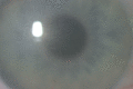
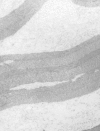
Corneal transparency requires that collagen fibrils be properly organized with a uniform diameter and a regular interfibrillar space. Congenital stromal corneal dystrophy (CSCD) is characterized by stromal opacities throughout the cornea. By transmission electron microscopy these opacities are seen as layers of amorphous material with thin filaments. The reported DCN pathogenic variants all lead to formation of a truncated decorin that has a tendency to aggregate in vitro. Decorin is found to accumulate in the amorphous areas. The authors hypothesize that truncated decorin accumulates in CSCD, thus causing the opacities [Bredrup et al 2010]. Studies in mice have shown that extracellular export of truncated decorin is necessary for the development of corneal opacities [Mellgren et al 2015].

Gene structure. DCN spans 3,777 kb. The full-length gene consists of eight exons with the AUG start codon in exon 2. For a detailed summary of gene and protein information, see Table A, Gene.

Benign variants. An imperfect dinucleotide repeat variation is in intron 1. In a small cohort of individuals with type 1 diabetes, one of these variants was associated with slower progression of renal disease [De Cosmo et al 2002] (see Table A, HGMD).

Pathogenic variants. Four variants associated with CSCD have been detected in DCN (Table 3) [Bredrup et al 2005, Rødahl et al 2006, Kim et al 2011, Jing et al 2014]. They are all frameshift variants located in the last coding exon. The resulting proteins are predicted to have a few altered terminal amino acid residues and a deletion of the 33 C-terminal amino acids.

Lee et al [2012] have reported a c.1036T>G (p.Cys346Gly) variant in a family where affected individuals developed corneal opacities in adult life. The functional consequences of this variant at the protein level have not yet been studied. The authors suggest that this could represent a mild form of CSCD.

Table 3.

DCN Pathogenic Variants Discussed in This GeneReview

DNA Nucleotide ChangePredicted Protein ChangeReference Sequences
c.1036T>Gp.Cys346Gly NM_001920​.3
NP_001911​.1
c.967delTp.Ser323LeufsTer5
c.941delCp.Pro314HisfsTer14
c.947delGp.Gly316AspfsTer12
c.962delAp.Lys321ArgfsTer7

Variants listed in the table have been provided by the authors. GeneReviews staff have not independently verified the classification of variants.

GeneReviews follows the standard naming conventions of the Human Genome Variation Society (varnomen​.hgvs.org). See Quick Reference for an explanation of nomenclature.

Normal gene product. Decorin is a member of the class I family of small leucine-rich repeat proteoglycans; other members of this class include biglycan and asporin. A 14-amino-acid propeptide is cleaved from the N-terminal part to make the mature core protein of 329 amino acids. The mature core protein is substituted with one chondroitin/dermatan sulphate glycosaminoglycan chain at p.Ser4 and two or three N-linked oligosaccharides at residues p.Asn181, p.Asn232, and p.Asn273.

Decorin is expressed in a wide range of connective tissues and can bind to several biologically important molecules including collagen I, collagen VI, fibronectin, thrombospondin, epidermal growth factor receptor, insulin-like growth factor 1 receptor, and transforming growth factor beta. It has been implicated in a number of biologic processes, primarily in the regulation of collagen fibril morphology, where there is evidence suggesting that decorin is the main inhibitor of lateral growth of collagen fibrils [Zhang et al 2009]. In addition, decorin may play an important role as a regulatory protein in processes that include cell adhesion, cell proliferation, angiogenesis, cell matrix formation, autophagy, and carbohydrate metabolism [Schaefer & Iozzo 2008, Gubbiotti et al 2018].

Abnormal gene product. Pathogenic frameshift variants so far detected in DCN are predicted to result in alteration of a few amino acids and premature protein truncation (i.e., of the 33 carboxy-terminal amino acids) [Bredrup et al 2005, Rødahl et al 2006, Kim et al 2011, Jing et al 2014]. The predicted truncation is hypothesized to cause decorin to accumulate in the cornea, causing corneal opacities. The mechanism of accumulation may be due to aggregation of truncated decorin [Bredrup et al 2010].

Note: Decorin has attracted particular attention in malignancies where the decorin protein has been shown to be a strong inhibitor of cell growth and to act as a pro-apoptotic agent. None of these studies concerns germline variants in humans.

References

Literature Cited

  • Acar BT, Bozkurt KT, Duman E, Acar S. Bilateral cloudy cornea: is the usual suspect congenital hereditary endothelial dystrophy or stromal dystrophy? BMJ Case Rep. 2016:2016. [PMC free article: PMC4854138] [PubMed: 27107055]
  • Bredrup C, Knappskog PM, Majewski J, Rødahl E, Boman H. Congenital stromal dystrophy of the cornea caused by a mutation in the decorin gene. Invest Ophthalmol Vis Sci. 2005;46:420–6. [PubMed: 15671264]
  • Bredrup C, Stang E, Bruland O, Palka BP, Young RD, Haavik J, Knappskog PM, Rødahl E. Decorin accumulation contributes to the stromal opacities found in congenital stromal corneal dystrophy. Invest Ophthalmol Vis Sci. 2010;51:5578–82. [PubMed: 20484579]
  • De Cosmo S, Tassi V, Thomas S, Piras GP, Trevisan R, Cavallo Perin P, Bacci S, Zucaro L, Cisternino C, Trischitta V, Viberti GC. The decorin gene 179 allelic variant is associated with a slower progression of renal disease in patients with type 1 diabetes. Nephron. 2002;92:72–6. [PubMed: 12187087]
  • Gubbiotti MA, Seifert E, Rodeck U, Hoek JB, Iozzo RV. Metabolic reprogramming of murine cardiomyocytes during autophagy requires the extracellular nutrient sensor decorin. J Biol Chem. 2018;293:16940–50. [PMC free article: PMC6204907] [PubMed: 30049794]
  • Jing Y, Kumar PR, Zhu L, Edward DP, Tao S, Wang L, Chuck R, Zhang C. Novel decorin mutation in a Chinese family with congenital stromal corneal dystrophy. Cornea. 2014;33:288–93. [PubMed: 24413633]
  • Kim JH, Ko JM, Lee I, Kim JY, Kim MJ, Tchah H. A novel mutation of the decorin gene identified in a Korean family with congenital hereditary stromal dystrophy. Cornea. 2011;30:1473–7. [PubMed: 21993463]
  • Lee JH, Ki CS, Chung ES, Chung TY. A novel decorin gene mutation in congenital hereditary stromal dystrophy: a Korean family. Korean J Ophthalmol. 2012;26:301–5. [PMC free article: PMC3408537] [PubMed: 22870031]
  • Mellgren AE, Bruland O, Vedeler A, Saraste J, Schönheit J, Bredrup C, Knappskog PM, Rødahl E. Development of congenital stromal corneal dystrophy is dependent on export and extracellular deposition of truncated decorin. Invest Ophthalmol Vis Sci. 2015;56:2909–15. [PubMed: 26029887]
  • Odland M. Dystrophia corneae parenchymatosa congenita. A clinical, morphological and histochemical examination. Acta Ophthalmol (Copenh). 1968;46:477–85. [PubMed: 5304426]
  • Pouliquen Y, Lacombe E, Schreinzer C, Giraud JP, Savoldelli M. Familial congenital dystrophy of the corneal stroma: Turpin's syndrome (author's transl). J Fr Ophtalmol. 1979;2:115–25. [PubMed: 312637]
  • Rahbari R, Wuster A, Lindsay SJ, Hardwick RJ, Alexandrov LB, Turki SA, Dominiczak A, Morris A, Porteous D, Smith B, Stratton MR, Hurles ME, et al. Timing, rates and spectra of human germline mutation. Nat Genet. 2016;48:126–33. [PMC free article: PMC4731925] [PubMed: 26656846]
  • Rødahl E, Van Ginderdeuren R, Knappskog PM, Bredrup C, Boman H. A second decorin frame shift mutation in a family with congenital stromal corneal dystrophy. Am J Ophthalmol. 2006;142:520–1. [PubMed: 16935612]
  • Schaefer L, Iozzo RV. Biological functions of the small leucine-rich proteoglycans: from genetics to signal transduction. J Biol Chem. 2008;283:21305–9. [PMC free article: PMC2490788] [PubMed: 18463092]
  • Turpin R, Tisserand M, Sérane J. Opacités cornéennes héréditaires et congénitales réparties sur trois générations et atteignant deux jumelles monozygotes. Article in French. Arch Ophthalmol (Paris). 1939;3:109–11.
  • Van Ginderdeuren R, De Vos R, Casteels I, Foets B. Report of a new family with dominant congenital heredity stromal dystrophy of the cornea. Cornea. 2002;21:118–20. [PubMed: 11805522]
  • Weiss JS, Møller HU, Aldave AJ, Seitz B, Bredrup C, Kivelä T, Munier FL, Rapuano CJ, Nischal KK, Kim EK, Sutphin J, Busin M, Labbé A, Kenyon KR, Kinoshita S, Lisch W (2015) IC3D classification of corneal dystrophies--edition 2. Cornea 34:117-59. [PubMed: 25564336]
  • Witschel H, Fine BS, Grützner P, McTigue JW. Congenital hereditary stromal dystrophy of the cornea. Arch Ophthalmol. 1978;96:1043–51. [PubMed: 350201]
  • Zhang G, Chen S, Goldoni S, Calder BW, Simpson HC, Owens RT, McQuillan DJ, Young MF, Iozzo RV, Birk DE. Genetic evidence for the coordinated regulation of collagen fibrillogenesis in the cornea by decorin and biglycan. J Biol Chem. 2009;284:8888–97. [PMC free article: PMC2659246] [PubMed: 19136671]

Chapter Notes

Revision History

  • 29 November 2018 (sw) Comprehensive update posted live
  • 2 February 2012 (me) Comprehensive update posted live
  • 25 November 2008 (me) Review posted live
  • 10 September 2008 (er) Original submission
Copyright © 1993-2024, University of Washington, Seattle. GeneReviews is a registered trademark of the University of Washington, Seattle. All rights reserved.

GeneReviews® chapters are owned by the University of Washington. Permission is hereby granted to reproduce, distribute, and translate copies of content materials for noncommercial research purposes only, provided that (i) credit for source (http://www.genereviews.org/) and copyright (© 1993-2024 University of Washington) are included with each copy; (ii) a link to the original material is provided whenever the material is published elsewhere on the Web; and (iii) reproducers, distributors, and/or translators comply with the GeneReviews® Copyright Notice and Usage Disclaimer. No further modifications are allowed. For clarity, excerpts of GeneReviews chapters for use in lab reports and clinic notes are a permitted use.

For more information, see the GeneReviews® Copyright Notice and Usage Disclaimer.

For questions regarding permissions or whether a specified use is allowed, contact: ude.wu@tssamda.

Bookshelf ID: NBK2690PMID: 20301741

Views

Tests in GTR by Gene

Related information

  • MedGen
    Related information in MedGen
  • OMIM
    Related OMIM records
  • PMC
    PubMed Central citations
  • PubMed
    Links to PubMed
  • Gene
    Locus Links

Similar articles in PubMed

See reviews...See all...

Recent Activity

Your browsing activity is empty.

Activity recording is turned off.

Turn recording back on

See more...