U.S. flag

An official website of the United States government

NCBI Bookshelf. A service of the National Library of Medicine, National Institutes of Health.

Adam MP, Feldman J, Mirzaa GM, et al., editors. GeneReviews® [Internet]. Seattle (WA): University of Washington, Seattle; 1993-2024.

Cover of GeneReviews®

GeneReviews® [Internet].

Show details

Epidermolysis Bullosa with Pyloric Atresia

Synonym: EB-PA

, MD and , DO, MS.

Author Information and Affiliations

Initial Posting: ; Last Update: January 26, 2023.

Estimated reading time: 30 minutes

Summary

Clinical characteristics.

Epidermolysis bullosa with pyloric atresia (EB-PA) is characterized by fragility of the skin and mucous membranes, manifested by blistering with little or no trauma; congenital pyloric atresia; renal and/or ureteral anomalies; and protein-losing enteropathy. The course of EB-PA is usually severe and most often lethal in the neonatal period. Those who survive may have severe blistering with formation of granulation tissue on the skin around the mouth, nose, diaper area, fingers, and toes, and internally around the trachea. However, some affected individuals have little or no blistering later in life. Additional features shared by EB-PA and the other major forms of epidermolysis bullosa (EB) include congenital localized absence of skin (aplasia cutis congenita) affecting the extremities and/or head, milia, nail dystrophy, scarring alopecia, hypotrichosis, and corneal abnormalities.

Diagnosis/testing.

The diagnosis of EB-PA is established in a proband with characteristic clinical findings by identification of biallelic pathogenic variants in ITGA6, ITGB4, or PLEC. Skin biopsy using transmission electron microscopy and/or immunofluorescent antibody/antigen mapping can be considered in those with inconclusive molecular genetic testing.

Management.

Treatment of manifestations: Tracheostomy when indicated for respiratory failure; nutrition consult to address oral intake and nutritional needs; minimization of new blister formation by teaching caretakers proper handling of infants and children to protect skin from shearing forces, wrapping and padding of extremities, and use of soft and properly fitted clothing and footwear; lance and drain new blisters and dress with three layers (primary: nonadherent; secondary: for stability and protection; tertiary: elastic properties to insure integrity); antibiotics and antiseptics to treat and prevent wound infections; surgical intervention to correct pyloric atresia; gastrostomy if indicated; treatment of protein-losing enteropathy by gastroenterologist; referral to urologist and/or nephrologist for renal anomalies, abnormal voiding, and/or abnormal renal function; calcium, vitamin D, vitamin A, zinc, carnitine, selenium, and iron supplements as indicated by laboratory studies; treatment of corneal abnormalities by ophthalmologist; psychosocial support; palliative care consultation.

Surveillance: Assessment of oral mucosa, feeding, and nutritional status at each visit; assessment of tracheal involvement at each visit; assessment of skin for blisters and infection at each visit; assessment of renal function per nephrologist; assessment for gastrointestinal involvement at each visit; assessment of urinary involvement per urologist; annual CBC, iron studies, zinc, vitamin D, selenium, carnitine, and vitamin A; periodic DXA scan for risk of osteopenia; assessment for corneal abnormalities at each visit; assessment of family needs at each visit.

Agents/circumstances to avoid: Ordinary medical tape or Band-Aids®; EKG leads with adhesive; poorly fitting or coarse-textured clothing and footwear; activities that traumatize the skin.

Pregnancy management: Consider cesarean section to reduce trauma to the skin of an affected fetus during delivery.

Genetic counseling.

EB-PA is inherited in an autosomal recessive manner. If both parents are known to be heterozygous for an EB-PA-related pathogenic variant, each sib of an affected individual has at conception a 25% chance of being affected, a 50% chance of being an asymptomatic carrier, and a 25% chance of being unaffected and not a carrier. Once the EB-PA-causing pathogenic variants have been identified in an affected family member, carrier testing for at-risk relatives and prenatal/preimplantation genetic testing for EB-PA are possible.

Diagnosis

Suggestive Findings

Epidermolysis bullosa with pyloric atresia (EB-PA) should be suspected in newborns with the following clinical features:

  • Fragility of the skin with:
    • Blistering with little or no trauma. Blistering may be mild or severe. Blisters generally heal with no significant scarring.
    • Significant oral and mucous membrane involvement
    • Large areas of absent skin (aplasia cutis congenita), often with a thin membranous covering, affecting the extremities or head
  • Congenital pyloric atresia with vomiting and abdominal distention resulting from complete obstruction of the gastric outlet. Radiographs reveal that the stomach is distended and filled with air (see Figure 1).
  • Renal and/or ureteral anomalies, including dysplastic/multicystic kidney, hydronephrosis/hydroureter, acute renal tubular necrosis, obstructive uropathy, ureterocele, duplicated renal collecting system, vesicoureteral reflux, interstitial nephritis, and/or absent bladder
  • Protein-losing enteropathy
Figure 1.

Figure 1.

Single gastric bubble (white arrow) in a newborn with epidermolysis bullosa with pyloric atresia

Family history is consistent with autosomal recessive inheritance (e.g., affected sibs and/or parental consanguinity). Absence of a known family history does not preclude the diagnosis.

Establishing the Diagnosis

The diagnosis of EB-PA is established in a proband with suggestive findings and one of the following:

Note: (1) Molecular genetic testing is the preferred diagnostic method; skin biopsy for diagnostic purposes should only be considered if molecular results are inconclusive. (2) Per ACMG/AMP variant interpretation guidelines, the terms "pathogenic variants" and "likely pathogenic variants" are synonymous in a clinical setting, meaning that both are considered diagnostic, and both can be used for clinical decision making [Richards et al 2015]. Reference to "pathogenic variants" in this section is understood to include any likely pathogenic variants. (3) Identification of biallelic variants of uncertain significance (or of one known pathogenic variant and one variant of uncertain significance) does not establish or rule out the diagnosis.

Molecular Genetic Testing

Molecular testing approaches can include a combination of a multigene panel and comprehensive genomic testing (exome sequencing, genome sequencing) depending on the phenotype.

Gene-targeted testing requires that the clinician determine which gene(s) are likely involved, whereas genomic testing does not. Individuals with the distinctive findings described in Suggestive Findings are likely to be diagnosed using gene-targeted testing (see Option 1), whereas those with a phenotype indistinguishable from many other inherited disorders with epidermolysis bullosa are more likely to be diagnosed using genomic testing (see Option 2).

Option 1

A multigene panel that includes ITGA6, ITGB4, PLEC, and other genes of interest (see Differential Diagnosis) is most likely to identify the genetic cause of the condition while limiting identification of variants of uncertain significance and pathogenic variants in genes that do not explain the underlying phenotype. Note: (1) The genes included in the panel and the diagnostic sensitivity of the testing used for each gene vary by laboratory and are likely to change over time. (2) Some multigene panels may include genes not associated with the condition discussed in this GeneReview. (3) In some laboratories, panel options may include a custom laboratory-designed panel and/or custom phenotype-focused exome analysis that includes genes specified by the clinician. (4) Methods used in a panel may include sequence analysis, deletion/duplication analysis, and/or other non-sequencing-based tests.

For an introduction to multigene panels click here. More detailed information for clinicians ordering genetic tests can be found here.

Option 2

Comprehensive genomic testing does not require the clinician to determine which gene(s) are likely involved. Exome sequencing is most commonly used; genome sequencing is also possible.

For an introduction to comprehensive genomic testing click here. More detailed information for clinicians ordering genomic testing can be found here.

Table 1.

Molecular Genetic Testing Used in Epidermolysis Bullosa with Pyloric Atresia

Gene 1, 2Proportion of EB-PA Attributed to Pathogenic Variants in Gene 3Proportion of Pathogenic Variants 4 Detectable by Method
Sequence analysis 5Gene-targeted deletion/duplication analysis 6
ITGA6 5%100%None reported 7
ITGB4 60%>95% 8<5% 9
PLEC 15%100%None reported 7
Unknown20%NANA

EB-PA = epidermolysis bullosa with pyloric atresia; NA = not applicable

1.

Genes are listed in alphabetic order.

2.
3.
4.

See Molecular Genetics for information on variants detected in this gene.

5.

Sequence analysis detects variants that are benign, likely benign, of uncertain significance, likely pathogenic, or pathogenic. Variants may include small intragenic deletions/insertions and missense, nonsense, and splice site variants; typically, exon or whole-gene deletions/duplications are not detected. For issues to consider in interpretation of sequence analysis results, click here.

6.

Gene-targeted deletion/duplication analysis detects intragenic deletions or duplications. Methods used may include a range of techniques such as quantitative PCR, long-range PCR, multiplex ligation-dependent probe amplification (MLPA), and a gene-targeted microarray designed to detect single-exon deletions or duplications.

7.

To date, large exon or multiexon ITGA6 or PLEC deletions have not been reported in individuals with EB-PA [Stenson et al 2020].

8.

In one report, 50% of persons of Hispanic heritage with EB-PA in the United States had the ITGB4 pathogenic variant p.Cys61Tyr [Varki et al 2006].

9.

At least five large single-exon or multiexon ITGB4 deletions have been reported [Birnbaum et al 2008, Mencía et al 2016, Stenson et al 2020]

Skin Biopsy

Examination of a skin biopsy by transmission electron microscopy (TEM) and/or immunofluorescent antibody/antigen mapping is sometimes performed to establish the diagnosis of EB-PA, although molecular genetic testing is preferred.

If skin biopsy is pursued, a punch biopsy that includes the full basement membrane zone should be performed. The biopsy should be taken from the leading edge of a fresh (<12 hours old) blister or from a mechanically induced blister (with a pencil eraser rubbed on the skin). Older blisters undergo change that may obscure the diagnostic morphology.

Light microscopy is inadequate and unacceptable for the accurate diagnosis of any subtypes of epidermolysis bullosa (EB).

Transmission electron microscopy (TEM)

  • TEM is used to examine the number and morphology of the basement membrane zone structures – in particular, the presence and morphology of hemidesmosomes. TEM also shows the condition of anchoring filaments, anchoring fibrils, and keratin intermediate filaments. TEM also allows examination of microvesicles that show the tissue cleavage plane.
  • Specimens must be placed in a special fixation medium (e.g., glutaraldehyde) as designated by the laboratory performing the test.
  • Formaldehyde-fixed samples cannot be used for electron microscopy.

Findings on TEM in EB-PA include the following:

Immunofluorescent antibody/antigen mapping

  • Specimens should be sent in a sterile carrying medium (e.g., Michel's or Zeus's) as specified by the laboratory performing the test.
  • Some laboratories prefer flash-frozen tissue.
  • In some laboratories the mapping only designates the level of the cleavage by using various marker antibodies of different layers of the basement membrane. A laboratory that has the antigens for the proteins of interest in EB is preferred because both the level of cleavage and the presence or absence of the specific mutated gene products can be assessed.
  • Direct immunofluorescence for immunoglobulins is not appropriate or useful in individuals with EB-PA.

Findings on immunofluorescent antibody/antigen mapping in EB-PA include the following:

  • Abnormal or absent staining with antibodies to α6β4 integrin in EB-PA and other rare forms of junctional epidermolysis bullosa (JEB) as a result of pathogenic variants in either ITGA6 or ITGB4
  • Abnormal or absent staining with antibodies to plectin in EB-PA as a result of pathogenic variants in PLEC

Normal staining for other antigens (e.g., collagen VII, laminin 332, keratins 5 and 14) confirms the diagnosis of EB-PA.

Note: (1) Especially in milder forms of EB, immunofluorescent studies are often not sufficient to make the diagnosis because near-normal antigen levels are detected, and no cleavage plane is observed. (2) Absence of one hemidesmosomal component (e.g., ITGA6 or ITGB4) may reduce the staining of other hemidesmosomal components as well (e.g., PLEC, COL17). In these cases, electron microscopic examination of a skin biopsy must be performed.

Clinical Characteristics

Clinical Description

The course of epidermolysis bullosa with pyloric atresia (EB-PA) is usually severe and often lethal in the neonatal period. Most affected children die as neonates due to mucosal erosions and blistering, pyloric stenosis or atresia, respiratory failure, or overwhelming infection.

Cutaneous manifestations. Those who survive the neonatal period may have severe blistering with formation of granulation tissue on the skin around the mouth, diaper area, nose, fingers, and toes, and internally around the trachea. However, some affected individuals have little or no blistering later in life.

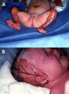
  • Congenital localized absence of skin (aplasia cutis congenita). Infants with extensive aplasia cutis congenita and blistering or erosions may have fatal infections with sepsis and severe electrolyte imbalance in the first weeks to months of life. Aplasia cutis congenita can be extensive in these neonates, especially on the extremities and sides of the scalp (see Figure 2). One review reported that 50% of individuals with EB-PA and aplasia cutis congenita died during the first months of life [Martinez-Moreno et al 2020].
  • Mucosal erosions are predominantly oropharyngeal, but erosions have also been reported of the bladder, trachea, bowel, stomach, and esophagus [Mylonas et al 2019].
  • Deformities of the ear and/or nose, such as ear hypoplasia
  • Milia
  • Nail dystrophy and granulation tissue of the nail, including changes in size, color, shape, or texture and subsequent loss of nails
  • Scarring alopecia. Complete loss of scalp hair follicles as a result of scarring
  • Hypotrichosis. Reduction in the number of hair follicles in a given area compared to the number of hair follicles in the same area of an unaffected individual of the same sex
  • Exuberant granulation tissue does not usually appear until early childhood, and most children with EB-PA do not survive beyond infancy.
  • Wound infections. Treatment of chronic wound infections is a challenge. Many affected individuals become infected with resistant bacteria, most often methicillin-resistant Staphylococcus aureus (MRSA), multidrug-resistant Pseudomonas aeruginosa, and group A beta-hemolytic Streptococci.
Figure 2. . Child with epidermolysis bullosa with pyloric atresia and extensive aplasia cutis congenita of the extremities (A) and scalp (B).

Figure 2.

Child with epidermolysis bullosa with pyloric atresia and extensive aplasia cutis congenita of the extremities (A) and scalp (B). Reproduced with permission from Lucky et al [2021]

Pyloric atresia may be detected in utero as polyhydramnios via ultrasound or MRI (see Genetic Counseling, Prenatal Testing and Preimplantation Genetic Testing) or becomes evident at birth. It is characterized by vomiting, failure to tolerate any feeding or to pass stool, and a distended abdomen with a large stomach bubble (see Figure 1). Surgical repair of the pyloric atresia is necessary for survival. Surgical repair is most commonly performed with a Heineke-Mikulicz pyloroplasty (for type 1 pyloric atresia) or gastroduodenostomy (for type 2 pyloric atresia) [Mylonas et al 2019].

Renal and ureteral anomalies can include dysplastic/multicystic kidney, hydronephrosis/hydroureter, acute renal tubular necrosis, obstructive uropathy, ureterocele, duplicated renal collecting system, vesicoureteral reflux, interstitial nephritis, and/or absent bladder [Puvabanditsin et al 1997, Kambham et al 2000, Wallerstein et al 2000, Nakano et al 2001, Varki et al 2006, Pfendner et al 2007, Walker et al 2017, Mylonas et al 2019].

Protein-losing enteropathy has been reported in some individuals with EB-PA, resulting in large-volume diarrhea and poor weight gain [Verma et al 2020, Wee et al 2021, DeMaria et al 2022, Kaneyasu et al 2022]. Bowel biopsies show decreased integrin B4 [Wee et al 2021] as well as lamina lucida separation with hypoplastic hemidesmosomes [DeMaria et al 2022].

Ocular findings are seen in a minority of individuals but can include corneal blisters/erosions, conjunctivitis, and corneal opacification [Mylonas et al 2019].

Fluids/electrolytes/nutrition. Fluid and electrolyte issues can be significant and life threatening in the neonatal period. Infants with widespread disease require careful management.

In children who survive the newborn period, nutritional deficiencies can include the following:

  • Low calcium and/or vitamin D resulting in osteopenia and osteoporosis
  • Zinc deficiency, which can impair wound healing [Mellerio et al 2007]
  • Chronic iron deficiency anemia

Phenotype Correlations by Gene

ITGA6 pathogenic variants are associated with a poor prognosis [Masunaga et al 2017, Mylonas et al 2019].

ITGB4 pathogenic variants are associated with a more variable prognosis than either ITGA6- or PLEC-related EB-PA.

PLEC pathogenic variants are associated with a poor prognosis, and the majority of infants die as neonates or in the first year [Kaneyasu et al 2022, Pongmee et al 2022]. Individuals with PLEC pathogenic variants have been reported to have cardiomyopathy [Vahidnezhad et al 2022], but there are no reports of cardiomyopathy occurring in an individual with pyloric atresia.

Genotype-Phenotype Correlations

ITGB4. The most severe cutaneous manifestations are caused by biallelic pathogenic variants that result in a premature termination codon, although pathogenic variants between exon 3 and intron 11 are also associated with a poor prognosis. A more favorable prognosis is typically associated with missense variants and in-frame insertions or deletions. However, missense variants in the plectin-binding region or in cysteine-rich domains are associated with a poor prognosis. Several additional missense variants result in a severe phenotype, such as the recurrent ITGB4 variant p.Cys61Tyr, which is common in Hispanic individuals with EB-PA [Varki et al 2006, Masunaga et al 2015, Mutlu et al 2015, Mencía et al 2016, Mylonas et al 2019].

Nomenclature

Although ITGA6-, ITGB4-, and PLEC-related EB-PA are clinically quite similar, in the 2020 epidermolysis bullosa (EB) classification system [Has et al 2020], individuals with EB-PA due to PLEC pathogenic variants are considered to have EB simplex and those with EB-PA due to ITGB4 or ITGA6 pathogenic variants are considered to have junctional EB.

ITGA6- and ITGB4-related EB-PA may also be referred to as Carmi syndrome or junctional epidermolysis bullosa with pyloric atresia (JEB-PA).

Prevalence

EB-PA is rare, and its prevalence has not been determined. However, it can be conservatively estimated at fewer than one in 5,000 (~10 times rarer than severe junctional EB [previously called Herlitz junctional EB], the carrier frequency of which is ~1:700).

Differential Diagnosis

Pyloric Atresia

In contrast to pyloric stenosis, which presents insidiously with vomiting, pyloric atresia is present at birth and causes complete obstruction of the gastric outlet. The diagnosis of epidermolysis bullosa with pyloric atresia (EB-PA) should be considered in every neonate with pyloric atresia regardless of the degree of skin blistering.

Epidermolysis Bullosa

The 2020 classification system [Has et al 2020] names four major types of epidermolysis bullosa (EB). Classification into major type is based on the location of blistering in relation to the dermal-epidermal junction of the skin.

  • EB simplex (EBS). Blistering occurs within basal keratinocytes.
  • Junctional EB (JEB). Splitting is seen in the lamina lucida of the basement membrane of the epidermis or just above the basement membrane at the level of the hemidesmosomes in the lowest level of the keratinocytes layer.
  • Dystrophic EB (DEB). Blisters form below the basement membrane, in the superficial dermis.
  • Kindler EB. The level of cleavage of blisters can be variable: intradermal, junctional, and dermal cleavage planes have been reported in a single biopsy from one individual.

All forms of EB are characterized by increased skin (and often mucosa) fragility and blistering with little or no trauma. Subtypes are predominantly determined by clinical features (see Table 3) and supported by molecular diagnosis (see Table 4). In the neonatal period, it is almost impossible to determine a subtype based on clinical phenotype alone. Subtle clues include hypergranulation tissue around the nails in JEB and absence of lingual papillae in DEB, though definitive diagnosis requires molecular testing [Lucky et al 2021].

Table 3.

Clinical Features Observed in the Four Major Types of Epidermolysis Bullosa

FeatureComment
Easy fragility of skin (& often mucosa) manifested by blistering w/little or no traumaShared by 4 major EB types
Blisters can be induced w/friction (amount of friction can vary) & enlarged by applying pressure to blister edge
Mucosal involvementMay not be helpful discriminator
Nail involvement
Presence or absence of milia
Scarring 1In DEB, wounds heal w/scarring. However, scarring can occur in EBS & JEB as a result of infection of erosions or scratching, which further damages exposed surfaces.
Congenital absence of skin (congenital aplasia cutis)Can be seen in all forms of EB & may not be distinguishing feature of any particular form of EB
Corneal erosionsMay indicate either DEB or JEB
Esophageal stricturesMay indicate DEB, less likely JEB
Nail & tooth enamel involvementGenerally, indicates JEB; in rare instances can be seen in DEB or EBS (e.g., in EBS, intermediate w/muscular dystrophy)
Pseudosyndactyly (mitten deformities) 2Usually suggests DEB; typically does not develop until early to mid-childhood

DEB = dystrophic epidermolysis bullosa; EB = epidermolysis bullosa; EBS = epidermolysis bullosa simplex; JEB = junctional epidermolysis bullosa

1.

Postinflammatory changes, such as those seen in severe EBS, are often mistaken for scarring or mottled pigmentation.

2.

Results from extensive scarring of the hands and feet in older children and adults

Although clinical examination is useful in determining the extent of blistering and the presence of oral and other mucous membrane lesions, defining characteristics such as the presence and extent of scarring – especially in young children and neonates – may not be established or significant enough to allow identification of EB type. Thus, molecular genetic testing (or less commonly skin biopsy) is usually required to establish the most precise diagnosis.

Table 4.

Genes Associated with the Four Major Epidermolysis Bullosa Types

GeneEB TypeMOI
CD151 EBS AR
COL17A1 JEB AR
COL7A1 DEB AD, AR
DST EBS AR
EXPH5 EBS AR
FERMT1 Kindler EB AR
ITGA3 JEB AR
ITGA6 JEB AR
ITGB4 JEB AR
KLHL24 EBS AD
KRT5 EBS AR, AD
KRT14 EBS AR, AD
LAMA3 JEB AR
LAMB3 JEB AR
LAMC2 JEB AR
PLEC EBS AR, AD

Adapted from Has et al [2020], Table 1

AD = autosomal dominant; AR = autosomal recessive; DEB = dystrophic epidermolysis bullosa; EB = epidermolysis bullosa; EBS = epidermolysis bullosa simplex; JEB = junctional epidermolysis bullosa; MOI = mode of inheritance

Management

No clinical practice guidelines for epidermolysis bullosa with pyloric atresia (EB-PA) have been published.

Evaluations Following Initial Diagnosis

To establish the extent of disease and needs in an individual diagnosed with EB-PA, the evaluations in Table 5 (if not performed as part of the evaluation that led to the diagnosis) are recommended.

Table 5.

Recommended Evaluations Following Initial Diagnosis in Individuals with Epidermolysis Bullosa with Pyloric Atresia

System/ConcernEvaluationComment
Cutaneous & mucosal manifestations Eval of blister formation sites incl skin & oral mucosaEndoscopy can be traumatic & should be avoided if possible.
Tracheal manifestations Assess for hoarse cry in infantMay be caused by airway obstruction w/granulation tissue or tracheomalacia 1
Pyloric atresia Surgical referral for those w/manifestations of pyloric atresia
Renal & ureteral anomalies
  • Assess renal function w/serum BUN & creatinine
  • Urinalysis
  • Renal ultrasound
Genetic counseling By genetics professionals 2To inform affected persons & their families about nature, MOI, & implications of EB-PA to facilitate medical & personal decision making
Family support
& resources
Assess need for:

BUN = blood urea nitrogen; EB-PA = epidermolysis bullosa with pyloric atresia; MOI = mode of inheritance

1.
2.

Medical geneticist, certified genetic counselor, certified advanced genetic nurse

Treatment of Manifestations

There is no cure for EB-PA, and most infants die in the first years of life from complications secondary to mucosal erosions and blistering; esophageal erosions, stenosis, and/or atresia; pyloric stenosis or atresia; or overwhelming infection. Supportive care to improve quality of life, maximize function, and reduce complications is recommended [Lucky et al 2021]. This ideally involves multidisciplinary care by specialists in relevant fields (see Table 6).

There has been one report of Integra® skin grafting to treat aplasia cutis congenita in an infant with EB-PA [Trah et al 2018]. The skin graft was successful but unfortunately the neonate did not survive.

Table 6.

Treatment of Manifestations in Individuals with Epidermolysis Bullosa with Pyloric Atresia

Manifestation/ConcernTreatmentConsiderations/Other
Mucosal involvement (incl tracheal & gastrointestinal) Decisions about tracheostomy should involve family & consider medical condition of infant.Poor prognosis & severe pain warrants discussion w/family & hospital ethics committee to determine type of intervention & comfort care to provide. 1
  • Consult w/dietitian or nutritionist if there is significant mucosal blistering in mouth preventing adequate oral intake.
  • Consider placement of gastrostomy.
  • Evaluate & treat protein-losing enteropathy.
  • Additional nutritional support incl gastrostomy tube feeding when necessary
  • Maintenance of tracheostomy & gastrostomy can be difficult due to fragile skin.
Skin Minimize new blister formation by:
  • Teaching caretakers proper handling of infants & children to protect skin from shearing forces;
  • Wrapping & padding extremities;
  • Use of soft & properly fitted clothing & footwear.
New blisters should be lanced & drained to prevent further spread from fluid pressure. In most cases, dressings for blisters involve three layers: 2
  • A primary nonadherent contact layer that does not strip top layers of epidermis. Tolerance to different primary layers varies. Primary layers include: dressings impregnated w/emollient (e.g., petrolatum, topical antiseptic); nonstick products (e.g., Telfa®, N-Terface®); silicone-based products w/o adhesive (e.g., Mepitel®, Mepilex®); additional topical antibiotic or antiseptic (e.g., bacitracin, mupirocin, silver, honey).
  • A secondary layer that provides stability for primary layer & adds padding to allow more activity, such as rolls of gauze (e.g., Kerlix®, Conform®)
  • A tertiary layer that usually has some elastic properties & ensures integrity of dressing (e.g., Coban® or elasticized tube gauze of varying diameters such as BandNet® or Tubifast®).
  • Treatment of wound infection using antibiotics & antiseptics
  • Appropriate footwear & physical therapy are essential to preserve ambulation in children w/delays or difficulty walking due to blistering &/or hyperkeratosis.
Gastrointestinal
  • Surgical intervention to correct pyloric atresia
  • Gastrostomy if indicated
  • Evaluate & treat protein-losing enteropathy.
Renal & ureteral anomalies
  • Referral to urologist if there are symptoms of difficulty or discomfort w/voiding
  • Referral to nephrologist if renal function studies &/or urinalysis are abnormal
Fluid/electrolyte/
nutritional deficiencies
  • Fluid replacement as needed
  • Calcium & vitamin D supplementation to prevent osteopenia
  • Zinc supplementation for wound healing 3
  • Supplementation of carnitine, selenium, vitamin A as indicated based on laboratory studies
  • Oral or intravenous iron infusions & red blood cell transfusions as needed for chronic anemia
Ocular symptoms Treatment of corneal abrasions per ophthalmologist
Social/Family
  • Psychosocial support, incl social services & psychological counseling 4
  • Palliative care consultation 5

Surveillance

To monitor existing manifestations in survivors, the individual's response to supportive care, and the emergence of new manifestations, the evaluations in Table 7 are recommended.

Table 7.

Recommended Surveillance for Individuals with Epidermolysis Bullosa with Pyloric Atresia

System/ConcernEvaluationFrequency
Mucosal involvement
  • Assessment of oral mucosa, feeding, & esophageal involvement
  • Assessment for hoarse cry (tracheal involvement)
At each visit & per ENT
Skin Assessment of blisters & skin infectionAt each visit & per dermatologist
Gastrointestinal Assessment of gastrointestinal involvementAt each visit
Renal & ureteral anomalies
  • Assess renal function w/serum BUN & creatinine
  • Urinalysis
Per nephrologist
Fluid/electrolyte/
nutritional deficiencies
  • CBC
  • Iron studies
  • Serum zinc
  • Serum vitamin D
  • Selenium, carnitine, vitamin A
Annually
DXA scan for osteopenia/osteoporosisNo guidelines have been established regarding age at which this should begin & frequency.
Ocular Assessment of corneal abnormalitiesAt each visit
Social/Family Assessment of family needs

BUN = blood urea nitrogen; DXA = dual-energy x-ray absorptiometry

Agents/Circumstances to Avoid

Most persons with EB-PA cannot use ordinary medical tape or Band-Aids®. EKG leads should be applied without adhesive.

Poor-fitting or coarse-textured clothing and footwear should be avoided as they can cause trauma.

In general, activities that traumatize the skin should be avoided. Affected individuals who are determined to participate in such activities should be encouraged to find creative ways to protect their skin.

Evaluation of Relatives at Risk

See Genetic Counseling for issues related to testing of at-risk relatives for genetic counseling purposes.

Pregnancy Management

Cesarean section is recommended by some obstetricians to reduce trauma to the skin of an affected fetus during delivery.

Therapies Under Investigation

Several therapies are being investigated in treatment of junctional epidermolysis bullosa (JEB) (see Junctional Epidermolysis Bullosa, Therapies Under Investigation). There are no therapies under investigation specifically for EB-PA due to the rarity of the condition.

The antibiotic gentamicin can induce read-through of premature termination codons. Topical and intravenous gentamicin is being investigated as a treatment for JEB [Kwong et al 2020, Mosallaei et al 2022] in individuals with LAMA3, LAMB3, and LAMC2 pathogenic variants; it has not been studied in genes associated with EB-PA. Systemic gentamicin has also been studied for treatment of PLEC-related epidermolysis bullosa simplex with muscular dystrophy due to nonsense variants [Martínez-Santamaría et al 2022].

Topical Oleogel-S10, made from birch bark extract, is approved for treatment of dystrophic epidermolysis bullosa (DEB) and JEB in individuals age six months or older in the European Union under the brand name Filsuvez® (see European Medicines Agency). A Phase III clinical trial has been completed in the United States, which enrolled individuals with JEB and DEB. However, wound healing in individuals with JEB treated with Oleogel-S10 was not significantly different than those treated with placebo [Murrell et al 2020].

Search ClinicalTrials.gov in the US and EU Clinical Trials Register in Europe for access to information on clinical studies for a wide range of diseases and conditions.

Genetic Counseling

Genetic counseling is the process of providing individuals and families with information on the nature, mode(s) of inheritance, and implications of genetic disorders to help them make informed medical and personal decisions. The following section deals with genetic risk assessment and the use of family history and genetic testing to clarify genetic status for family members; it is not meant to address all personal, cultural, or ethical issues that may arise or to substitute for consultation with a genetics professional. —ED.

Mode of Inheritance

Epidermolysis bullosa with pyloric atresia (EB-PA) is inherited in an autosomal recessive manner.

Risk to Family Members

Parents of a proband

Sibs of a proband

  • If both parents are known to be heterozygous for an EB-PA-related pathogenic variant, each sib of an affected individual has at conception a 25% chance of being affected, a 50% chance of being an asymptomatic carrier, and a 25% chance of being unaffected and not a carrier.
  • Heterozygotes (carriers) are asymptomatic. To date, there is no evidence to indicate that a heterozygous pathogenic variant in ITGA6, ITGB4, or PLEC results in EB-PA.

Offspring of a proband. The offspring of an individual with EB-PA are obligate heterozygotes (carriers) for an EB-PA-related pathogenic variant.

Other family members. Each sib of the proband's carrier parents is at a 50% risk of being a carrier of an EB-PA-related pathogenic variant.

Carrier Detection

Carrier testing for at-risk relatives requires prior identification of the EB-PA-related pathogenic variants in the family.

Related Genetic Counseling Issues

Family planning

  • The optimal time for determination of genetic risk and discussion of the availability of prenatal/preimplantation genetic testing is prior to pregnancy.
  • It is appropriate to offer genetic counseling (including discussion of potential risks to offspring and reproductive options) to young adults who are affected or at risk of being carriers.
  • Carrier testing for the reproductive partners of known carriers should be considered. Carrier testing for the reproductive partners individuals affected with EB-PA should also be considered, particularly if both partners are of the same ethnic background. The ITGB4 variant p.Cys61Tyr is common in individuals of Hispanic heritage with EB-PA in the United States [Varki et al 2006].

DNA banking. Because it is likely that testing methodology and our understanding of genes, pathogenic mechanisms, and diseases will improve in the future, consideration should be given to banking DNA from probands in whom a molecular diagnosis has not been confirmed (i.e., the causative pathogenic mechanism is unknown). For more information, see Huang et al [2022].

Prenatal Testing and Preimplantation Genetic Testing

Molecular genetic testing. Once the EB-PA-causing pathogenic variants have been identified in an affected family member, prenatal and preimplantation genetic testing for EB-PA are possible.

Ultrasound examination. Occasionally pyloric atresia may be suspected because of polyhydramnios, with or without elevated concentration of alpha-fetoprotein and acetylcholinesterase, and echogenic material in the amniotic fluid [Azarian et al 2006]. Gastric blockage may also be observed on fetal MRI [Maurice et al 2013, Merrow et al 2013]. Complete chorioamniotic membrane separation detected through ultrasound can also be suggestive of EB-PA [Dural et al 2014].

Differences in perspective may exist among medical professionals and within families regarding the use of prenatal testing. While most centers would consider use of prenatal testing to be a personal decision, discussion of these issues may be helpful.

Resources

GeneReviews staff has selected the following disease-specific and/or umbrella support organizations and/or registries for the benefit of individuals with this disorder and their families. GeneReviews is not responsible for the information provided by other organizations. For information on selection criteria, click here.

Molecular Genetics

Information in the Molecular Genetics and OMIM tables may differ from that elsewhere in the GeneReview: tables may contain more recent information. —ED.

Table A.

Epidermolysis Bullosa with Pyloric Atresia: Genes and Databases

Data are compiled from the following standard references: gene from HGNC; chromosome locus from OMIM; protein from UniProt. For a description of databases (Locus Specific, HGMD, ClinVar) to which links are provided, click here.

Table B.

OMIM Entries for Epidermolysis Bullosa with Pyloric Atresia (View All in OMIM)

147556INTEGRIN, ALPHA-6; ITGA6
147557INTEGRIN, BETA-4; ITGB4
226730EPIDERMOLYSIS BULLOSA, JUNCTIONAL 5B, WITH PYLORIC ATRESIA; JEB5B
601282PLECTIN; PLEC
612138EPIDERMOLYSIS BULLOSA SIMPLEX 5C, WITH PYLORIC ATRESIA; EBS5C
619817EPIDERMOLYSIS BULLOSA, JUNCTIONAL 6, WITH PYLORIC ATRESIA; JEB6

Molecular Pathogenesis

Integrins associate in pairs containing one alpha (α) and one beta (β) chain. The α6β4 integrin comprises one α6 (encoded by ITGA6) and one β4 integrin (encoded by ITGB4) from the integrin family of proteins and is a component of the hemidesmosomes of the epidermis. Plectin (encoded by PLEC) is a large cytolinker protein expressed in the epidermis, muscle, and other tissues. Together, along with collagen XVII, CD151 (which are transmembrane proteins), and BP230, plectin and α6β4 integrin are critical structural components of the hemidesmosomes in the epidermis. The α6β4 integrin and plectin associate, causing a conformational change to a stable complex. This stabilization is thought to cause the recruitment of the other proteins into the hemidesmosomes. The α6β4 integrin also binds laminin 332, located in the lamina lucida of the basement membrane, and plectin binds the cytokeratins (as well as BP230), forming a continuous network of proteins that anchor the basement membrane complex to the cytokeratin layers of the epidermis. The result is a structurally resilient network of proteins binding the layers of the epidermis together [Chung et al 2004, Guo et al 2006, Yoon et al 2006, Folgiero et al 2007, de Pereda et al 2009].

Mechanism of disease causation. Loss of function

Table 8.

Epidermolysis Bullosa with Pyloric Atresia: Gene-Specific Laboratory Considerations

Gene 1Special Consideration
ITGA6 2 transcript variants encoding 2 different isoforms have been reported. The cDNA of variant 1 comprises 5,680 bp w/an open reading frame of 3,273 nucleotides encoding 1,091 amino acids in 26 exons. Transcript variant 2 contains an alternate coding exon from variant 1 that results in a frameshift & is encoded in a 5,810-bp cDNA. The resulting protein isoform B is shorter (1,073 amino acids) than isoform A & has a distinct C terminus.
ITGB4 2 splicing variants express different isoforms of the protein [Pulkkinen et al 1997]. The most common epidermal variant does not express exon 33.
PLEC Expression of the different isoforms results from alternative splicing of exon 1 as well as use of different 5' untranslated regions. At least 11 variant transcripts have been described. There is also a transcript lacking exon 31.
1.

Genes from Table 1 in alphabetic order

Table 9.

Epidermolysis Bullosa with Pyloric Atresia: Notable Pathogenic Variants by Gene

Gene 1Reference SequencesDNA Nucleotide Change

(Alias 2)

Predicted Protein Change

(Alias 2)

Comment [Reference]
ITGB4 NM_000213​.3
NP_000204​.3
c.182G>Ap.Cys61TyrCommon pathogenic variant in Hispanic persons w/EB-PA in the US [Varki et al 2006]
PLEC NM_000445​.5
NP_000436​.2
c.5998C>T
(5917C>T)
p.Arg2000Trp
(Arg1973Trp)
See Table 2.

Variants listed in the table have been provided by the authors. GeneReviews staff have not independently verified the classification of variants.

GeneReviews follows the standard naming conventions of the Human Genome Variation Society (varnomen​.hgvs.org). See Quick Reference for an explanation of nomenclature.

1.

Genes from Table 1 in alphabetic order

2.

Variant designation that does not conform to current naming conventions

Chapter Notes

Author Notes

Cincinnati Children's Epidermolysis Bullosa Center

Acknowledgments

We acknowledge support from the Cooperative Society of the Cincinnati Children's Hospital and the Epidermolysis Bullosa Research Partnership (EBRP), www.ebresearch.org.

Author History

Emily Gorell, DO, MS (2023-present)
Anne W Lucky, MD (2008-present)
Ellen G Pfendner, PhD; GeneDx, Inc (2008-2023)

Revision History

  • 26 January 2023 (sw) Comprehensive update posted live
  • 7 September 2017 (ma) Comprehensive update posted live
  • 14 February 2013 (me) Comprehensive update posted live
  • 28 April 2009 (cd) Revision: PLEC testing clinically available; prenatal testing available
  • 22 February 2008 (me) Review posted live
  • 10 May 2007 (egp) Original submission

References

Literature Cited

  • Azarian M, Dreux S, Vuillard E, Meneguzzi G, Haber S, Guimiot F, Muller F. Prenatal diagnosis of inherited epidermolysis bullosa in a patient with no family history: a case report and literature review. Prenat Diagn. 2006;26:57–9. [PubMed: 16378325]
  • Birnbaum RY, Landau D, Elbedour K, Ofir R, Birk OS, Carmi R. Deletion of the first pair of fibronectin type III repeats of the integrin beta-4 gene is associated with epidermolysis bullosa, pyloric atresia and aplasia cutis congenita in the original Carmi syndrome patients. Am J Med Genet A. 2008;146A:1063–6. [PubMed: 18348258]
  • Charlesworth A, Gagnoux-Palacios L, Bonduelle M, Ortonne JP, De Raeve L, Meneguzzi G. Identification of a lethal form of epidermolysis bullosa simplex associated with a homozygous genetic mutation in plectin. J Invest Dermatol. 2003;121:1344–8. [PubMed: 14675180]
  • Chung J, Yoon SO, Lipscomb EA, Mercurio AM. The Met receptor and alpha 6 beta 4 integrin can function independently to promote carcinoma invasion. J Biol Chem. 2004;279:32287–93. [PubMed: 15161909]
  • DeMaria KA, Fink C, Pepper M, Rieger K, Tan SY, Namjoshi SS. Intestinal failure in junctional epidermolysis bullosa: mild skin disease, severe diarrhea. Dig Dis Sci. 2022;67:1740–3. [PubMed: 35147818]
  • de Pereda JM, Lillo MP, Sonnenberg A. Structural basis of the interaction between integrin alpha6beta4 and plectin at the hemidesmosomes. EMBO J. 2009;28:1180–90. [PMC free article: PMC2683700] [PubMed: 19242489]
  • Dural O, Acar DK, Ekiz A, Aslan H, Polat İ, Yildirim G, Gulac B, Erdemoglu Y, Cay A, Hacıhasanoglu O. (2014) Prenatal ultrasound findings and a new ultrasonographic sign of epidermolysis bullosa with congenital pyloric atresia: a report of three cases. J Med Ultrason (2001). 41:495-8. [PubMed: 27278032]
  • Folgiero V, Bachelder RE, Bon G, Sacchi A, Falcioni R, Mercurio AM. The alpha6beta4 integrin can regulate ErbB-3 expression: implications for alpha6beta4 signaling and function. Cancer Res. 2007;67:1645–52. [PubMed: 17308105]
  • Gundesli H, Talim B, Korkusuz P, Balci-Hayta B, Cirak S, Akarsu NA, Topaloglu H, Dincer P. Mutation in exon 1f of PLEC, leading to disruption of plectin isoform 1f, causes autosomal-recessive limb-girdle muscular dystrophy. Am J Hum Genet. 2010;87:834–41. [PMC free article: PMC2997373] [PubMed: 21109228]
  • Guo W, Pylayeva Y, Pepe A, Yoshioka T, Muller WJ, Inghirami G, Giancotti FG. Beta 4 integrin amplifies ErbB2 signaling to promote mammary tumorigenesis. Cell. 2006;126:489–502. [PubMed: 16901783]
  • Has C, Bauer JW, Bodemer C, Bolling MC, Bruckner-Tuderman L, Diem A, Fine JD, Heagerty A, Hovnanian A, Marinkovich MP, Martinez AE, McGrath JA, Moss C, Murrell DF, Palisson F, Schwieger-Briel A, Sprecher E, Tamai K, Uitto J, Woodley DT, Zambruno G, Mellerio JE. Consensus reclassification of inherited epidermolysis bullosa and other disorders with skin fragility. Br J Dermatol. 2020;183:614–27. [PubMed: 32017015]
  • Hicks TD, Singh H, Mikhael M, Shah AR. Carmi syndrome in a preterm neonate: a multidisciplinary approach and ethical challenge. Case Rep Pediatr. 2018;2018:4548194. [PMC free article: PMC6317128] [PubMed: 30671273]
  • Huang SJ, Amendola LM, Sternen DL. Variation among DNA banking consent forms: points for clinicians to bank on. J Community Genet. 2022;13:389–97. [PMC free article: PMC9314484] [PubMed: 35834113]
  • Ida JB, Livshitz I, Azizkhan RG, Lucky AW, Elluru RG. Upper airway complications of junctional epidermolysis bullosa. J Pediatr. 2012;160:657–61.e1. [PubMed: 22050875]
  • Jonkman MF, Pas HH, Nijenhuis M, Kloosterhuis G, Steege G. Deletion of a cytoplasmic domain of integrin beta4 causes epidermolysis bullosa simplex. J Invest Dermatol. 2002;119:1275–81. [PubMed: 12485428]
  • Kambham N, Tanji N, Seigle RL, Markowitz GS, Pulkkinen L, Uitto J, D'Agati VD. Congenital focal segmental glomerulosclerosis associated with beta4 integrin mutation and epidermolysis bullosa. Am J Kidney Dis. 2000;36:190–6. [PubMed: 10873890]
  • Kaneyasu H, Takahashi K, Ohta N, Okada S, Kimura S, Yasuno S, Murata S, Katsura S, Yamada N, Shiraishi K, Tsuda J, Miyai S, Kurahashi H, Hasegawa S, Shimomura Y. Novel compound heterozygous mutations in the PLEC gene in a neonate with epidermolysis bullosa simplex with pyloric atresia. J Dermatol. 2022. Epub ahead of print. [PubMed: 35996939]
  • Kwong A, Cogan J, Hou Y, Antaya R, Hao M, Kim G, Lincoln V, Chen Q, Woodley DT, Chen M. Gentamicin induces laminin 332 and improves wound healing in junctional epidermolysis bullosa patients with nonsense mutations. Mol Ther. 2020;28:1327–38. [PMC free article: PMC7210719] [PubMed: 32222156]
  • Lucky AW, Pfendner E, Pillay E, Paskel J, Weiner M, Palisson F. Psychosocial aspects of epidermolysis bullosa: proceedings of the IInd International Symposium on Epidermolysis Bullosa, Santiago, Chile, 2005. Int J Dermatol. 2007;46:809–14. [PubMed: 17651161]
  • Lucky AW, Whalen J, Rowe S, Marathe KS, Gorell E. Diagnosis and care of the newborn with epidermolysis bullosa. Neoreviews. 2021;22:e438–e451. [PubMed: 34210808]
  • Martinez-Moreno A, Ocampo-Candiani J, Alba-Rojas E. Epidermolysis bullosa with congenital absence of skin: review of the literature. Pediatr Dermatol. 2020;37:821–6. [PubMed: 32686866]
  • Martínez-Santamaría L, Maseda R, de Arriba MDC, Membrilla JA, Sigüenza AI, Mascías J, García M, Quintana L, Esteban-Rodríguez I, Hernández-Fernández CP, Illera N, Duarte B, Guerrero-Aspizúa S, Woodley DT, Del Río M, de Lucas R, Larcher F, Escámez MJ. Evaluation of systemic gentamicin as translational readthrough therapy for a patient with epidermolysis bullosa simplex with muscular dystrophy owing to PLEC1 pathogenic nonsense variants. JAMA Dermatol. 2022;158:439–43. [PMC free article: PMC8892370] [PubMed: 35234827]
  • Masunaga T, Niizeki H, Yasuda F, Yoshida K, Amagai M, Ishiko A. Splicing abnormality of integrin β4 gene (ITGB4) due to nucleotide substitutions far from splice site underlies pyloric atresia-junctional epidermolysis bullosa syndrome. J Dermatol Sci. 2015;78:61–6. [PubMed: 25728941]
  • Masunaga T, Ogawa J, Akiyama M, Nishikawa T, Shimizu H, Ishiko A. Compound heterozygosity for novel splice site mutations of ITGA6 in lethal junctional epidermolysis bullosa with pyloric atresia. J Dermatol. 2017;44:160–6. [PubMed: 27607025]
  • Mattioli G, Diociaiuti A, Rossi S, Zambruno G, Carlucci M, Pisaneschi E, El Hachem M. ITGB4-mutated junctional epidermolysis bullosa without pyloric atresia presenting with severe urinary involvement and late-onset minimal skin fragility: diagnostic and therapeutic challenges. Acta Derm Venereol. 2022;102:adv00706. [PMC free article: PMC9631280] [PubMed: 35312019]
  • Maurice P, Eyrolle-Guignot D, Dhombres F, Garel C, Gonzales M, Muller F, Jouannic JM. The key role of ultrasound examination in the prenatal diagnosis of epidermolysis bullosa with pyloric atresia. Prenat Diagn. 2013;33:908–9. [PubMed: 23613220]
  • Mellerio JE, Weiner M, Denyer JE, Pillay EI, Lucky AW, Bruckner A, Palisson F. Medical management of epidermolysis bullosa: proceedings of the IInd International Symposium on Epidermolysis Bullosa, Santiago, Chile, 2005. Int J Dermatol. 2007;46:795–800. [PubMed: 17651159]
  • Mencía Á, García M, García E, Llames S, Charlesworth A, de Lucas R, Vicente A, Trujillo-Tiebas MJ, Coto P, Costa M, Vera Á, López-Pestaña A, Murillas R, Meneguzzi G, Jorcano JL, Conti CJ, Escámez Toledano MJ, del Río Nechaevsky M. Identification of two rare and novel large deletions in ITGB4 gene causing epidermolysis bullosa with pyloric atresia. Exp Dermatol. 2016;25:269–74. [PubMed: 26739954]
  • Merrow AC, Frischer JS, Lucky AW. Pyloric atresia with epidermolysis bullosa: fetal MRI diagnosis with postnatal correlation. Pediatr Radiol. 2013;43:1656–61. [PubMed: 23832020]
  • Mosallaei D, Hao M, Antaya RJ, Levian B, Kwong A, Cogan J, Hamilton C, Schwieger-Briel A, Tan C, Tang X, Woodley DT, Chen M. Molecular and clinical outcomes after intravenous gentamicin treatment for patients with junctional epidermolysis bullosa caused by nonsense variants. JAMA Dermatol. 2022;158:366–74. [PMC free article: PMC8892363] [PubMed: 35234826]
  • Murrell DF, Sprecher E, Bruckner AL, Davis C, Sumeray M, Kern JS. Efficacy and safety of Oleogel-S10 (birch triterpenes) for epidermolysis bullosa – results of 3 months double-blind treatment during the Phase 3 study "EASE." D3T03.3B, EADV Virtual Congress, 29-31 October 2020.
  • Mutlu M, Kalay E, Dilber B, Aslan Y, Dilber E, Almaani N, McGrath JA. Pyloric atresia-junctional epidermolysis bullosa syndrome showing novel c.4505-4508insACTC mutations in integrin b4 gene (ITGB4). Turk J Pediatr. 2015;57:385–7. [PubMed: 27186702]
  • Mylonas KS, Hayes M, Ko LN, Griggs CL, Kroshinsky D, Masiakos PT. Clinical outcomes and molecular profile of patients with Carmi syndrome: a systematic review and evidence quality assessment. J Pediatr Surg. 2019;54:1351–58. [PubMed: 29935895]
  • Nakano A, Pulkkinen L, Murrell D, Rico J, Lucky AW, Garzon M, Stevens CA, Robertson S, Pfendner E, Uitto J. Epidermolysis bullosa with congenital pyloric atresia: novel mutations in the beta 4 integrin gene (ITGB4) and genotype/phenotype correlations. Pediatr Res. 2001;49:618–26. [PubMed: 11328943]
  • Natsuga K, Nishie W, Arita K, Shinkuma S, Nakamura H, Kubota S, Imakado S, Akiyama M, Shimizu H. Complete paternal isodisomy of chromosome 17 in junctional epidermolysis bullosa with pyloric atresia. J Invest Dermatol. 2010;130:2671–4. [PubMed: 20596088]
  • Pfendner EG, Bruckner A, Conget P, Mellerio J, Palisson F, Lucky AW. Basic science of epidermolysis bullosa and diagnostic and molecular characterization: proceedings of the IInd International Symposium on Epidermolysis Bullosa, Santiago, Chile, 2005. Int J Dermatol. 2007;46:781–94. [PubMed: 17651158]
  • Pongmee P, Wittayakornrerk S, Lekwuttikarn R, Pakdeeto S, Watcharakuldilok P, Prempunpong C, Tim-Aroon T, Puttanapitak C, Wattanasoontornsakul P, Junhasavasdikul T, Wongkittichote P, Noojarern S, Wattanasirichaigoon D. Epidermolysis bullosa with congenital absence of skin: congenital corneal cloudiness and esophagogastric obstruction including extended genotypic spectrum of PLEC, LAMC2, ITGB4 and COL7A1. Front Genet. 2022;13:847150. [PMC free article: PMC9010945] [PubMed: 35432467]
  • Pulkkinen L, Kurtz K, Xu Y, Bruckner-Tuderman L, Uitto J. Genomic organization of the integrin beta 4 gene (ITGB4): a homozygous splice-site mutation in a patient with junctional epidermolysis bullosa associated with pyloric atresia. Lab Invest. 1997;76:823–33. [PubMed: 9194858]
  • Puvabanditsin S, Garrow E, Samransamraujkit R, Lopez LA, Lambert WC. Epidermolysis bullosa associated with congenital localized absence of skin, fetal abdominal mass, and pyloric atresia. Pediatr Dermatol. 1997;14:359–62. [PubMed: 9336805]
  • Richards S, Aziz N, Bale S, Bick D, Das S, Gastier-Foster J, Grody WW, Hegde M, Lyon E, Spector E, Voelkerding K, Rehm HL, et al. Standards and guidelines for the interpretation of sequence variants: a joint consensus recommendation of the American College of Medical Genetics and Genomics and the Association for Molecular Pathology. Genet Med. 2015;17:405–24. [PMC free article: PMC4544753] [PubMed: 25741868]
  • Salvestrini C, McGrath JA, Ozoemena L, Husain K, Buhamrah E, Sabery N, Leichtner A, Rufo PA, Perez-Atayde A, Orteu CH, Torrente F, Heuschkel RB, Thomson MA, Murch SH. Desquamative enteropathy and pyloric atresia without skin disease caused by a novel intracellular beta4 integrin mutation. J Pediatr Gastroenterol Nutr. 2008;47:585–91. [PubMed: 18955862]
  • Stenson PD, Mort M, Ball EV, Chapman M, Evans K, Azevedo L, Hayden M, Heywood S, Millar DS, Phillips AD, Cooper DN. The Human Gene Mutation Database (HGMD®): optimizing its use in a clinical diagnostic or research setting. Hum Genet. 2020;139:1197–207. [PMC free article: PMC7497289] [PubMed: 32596782]
  • Soyer T, Karaosmanoglu B, Taskiran EZ, Kiper PÖŞ, Karnak İ, Boduroğlu K, Utine GE. Biallelic ITGB4 variants in familial pyloric atresia without epidermolysis bullosa: Report of two families with five siblings. Am J Med Genet A. 2021;185:3427–32. [PubMed: 34403180]
  • Theodoro MF, Hays J, DiBartolomeo M, Carter B. Ethics roundtable: how much is too much? Am J Hosp Palliat Care. 2023;40:106–10. [PubMed: 35466683]
  • Trah J, Has C, Hausser I, Kutzner H, Reinshagen K, Königs I. Integra®-dermal regeneration template and split-thickness skin grafting: a therapy approach to correct aplasia cutis congenita and epidermolysis bullosa in Carmi syndrome. Dermatol Ther (Heidelb). 2018;8:313–21. [PMC free article: PMC6002319] [PubMed: 29777447]
  • Vahidnezhad H, Youssefian L, Harvey N, Tavasoli AR, Saeidian AH, Sotoudeh S, Varghaei A, Mahmoudi H, Mansouri P, Mozafari N, Zargari O, Zeinali S, Uitto J. Mutation update: the spectra of PLEC sequence variants and related plectinopathies. Hum Mutat. 2022;43:1706–31. [PMC free article: PMC9771971] [PubMed: 35815343]
  • Valari M, Theodoraki M, Loukas I, Gkantseva-Patsoura S, Karavana G, Falaina V, Lykopoulou L, Pons R, Athanasiou I, Wertheim-Tysarowska K, Kanaka-Gantenbein C, Kiritsi D. Novel PLEC variant causes mild skin fragility, pyloric atresia, muscular dystrophy and urological manifestations. Acta Derm Venereol. 2019;99:1309–10. [PubMed: 31513275]
  • Varki R, Sadowski S, Pfendner E, Uitto J. Epidermolysis bullosa. I. Molecular genetics of the junctional and hemidesmosomal variants. J Med Genet. 2006;43:641–52. [PMC free article: PMC2564586] [PubMed: 16473856]
  • Verma KP, Robertson SJ, Winship IM. Phenotypic discordance between siblings with junctional epidermolysis bullosa-pyloric atresia. Clin Exp Dermatol. 2020;45:793–5. [PubMed: 32359087]
  • Walker GD, Woody M, Orrin E, Mellerio JE, Levy ML. Epidermolysis bullosa with pyloric atresia and significant urologic involvement. Pediatr Dermatol. 2017;34:e61–e64. [PubMed: 27813154]
  • Wallerstein R, Klein ML, Genieser N, Pulkkinen L, Uitto J. Epidermolysis bullosa, pyloric atresia, and obstructive uropathy: a report of two case reports with molecular correlation and clinical management. Pediatr Dermatol. 2000;17:286–9. [PubMed: 10990577]
  • Wang JY, Marinkovich MP, Rieger KE. Epidermolysis bullosa with pyloric atresia consistently demonstrates concurrent low intra-basal epidermal and lamina lucida cleavage planes: a survey of six cases. J Eur Acad Dermatol Venereol. 2020;34:e200–e203. [PubMed: 31851393]
  • Wee LWY, Tan EC, Bishnoi P, Ng YZ, Lunny DP, Lim HW, Lee SP, Ong C, Yap TL, Mok YH, Low MY, Chu-Tian Chow C, Derrick L, Common JEA, Birgitte Lane E, Koh MJA. Epidermolysis bullosa with pyloric atresia associated with compound heterozygous ITGB4 pathogenic variants: Minimal skin involvement but severe mucocutaneous disease. Pediatr Dermatol. 2021;38:908–12. [PubMed: 34152038]
  • Yan EG, Paris JJ, Ahluwalia J, Lane AT, Bruckner AL. Treatment decision-making for patients with the Herlitz subtype of junctional epidermolysis bullosa. J Perinatol. 2007;27:307–11. [PubMed: 17363907]
  • Yoon SO, Shin S, Lipscomb EA. A novel mechanism for integrin-mediated ras activation in breast carcinoma cells: the alpha6beta4 integrin regulates ErbB2 translation and transactivates epidermal growth factor receptor/ErbB2 signaling. Cancer Res. 2006;66:2732–9. [PubMed: 16510594]
  • Yoshida K, Sadamoto M, Sasaki T, Kubo A, Ishiko A. Junctional epidermolysis bullosa without pyloric atresia due to a homozygous missense mutation in ITGB4. J Dermatol. 2019;46:e61–e63. [PubMed: 30079450]
  • Zhou X, Wang M, Wang S, Jiang X, Li W. Identification of novel compound heterozygous ITGB4 mutations in a Chinese woman with junctional epidermolysis bullosa without pylori atresia but profound urinary symptoms: a case report and review of the literature. J Dermatol. 2021;48:1780–5. [PubMed: 34462954]
Copyright © 1993-2024, University of Washington, Seattle. GeneReviews is a registered trademark of the University of Washington, Seattle. All rights reserved.

GeneReviews® chapters are owned by the University of Washington. Permission is hereby granted to reproduce, distribute, and translate copies of content materials for noncommercial research purposes only, provided that (i) credit for source (http://www.genereviews.org/) and copyright (© 1993-2024 University of Washington) are included with each copy; (ii) a link to the original material is provided whenever the material is published elsewhere on the Web; and (iii) reproducers, distributors, and/or translators comply with the GeneReviews® Copyright Notice and Usage Disclaimer. No further modifications are allowed. For clarity, excerpts of GeneReviews chapters for use in lab reports and clinic notes are a permitted use.

For more information, see the GeneReviews® Copyright Notice and Usage Disclaimer.

For questions regarding permissions or whether a specified use is allowed, contact: ude.wu@tssamda.

Bookshelf ID: NBK1157PMID: 20301336

Views

Tests in GTR by Gene

Related information

  • MedGen
    Related information in MedGen
  • OMIM
    Related OMIM records
  • PMC
    PubMed Central citations
  • PubMed
    Links to PubMed
  • Gene
    Locus Links

Similar articles in PubMed

See reviews...See all...

Recent Activity

Your browsing activity is empty.

Activity recording is turned off.

Turn recording back on

See more...