U.S. flag

An official website of the United States government

NCBI Bookshelf. A service of the National Library of Medicine, National Institutes of Health.

Adam MP, Feldman J, Mirzaa GM, et al., editors. GeneReviews® [Internet]. Seattle (WA): University of Washington, Seattle; 1993-2024.

Cover of GeneReviews®

GeneReviews® [Internet].

Show details

Periodontal Ehlers-Danlos Syndrome

Synonyms: EDS Type VIII, pEDS

, DMD, , MD, PhD, and , MD, PhD.

Author Information and Affiliations

Initial Posting: .

Estimated reading time: 25 minutes

Summary

Clinical characteristics.

Periodontal Ehlers-Danlos syndrome (pEDS) is characterized by distinct oral manifestations. Periodontal tissue breakdown beginning in the teens results in premature loss of teeth. Lack of attached gingiva and thin and fragile gums lead to gingival recession. Connective tissue abnormalities of pEDS typically include easy bruising, pretibial plaques, distal joint hypermobility, hoarse voice, and less commonly manifestations such as organ or vessel rupture. Since the first descriptions of pEDS in the 1970s, 148 individuals have been reported in the literature; however, future in-depth descriptions of non-oral manifestations in newly diagnosed individuals with a molecularly confirmed diagnosis of pEDS will be important to further define the clinical features.

Diagnosis/testing.

The diagnosis of pEDS is established in a proband with suggestive clinical findings and a heterozygous pathogenic gain-of-function variant in either C1R or C1S identified by molecular genetic testing.

Management.

Treatment of manifestations: Treatment is individualized based on the clinical manifestations present. Due to the characteristic features of early and severe periodontitis, all individuals should be regularly seen by a periodontist beginning in early childhood. Excellent oral hygiene is also a major element of the treatment of existing periodontitis. Prosthetic rehabilitation after tooth loss is challenging as most of the alveolar bone is destroyed. Joint hypermobility may benefit from physiotherapy, occupational therapy, pain management, and appropriate exercise.

Surveillance: Due to high risk of progression of the periodontal disease, supportive periodontal care including reevaluation of periodontal parameters, oral hygiene instructions (e.g., use of interdental cleaning devices and electric toothbrushes), and supra- and sub-gingival debridement is recommended every three to six months, according to the needs of the individual. Complications of joint hypermobility addressed by consultant rheumatologist, physical therapist, and occupational therapist as needed.

Evaluation of relatives at risk: It is appropriate to clarify the genetic status of apparently asymptomatic older and younger at-risk relatives of an affected individual in order to identify as early as possible those who would benefit from prompt initiation of preventive dental hygiene and routine dental care and surveillance.

Genetic counseling.

Periodontal EDS is inherited in an autosomal dominant manner. Most individuals with pEDS have the disorder as the result of a C1R or C1S pathogenic variant inherited from an affected parent. Each child of an affected individual has a 50% chance of inheriting the pathogenic variant and developing the disorder. Once the pEDS-causing pathogenic variant has been identified in an affected family member, prenatal and preimplantation genetic testing are technically possible.

Diagnosis

Formal clinical diagnostic criteria for periodontal Ehlers-Danlos syndrome (pEDS) were established in the 2017 revised Ehlers-Danlos syndrome nosology [Malfait et al 2017].

Suggestive Findings

Periodontal Ehlers-Danlos syndrome (pEDS) should be suspected in adults with a combination of the following major and minor criteria and family history [Malfait et al 2017].

Clinical Findings

Major criteria

  • Severe periodontitis of early onset (childhood or adolescence)
  • Generalized lack of attached gingiva (Figure 1) (Complete lack of attached gingiva is considered pathognomonic for pEDS [Kapferer-Seebacher et al 2021].)
  • Pretibial plaques (i.e., hemosiderin deposition) (Figure 2)
  • Family history of a first-degree relative who meets diagnostic criteria (Absence of a known family history does not preclude the diagnosis.)
Figure 1. . Generalized lack of attached gingiva is a major criterion of periodontal Ehlers-Danlos syndrome.

Figure 1.

Generalized lack of attached gingiva is a major criterion of periodontal Ehlers-Danlos syndrome. A. In healthy individuals a band of thick and keratinized gingiva (*) provides mechanical protection. It is tightly and unmovably bound to the periosteum (more...)

Figure 2. . Pretibial plaques have been described in 83% of individuals with periodontal EDS [Kapferer-Seebacher et al 2016].

Figure 2.

Pretibial plaques have been described in 83% of individuals with periodontal EDS [Kapferer-Seebacher et al 2016]. Histologic analysis from brownish pretibial skin demonstrated dermal fibrosis and hemosiderin deposition [Ronceray et al 2013]. Some affected (more...)

Minor criteria

  • Easy bruising
  • Prominent vasculature
  • Joint hypermobility, mostly distal joints
  • Hernias
  • Marfanoid facial features
  • Acrogeria
  • Skin hyperextensibility and fragility, abnormal scarring (wide or atrophic)
  • Increased rate of infections

Minimal criteria suggestive for pEDS

  • EITHER of the following major criteria:
    • Severe and intractable periodontitis of early onset (childhood or adolescence)
    • Lack of attached gingiva
  • Plus BOTH of the following:
    • At least two other major criteria
    • One minor criterion

Since the publication of the diagnostic criteria for pEDS [Malfait et al 2017] hoarse, high-pitched voice has also been reported in individuals with pEDS [George et al 2016].

Generalized lack of attached gingiva is the only clinical finding consistently present in affected children younger than age ten years. As such, the current combination of criteria will not be suitable to make a diagnosis in children. Other clinical manifestations apart from easy bruising and gum bleeding are mild or absent in early childhood. Lack of attached gingiva in combination with family history of a first-degree relative would be suggestive of a diagnosis of pEDS [Kapferer-Seebacher et al 2021].

Imaging Findings

Dental

  • Dental screening includes an orthopantomogram.
  • For periodontal diagnostics and treatment, panoramic radiographs may be supplemented by selected intra-oral radiographs such as vertical bite-wings or (a full-mouth series of) periapical radiographs.

Brain MRI in adults with pEDS can reveal white matter abnormalities in a pattern suggestive of underlying small-vessel disease that progresses with age.

Establishing the Diagnosis

The diagnosis of periodontal Ehlers-Danlos syndrome is established in a proband with suggestive findings and a heterozygous pathogenic gain-of-function variant in either C1R or C1S identified by sequence analysis (see Table 1).

Note: Identification of a heterozygous variant of uncertain significance in either C1R or C1S does not establish or rule out the diagnosis of this disorder.

Molecular genetic testing approaches can include a combination of gene-targeted testing (multigene panel) and comprehensive genomic testing (exome sequencing, genome sequencing) depending on the phenotype.

Gene-targeted testing requires that the clinician determine which gene(s) are likely involved, whereas genomic testing does not. Individuals with the distinctive findings described in Suggestive Findings are likely to be diagnosed using gene-targeted testing (see Option 1), whereas those in whom the diagnosis of pEDS has not been considered are more likely to be diagnosed using genomic testing (see Option 2).

Option 1

An Ehlers-Danlos multigene panel that includes C1R and C1S is most likely to identify the genetic cause of the condition while limiting identification of variants of uncertain significance and pathogenic variants in genes that do not explain the underlying phenotype. Note: (1) The genes included in the panel and the diagnostic sensitivity of the testing used for each gene vary by laboratory and are likely to change over time. (2) Some multigene panels may include genes not associated with the condition discussed in this GeneReview. (3) In some laboratories, panel options may include a custom laboratory-designed panel and/or custom phenotype-focused exome analysis that includes genes specified by the clinician. (4) Methods used in a panel may include sequence analysis, deletion/duplication analysis, and/or other non-sequencing-based tests.

For an introduction to multigene panels click here. More detailed information for clinicians ordering genetic tests can be found here.

Option 2

Comprehensive genomic testing by exome or genome sequencing should also lead to the diagnosis of pEDS.

For an introduction to comprehensive genomic testing click here. More detailed information for clinicians ordering genomic testing can be found here.

Table 1.

Molecular Genetic Testing Used in Periodontal Ehlers-Danlos Syndrome

Gene 1Proportion of pEDS Attributed to Pathogenic Variants in Gene 2Proportion of Pathogenic Variants 3 Detectable by Method
Sequence analysis 4Gene-targeted deletion/duplication analysis 5
C1R ~ 90%Up to 100% 6No such pathogenic variants expected 6
C1S ~10%Up to 100% 6No such pathogenic variants expected 6
1.
2.
3.

See Molecular Genetics for information on variants detected in this gene.

4.

Sequence analysis detects variants that are benign, likely benign, of uncertain significance, likely pathogenic, or pathogenic. Relevant variants include small in-frame intragenic deletions/insertions, missense variants, and potentially some in-frame splice site variants; exon or whole-gene deletions/duplications are not typically detected but (like nonsense and out-of-frame splice site variants) are not expected to cause pEDS. For issues to consider in interpretation of sequence analysis results, click here.

5.

Gene-targeted deletion/duplication analysis is not expected to detect variants that cause pEDS.

6.

Clinical Characteristics

Clinical Description

Periodontal EDS (pEDS) is characterized by distinct oral manifestations. Early and severe breakdown of the tooth-supporting tissues (i.e., alveolar bone, periodontal ligament, root cementum, and gingival attachment) result in premature loss of teeth. Lack of attached gingiva and thin and fragile gums lead to gingival recession. Connective tissue abnormalities of pEDS typically include easy bruising, pretibial plaques, distal joint hypermobility, hoarse voice, and less commonly manifestations such as organ or vessel rupture (see Table 2).

Since the first descriptions of pEDS in the 1970s [McKusick 1972, Stewart et al 1977], 148 individuals have been described in 34 case reports, seven pedigree analyses, and two cohort studies [Kapferer-Seebacher et al 2017, Wu et al 2018, Kapferer-Seebacher et al 2019, Cortés-Bretón Brinkmann et al 2021, Kapferer-Seebacher et al 2021]. The following description of the phenotypic features associated with this condition is based on these reports. However, in-depth description of non-oral manifestations in newly diagnosed individuals with a molecularly confirmed diagnosis of pEDS will be important to further define the clinical features.

Table 2.

Periodontal Ehlers-Danlos Syndrome: Frequency of Select Features

Feature% of Persons
with Feature
Comment
DentalSevere early-onset periodontitis99%Diagnosed in childhood or adolescence (mean onset age 14 yrs)
Receding gums98% of adults
Generalized lack of attached gingiva93%Identification requires clinical expertise; appears to be pathognomonic for pEDS but not assessed/reported in all persons w/pEDS.
Easy bruising96%Mainly on the shins, but also in unusual areas (e.g., cheeks & thighs)
Pretibial plaques83%Also reported as pretibial (hemosiderin) discolorations
Skin fragility, atrophic scarring, delayed wound healing50%
Joint hypermobility44%Previously reported as mainly affecting the small joints
Arterial aneurysms/dissections8/145Abdominal aorta (n=2), carotid artery (2), cerebral artery (3), unknown location (1)
Brain white matter abnormality8/8Reported in all adults who had a brain MRI
Organ rupture2/145Lung, diaphragm, intestine, small bowel, eardrum

Dental Findings

Periodontitis with early onset is the predominant feature of pEDS.

Periodontitis with early onset was reported in 98.4% of affected adults [Kapferer-Seebacher et al 2017]. To date, detailed descriptions of periodontal and dental characteristics of pEDS are lacking. Single adults with pEDS but without periodontitis were identified in pedigree analyses: they fulfilled the clinical criteria due to generalized lack of attached gingiva, pretibial plaques, and family history of pEDS.

Median age of onset of periodontitis is reported to be 14 years (range: 2-35 years) [Kapferer-Seebacher et al 2016]. This should be regarded as a rough estimate as the data are based either on retrospectively collected dental radiographs or on recollections by affected individuals (in some cases the remembered age of first tooth loss due to periodontal reasons).

Clinical investigations of 12 affected children (ages 8 to 13 years) revealed localized periodontal bone loss of up to 6 mm only in the oldest individual (age 13 years), an observation supporting the notion that onset of periodontitis is predominantly in adolescence [Kapferer-Seebacher et al 2021]. Additionally, premature loss of primary teeth at age three or four years was reported by parents who noted that the "teeth came out with the whole root," which can be attributed to periodontitis of primary teeth.

Although most individuals report frequent gum bleeding when brushing their teeth, frequent gum bleeding (a sign of gingival inflammation) is difficult to evaluate as it is subjective and very frequent in the general population due to insufficient plaque control.

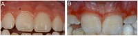
Generalized lack of attached gingiva. Absence or reduced amount of attached gingiva causing oral tissue fragility, first described in 1995 [Cunniff & Williamson-Kruse 1995], appears to be pathognomonic in pEDS.

In healthy individuals, the gums are classified as keratinized attached gingiva and non-keratinized movable mucosa. The keratinized attached gingiva is tightly and unmovably bound to the periosteum via collagen type I fibers as a thick band to provide mechanical protection. The non-keratinized oral mucosa is thin, more fragile, translucent, and only loosely connected to the periosteum. In pEDS, the thick band of gingiva is missing and the fragile mucosa extends to the gingival margin, causing tissue fragility and predisposing to gingival recession.

Generalized receding gums, reported in 87.1% of adults with pEDS, are attributable to the lack of attached gingiva and progressive periodontal attachment loss.

Ehlers-Danlos Syndrome Findings

Easy bruising. Almost all individuals with pEDS (96%) reported easy bruising from early childhood, especially in the pretibial area [Kapferer-Seebacher et al 2016]. Although easy bruising is difficult to evaluate as it is subjective and nonspecific, bruising may occur in atypical sites such as the cheeks or the thighs after taking a hot shower or doing sports [Kapferer-Seebacher et al 2019].

Pretibial plaques. Histologic analysis from brownish pretibial skin demonstrated dermal fibrosis and hemosiderin deposition [Ronceray et al 2013]. Affected individuals sometimes report a single pretibial trauma followed by hematomas that never resolve. In contrast to the hemosiderosis of the classic, vascular, and arthrochalastic EDS types, the pretibial plaques of pEDS are often necrobiotic, reflecting more severe connective tissue matrix inflammation and degradation [George et al 2016].

Skin fragility, atrophic scarring, and delayed wound healing. Skin fragility can manifest as easy bruising and "cigarette paper" scars mainly on the shins [George et al 2016]. Chronic leg ulcers may occur within the atrophic pretibial plaques [Ronceray et al 2013]. Single individuals reported open wounds in the pretibial area that took months or even years to heal or even required skin grafting [George et al 2016, Kapferer-Seebacher et al 2016].

Joint hypermobility often only affects the distal joints [Kapferer-Seebacher et al 2016].

Arterial aneurysms/dissections, documented in 8/145 individuals with pEDS (5.5%), may cluster in families. Four clinically affected individuals in one family had diverse cardiovascular events: aortic dissection (age 46 years), sudden death (23 years), hemorrhage of cerebral aneurysm (42 years), and heart attack (43 years) [Kapferer-Seebacher et al 2016]. Cerebral aneurysms were observed in individuals from unrelated families [Cıkla et al 2014, Kapferer-Seebacher et al 2016]. More data over a longer period of time are needed to clarify the risk for arterial aneurysms or dissections. This risk appears lower than in people with vascular EDS, in which 72% of individuals with one or more vascular complications have been reported [Frank et al 2015].

Organ rupture. Multiple organ ruptures were reported in two of 148 affected individuals (1.4%). One individual, age 67 years, had a history of recurrent pneumothoraces, an intestinal perforation, and an inguinal hernia [Kapferer-Seebacher et al 2016]. The other individual, age 57 years, had a rupture of the lung and diaphragm [Kapferer-Seebacher et al 2019].

Other

Acrogeric changes of the hands and feet seen in pEDS [George et al 2016] are reminiscent of those observed in vascular EDS.

Hoarse high-pitched voice has been reported in two individuals with pEDS to date [George et al 2016, Kapferer-Seebacher et al 2019]. Voice abnormalities have occasionally been described in other types of EDS; it has been hypothesized that they may be due to defects in the collagen of the vocal ligament [George et al 2016].

Brain white matter abnormalities. All adults investigated by brain MRI reported to date had brain white matter abnormalities [Spranger et al 1996, Kapferer-Seebacher et al 2019].

The MRI pattern was suggestive of an underlying small-vessel disease that is progressive with age. As observed in other leukoencephalopathies related to microangiopathies, the extent of the white matter changes was disproportionate to the paucity of neurologic features. Medical history revealed recurrent headaches or depression in single cases. Neurologic examination was unremarkable in all individuals except one who had mild cognitive decline, ataxia, and a history of one seizure.

Clinical manifestations in children. All but one child with pEDS reported to date were identified because of a positive family history. Generalized lack of attached gingiva is the only clinical finding that is consistently present in affected children younger than age ten years [Kapferer-Seebacher et al 2021].

Other clinical manifestations of pEDS are mild or absent in affected children, although the parents of all affected children reported easy bruising and gum bleeding. Median age of onset of periodontitis was reported to be 14 years.

Of note, a child with classic EDS, who also had periodontal bone loss in the primary dentition and a gingival phenotype specific for pEDS, had the familial COL5A1 pathogenic variant as well as a de novo C1R pathogenic variant [Stock et al 2021].

Genotype-Phenotype Correlations

No genotype-phenotype correlations in pEDs have been identified.

Penetrance

The penetrance in affected individuals identified to date is 100%. However, it is age-related: periodontitis may be absent in young individuals, particularly those with good dental hygiene [Kapferer-Seebacher et al 2021].

Nomenclature

Periodontal EDS was originally referred to as Ehlers Danlos syndrome type VIII [Stewart et al [1977]. The term EDS periodontitis was introduced by the Villefranche nomenclature [Beighton et al 1998].

Prevalence

There are no estimates of the prevalence of pEDS in any population.

To date, 148 individuals with pEDS have been described in 34 case reports, seven pedigree analyses, and two cohort studies [Kapferer-Seebacher et al 2017, Wu et al 2018, Kapferer-Seebacher et al 2019, Cortés-Bretón Brinkmann et al 2021, Kapferer-Seebacher et al 2021].

Due to the high prevalence of periodontitis in the general population [Chen et al 2021] and the sometimes only mild connective tissue manifestations of pEDS, this disorder is likely underdiagnosed.

Differential Diagnosis

Periodontal EDS (pEDS) must be distinguished from genetic disorders that include periodontitis and/or other features associated with pEDS and acquired periodontitis. The gingival phenotype as well as age of onset may be useful in differentiating pEDS from these conditions.

Note: The current classification of periodontal diseases distinguishes "periodontitis as a complex genetic disorder" (i.e., "acquired periodontitis") from "periodontitis as a manifestation of systemic diseases" – which includes periodontal EDS.

Genetic Disorders

Table 3.

Genetic Disorders Associated with Oral Manifestations in the Differential Diagnosis of Periodontal Ehlers-Danlos Syndrome

DisorderGene(s)MOIManifestationsOther Features
OralDermatologic
Vascular EDS (vEDS) COL3A1 AD
(AR)
Gingival recession, gingival fragility, & bleeding gums. 1 vEDS is not assoc w/early severe periodontitis.Bruising unrelated to identified trauma &/or in unusual sites. vEDS is not assoc w/pretibial plaques.Aneurysms/arterial rupture & organ ruptures (aneurysms & organ ruptures suggestive of vEDS also reported in pEDS but much less frequently; prematurely aged appearance, prominent eyes, attached earlobes, acrogeric changes of hands & feet, hypermobility of small joints
Hereditary angioedema (OMIM 106100)SERPING1
(C1INH)
AD
AR
Periodontal bone loss in conjunction w/episodes of acute gingival edemaAngioedematous attacks
Hypophosphatasia ALPL AD
AR
Premature loss of primary & secondary teethRecurrent fractures; osteomalacia; rickets, ↓ serum alkaline phosphatase, ↑ urinary phosphoethanolamin
Kindler syndrome FERMT1 AREarly severe periodontitisCongenital blistering, skin atrophy, photosensitivity, skin fragility
Odontohypophos-phatasia (See Hypophosphatasia.) ALPL AD
AR
Premature loss of primary & secondary teeth↓ serum alkaline phosphatase, ↑ urinary phosphoethanolamin
Papillon-Lefevre syndrome (OMIM 245000) CTSC ARSevere periodontitis w/early-childhood onsetHyperkeratosis of palms & soles
Severe congenital neutropenia (OMIM PS202700) CSF3R
ELANE
G6PC3
GFI1
HAX1
JAGN1
SRP54
VPS45
WAS
AD
AR
XL
Severe periodontitis w/early-childhood onsetRecurrent severe infections, hematologic findings
Singleton-Merten syndrome (OMIM 182250, 616298) DDX58
IFIH1
ADPremature loss of teeth, short tooth rootsShort stature, calcifications of the aorta & aortic & mitral valves, osteoporosis

In addition to the disorders summarized in Table 3, pEDS shares limited phenotypic overlap with other forms of EDS and disorders associated with vascular disease that can be clinically distinguished from pEDS by the absence of prominent oral manifestations. These disorders include the following:

Periodontitis is a multifactorial disorder caused by complex interactions between various pathogenic environmental and genetic factors that in combination are thought to determine the variability of disease onset, progression, and severity. Periodontitis is triggered by bacterial microorganisms and characterized by a chronic inflammatory destruction of the tooth-supporting tissues (alveolar bone, periodontal ligament, root cementum, and gingival attachment) in progredient cases leading to tooth loss. Key to periodontitis case definition is the notion of interdental clinical attachment loss [Tonetti et al 2018]. The typical individual with periodontitis is older than age 30 years and the amount of bone destruction is consistent with the presence of plaque and calculus. The rate of disease progression can be modified by local factors, systemic diseases, and extrinsic factors such as smoking or emotional stress.

According to the current classification of periodontal and peri-implant diseases and conditions, "periodontitis" is distinguished from "necrotizing periodontal diseases" and "periodontitis as a manifestation of systemic disease," the latter including rare systemic disorders like pEDS [Caton et al 2018].

In contrast to periodontitis as a manifestation of a rare disorder, periodontitis in general is a highly prevalent disease. In population-based studies periodontitis was diagnosed in up to 72.4% of individuals, with severe periodontitis (corresponding to stage III and IV) in up to 17.5% [Germen et al 2021, Stødle et al 2021]. Prevalence estimates for periodontitis in young individuals range from 0.1% (individuals of northern and central European origin) to 5% (individuals of African origin) [Albandar 2014]. In comparison, 98% of adults with pEDS present with periodontitis, clinical/radiologic attachment loss mainly according to stage III and IV periodontitis [Kapferer-Seebacher et al 2016, Kapferer-Seebacher et al 2017].

Management

At this point no clinical practice guidelines for periodontal Ehlers-Danlos syndrome (pEDS) have been published. However, some recommendations exist [Authors, personal observation].

Evaluations Following Initial Diagnosis

To establish the extent of disease and needs in an individual diagnosed with periodontal Ehlers-Danlos syndrome, the evaluations summarized in Table 4 (if not performed as part of the evaluation that led to the diagnosis) are recommended.

Table 4.

Recommended Evaluations Following Initial Diagnosis in Individuals with Periodontal Ehlers-Danlos Syndrome

System/ConcernEvaluationComment
Early-onset severe
periodontitis /
Receding gums
Clinical periodontal investigations, dental radiographsRequires a periodontist or a dentist specializing in periodontology
Easy bruising /
Scarring
Clinical assessment by dermatologist
Neurologic
manifestations
Clinical assessment by consultant neurologistBrain MRI, as white matter abnormalities of unknown clinical significance to date have been reported in persons w/pEDS
Joint hypermobility Clinical assessmentTypically by consultant who diagnosed pEDS
Aneurysms Imaging of the arterial tree in adults
Hoarse voice ENT consultationClinical assessment
Genetic
counseling
By genetics professionals 1To inform patients & families re nature, MOI, & implications of pEDS in order to facilitate medical & personal decision making
Family support
& resources
Assess:
1.

Clinical geneticist, certified genetic counselor, certified advanced genetic nurse

Preventive Measures

Periodontal breakdown with pEDS is most likely caused by a hyperinflammatory reaction [Gröbner et al 2019].

  • Early severe periodontitis may be ameliorated with strict oral hygiene measures starting in early childhood.
  • Professional tooth cleaning approximately every three months and frequent oral hygiene instructions to achieve livelong excellent personal oral hygiene appear to be essential.
  • Soft toothbrushes are recommended because of sensitive gums.

Treatment of Manifestations

Treatment is individualized based on the clinical manifestations present.

Given the characteristic features of early and severe periodontitis, all individuals should be regularly seen by a periodontist beginning in early childhood. Excellent oral hygiene is also a major element of the treatment of existing periodontitis. For example, affected individuals must be instructed frequently in order to achieve excellent personal oral hygiene with interdental cleaning devices and (electric) toothbrushes (see Surveillance).

Dental implants are often used to replace lost teeth. With pEDS there appears to be a high risk of peri-implantitis and consequently implant failure [Rinner et al 2018]. Dental implants affected by peri-implantitis clinically present with progressive inflammatory peri-implant bone loss representing a clinical phenotype similar to periodontitis.

Therefore, the primary objective in the oral treatment of pEDS is to help affected individuals retain their own teeth for as long as possible [Kapferer-Seebacher et al 2021].

Table 5.

Treatment of Manifestations in Individuals with Periodontal Ehlers-Danlos Syndrome

Manifestation/
Concern
TreatmentConsiderations/Other
Severe early-onset periodontitis Periodontal treatment
Receding gums No treatment
  • Surgical treatment of gingival recession in general is possible; however, no experience exists w/pEDS.
  • Oral hygiene instructions for gentle cleaning (e.g., w/soft toothbrushes) recommended
Easy bruising /
Scarring
  • Advice re avoidance of high-impact sports
  • For wounds that do not heal consult a dermatologist.
  • There is some evidence in classic EDS that ascorbic acid (vitamin C) may ↓ bruising, but no data on its use in pEDS.
Joint
hypermobility
Assessment by consultant rheumatologist to advise re need for PT, OT, pain management, & appropriate exerciseGeneral advice for those w/joint hypermobility can be followed w/moderate exercise & PT input when necessary
Arterial
aneurysms/
dissection
Depending on nature & location, surgical intervention is possible/required or treatment can be conservative (i.e., rpt imaging, anti-hypertensive medication, & advice re exercise).
Gastrointestinal
rupture
Surgical intervention
Hoarse voice Intervention depends on the primary cause.Surgical intervention for vocal cord stenosis has been reported.

OT = occupational therapy; PT = physical therapy

Surveillance

Table 6.

Recommended Surveillance for Individuals with Periodontal Ehlers-Danlos Syndrome

System/ConcernEvaluationFrequency
Early-onset severe
periodontitis
Detailed periodontal evalAt least annually
Supportive periodontal care incl reeval of periodontal parameters, oral hygiene instructions (e.g., use of interdental cleaning devices and electric toothbrushes), & supra- & sub-gingival debridementThe appropriate interval between periodontal maintenance visits should be ~3-6 mos based on the needs of the patient
Joint hypermobility
w/(sub)luxations
& joint pain
By consultant rheumatologist, PT, OTDepends on the specific concern(s)
Risk of arterial
aneurysm/
dissection
Some EDS services offer surveillance of the arterial tree in asymptomatic adults w/pEDS, w/multidisciplinary input in case of identified arterial aneurysm/dissection, but evidence for this approach is limited as the risk of arterial aneurysm/dissection in those w/pEDS is unknown but appears lower than in vEDS.Surveillance of the arterial tree in asymptomatic adults is offered w/a frequency of 1.5-2 yrs by some EDS services.

OT = occupational therapist; PT = physical therapist

Agents/Circumstances to Avoid

Currently, evidence does not support a general recommendation that persons with pEDS avoid any specific activities/treatment/medications. However, this may vary on an individual basis; for example, in an individual with pEDS and arterial aneurysms, activities such as high-impact sports and weight lifting would be actively discouraged.

Evaluation of Relatives at Risk

It is appropriate to clarify the genetic status of apparently asymptomatic older and younger at-risk relatives of an affected individual in order to identify as early as possible those who would benefit from prompt initiation of preventive dental hygiene and routine dental care and surveillance.

See Genetic Counseling for issues related to testing of at-risk relatives for genetic counseling purposes.

Therapies Under Investigation

Search ClinicalTrials.gov in the US and EU Clinical Trials Register in Europe for access to information on clinical studies for a wide range of diseases and conditions. Note: There may not be clinical trials for this disorder.

Genetic Counseling

Genetic counseling is the process of providing individuals and families with information on the nature, mode(s) of inheritance, and implications of genetic disorders to help them make informed medical and personal decisions. The following section deals with genetic risk assessment and the use of family history and genetic testing to clarify genetic status for family members; it is not meant to address all personal, cultural, or ethical issues that may arise or to substitute for consultation with a genetics professional. —ED.

Mode of Inheritance

Periodontal Ehlers-Danlos syndrome (pEDS) is inherited in an autosomal dominant manner.

Risk to Family Members

Parents of a proband

  • Most probands reported to date have pEDS as the result of a C1R or C1S pathogenic variant inherited from a heterozygous, affected parent.
  • Some individuals with pEDS may have the disorder as the result of a de novo C1R or C1S pathogenic variant; the relative frequency of de novo occurrence of pEDS is unknown.
  • If the proband appears to be the only affected family member (i.e., a simplex case), molecular genetic testing is recommended for the parents of the proband to confirm their genetic status and to allow reliable recurrence risk counseling.
  • If the pathogenic variant identified in the proband is not identified in either parent, the following possibilities should be considered:
    • The proband has a de novo pathogenic variant. Note: A pathogenic variant is reported as "de novo" if: (1) the pathogenic variant found in the proband is not detected in parental DNA; and (2) parental identity testing has confirmed biological maternity and paternity. If parental identity testing is not performed, the variant is reported as "assumed de novo" [Richards et al 2015].
    • The proband inherited a pathogenic variant from a parent with germline (or somatic and germline) mosaicism. Note: Testing of parental leukocyte DNA may not detect all instances of somatic mosaicism and will not detect a pathogenic variant that is present in the germ cells only.
      A parent with somatic and germline mosaicism for a C1R or C1S pathogenic variant may in theory be mildly/minimally affected.
  • The family history of some individuals diagnosed with pEDS may appear to be negative because of failure to recognize the disorder in family members, early death of the parent before the onset of symptoms, or late onset of the disease in the affected parent. Therefore, an apparently negative family history cannot be confirmed unless molecular genetic testing has demonstrated that neither parent is heterozygous for the pathogenic variant identified in the proband.

Sibs of a proband. The risk to the sibs of the proband depends on the clinical/genetic status of the proband's parents:

  • If a parent of the proband is affected and/or is known to have the pathogenic variant identified in the proband, the risk to the sibs of inheriting the pathogenic variant is 50%. All sibs who inherit a pEDS-related pathogenic variant are expected to develop manifestations of pEDS, although the age of onset may vary between heterozygous family members (see Penetrance).
  • If the proband has a known pEDS-related pathogenic variant that cannot be detected in the leukocyte DNA of either parent, the recurrence risk to sibs is estimated to be 1% because of the theoretic possibility of parental germline mosaicism [Rahbari et al 2016].
  • If the parents have not been tested for the C1R or C1S pathogenic variant but are clinically unaffected, the risk to the sibs of a proband appears to be low. However, sibs of a proband with clinically unaffected parents are still presumed to be at increased risk for pEDS because of the possibility of failure to diagnose pEDS in a heterozygous parent or the theoretic possibility of parental germline mosaicism.

Offspring of a proband. Each child of an individual with pEDS has a 50% chance of inheriting the C1R or C1S pathogenic variant and being affected by pEDS.

Other family members. The risk to other family members depends on the status of the proband's parents: if a parent has the C1R or C1S pathogenic variant, his or her family members may be at risk.

Related Genetic Counseling Issues

See Management, Evaluation of Relatives at Risk for information on evaluating at-risk relatives for the purpose of early diagnosis and treatment.

Family planning

  • The optimal time for determination of genetic risk and discussion of the availability of prenatal/preimplantation genetic testing is before pregnancy.
  • It is appropriate to offer genetic counseling (including discussion of potential risks to offspring and reproductive options) to young adults who are affected or at risk.

Prenatal Testing and Preimplantation Genetic Testing

Once the pEDS-causing pathogenic variant has been identified in an affected family member, prenatal and preimplantation genetic testing are technically possible.

Differences in perspective may exist among medical professionals and within families regarding the use of prenatal testing. While most centers would consider use of prenatal testing to be a personal decision, discussion of these issues may be helpful.

Resources

GeneReviews staff has selected the following disease-specific and/or umbrella support organizations and/or registries for the benefit of individuals with this disorder and their families. GeneReviews is not responsible for the information provided by other organizations. For information on selection criteria, click here.

Molecular Genetics

Information in the Molecular Genetics and OMIM tables may differ from that elsewhere in the GeneReview: tables may contain more recent information. —ED.

Table A.

Periodontal Ehlers-Danlos Syndrome: Genes and Databases

Data are compiled from the following standard references: gene from HGNC; chromosome locus from OMIM; protein from UniProt. For a description of databases (Locus Specific, HGMD, ClinVar) to which links are provided, click here.

Table B.

OMIM Entries for Periodontal Ehlers-Danlos Syndrome (View All in OMIM)

120580COMPLEMENT COMPONENT 1, s SUBCOMPONENT; C1S
130080EHLERS-DANLOS SYNDROME, PERIODONTAL TYPE, 1; EDSPD1
613785COMPLEMENT COMPONENT 1, r SUBCOMPONENT; C1R
617174EHLERS-DANLOS SYNDROME, PERIODONTAL TYPE, 2; EDSPD2

Molecular Pathogenesis

Periodontal Ehlers-Danlos syndrome (pEDS) results from specific heterozygous variants in C1R and C1S.

The central element in the pathogenesis of pEDS is the intracellular activation of C1r and/or C1s, and extracellular presence of activated C1s that – independent of microbial triggers – can activate the classic complement cascade. This mechanism is triggered by specific gain-of-function variants [Bally et al 2019, Gröbner et al 2019].

Mechanism of disease causation. The mechanism of pathogenesis of these variants differs from homozygous loss of function of these genes and from loss of the C1 esterase inhibitor.

Functional studies in transfected HEK cells and fibroblasts from affected individuals revealed that all 16 pEDS variants studied prevented normal integration into the C1 complex and caused abnormal extracellular C1r/C1s serine protease activation. C1R variants pathogenic for pEDS showed different, domain-specific abnormalities of intracellular processing and secretion such as retention of C1r fragments inside the cell, secretion of aggregates, or a new C1r cleavage site. One variant disabled a C1q binding site. All available fibroblasts from affected individuals exhibited activated C1s and activation of externally added C4 in the supernatant in contrast to control cell lines which secreted proenzyme C1s without C4 activation [Gröbner et al 2019]. Similarly, functional studies in C1S-related pEDS showed trypsin-like or C1s-like enzymatic cleavage at the end of the CCP1 domain of the C1s protein in both C1S pathogenic variants known at the time of study [Bally et al 2019].

Variants that remove C1r or C1s serine protease function – such as large genomic rearrangements, out-of-frame deletions or duplications, or nonsense variants – are not expected to cause pEDS. However, pEDS causation cannot be fully excluded for in-frame deletions or duplication.

Gene-specific laboratory technical considerations. The range of variants that cause pEDS is greater for C1R than C1S. This appears to be due to the autocatalytic activity of native C1r in the absence of C1 esterase inhibitor. C1R variants may be causative for pEDS when they interfere with the inhibition of autoactivation, whereas C1S variants may be causative for pEDS only if they trigger autoactivation. This is relevant for the interpretation of variants of uncertain significance in both genes. These considerations also explain why null variants in C1R or C1S are not associated with pEDS.

Chapter Notes

Author Notes

Further information is available at the dedicated website www.p-eds.org.

Acknowledgments

The authors wish to thank the affected individuals and their families who supported the detailed clinical characterization of the condition. The assistance of the many collaborative partners listed in the various publications is gratefully acknowledged.

Revision History

  • 29 July 2021 (bp) Review posted live
  • 5 May 2021 (jz) Original submission

References

Literature Cited

  • Albandar JM. Aggressive and acute periodontal diseases. Periodontol 2000. 2014;65:7–12. [PubMed: 24738583]
  • Bally I, Dalonneau F, Chouquet A, Grobner R, Amberger A, Kapferer-Seebacher I, Stoiber H, Zschocke J, Thielens NM, Rossi V, Gaboriaud C. Two different missense C1S mutations, associated to periodontal Ehlers-Danlos syndrome, lead to identical molecular outcomes. Front Immunol. 2019;10:2962. [PMC free article: PMC6930149] [PubMed: 31921203]
  • Beighton P, De Paepe A, Steinmann B, Tsipouras P, Wenstrup RJ. Ehlers-Danlos syndromes: revised nosology, Villefranche, 1997. Ehlers-Danlos National Foundation (USA) and Ehlers-Danlos Support Group (UK). Am J Med Genet. 1998;77:31–7. [PubMed: 9557891]
  • Caton JG, Armitage G, Berglundh T, Chapple ILC, Jepsen S, Kornman KS, Mealey BL, Papapanou PN, Sanz M, Tonetti MS. A new classification scheme for periodontal and peri-implant diseases and conditions - Introduction and key changes from the 1999 classification. J Periodontol. 2018;89 Suppl 1:S1–S8. [PubMed: 29926946]
  • Chen MX, Zhong YJ, Dong QQ, Wong HM, Wen YF. Global, regional, and national burden of severe periodontitis, 1990-2019: an analysis of the Global Burden of Disease Study 2019. J Clin Periodontol. 2021. Epub ahead of print. [PubMed: 34101223]
  • Cıkla U, Sadighi A, Bauer A, Başkaya MK. Fatal ruptured blood blister-like aneurysm of middle cerebral artery associated with Ehlers-Danlos syndrome type VIII (periodontitis type). J Neurol Surg Rep. 2014;75:e210–3. [PMC free article: PMC4242822] [PubMed: 25485215]
  • Cortés-Bretón Brinkmann J, García-Gil I, Lobato-Peña DM, Martínez-Mera C, Suárez-García MJ, Martínez-González JM, Rioboo M. The key role of the dental practitioner in early diagnosis of periodontal Ehlers-Danlos syndromes: a rare case report of siblings. Quintessence Int. 2021;52:166–74. [PubMed: 33433082]
  • Cunniff C, Williamson-Kruse L. Ehlers-Danlos syndrome, type VIII presenting with periodontitis and prolonged bleeding time. Clin Dysmorphol. 1995;4:145–9. [PubMed: 7606321]
  • Demirkaya E, Zhou Q, Smith CK, Ombrello MJ, Deuitch N, Tsai WL, Hoffmann P, Remmers EF, Takeuchi M, Park YH, Chae J, Barut K, Simsek D, Adrovic A, Sahin S, Caliskan S, Chandrasekharappa SC, Hasni SA, Ombrello AK, Gadina M, Kastner DL, Kaplan MJ, Kasapcopur O, Aksentijevich I. Brief report: deficiency of complement 1r subcomponent in early-onset systemic lupus erythematosus: the role of disease-modifying alleles in a monogenic disease. Arthritis Rheumatol. 2017;69:1832–9. [PMC free article: PMC5609811] [PubMed: 28544690]
  • Frank M, Albuisson J, Ranque B, Golmard L, Mazzella JM, Bal-Theoleyre L, Fauret AL, Mirault T, Denarié N, Mousseaux E, Boutouyrie P, Fiessinger JN, Emmerich J, Messas E, Jeunemaitre X. The type of variants at the COL3A1 gene associates with the phenotype and severity of vascular Ehlers-Danlos syndrome. Eur J Hum Genet. 2015;23:1657–64. [PMC free article: PMC4795191] [PubMed: 25758994]
  • Germen M, Baser U, Lacin CC, Fıratlı E, İşsever H, Yalcin F. Int J Environ Res Public Health. 2021;18:3459. [PMC free article: PMC8037399] [PubMed: 33810461]
  • George SM, Vandersteen A, Nigar E, Ferguson DJ, Topham EJ, Pope FM. Two patients with Ehlers-Danlos syndrome type VIII with unexpected hoarseness. Clin Exp Dermatol. 2016;41:771–4. [PubMed: 27663155]
  • Gröbner R, Kapferer-Seebacher I, Amberger A, Redolfi R, Dalonneau F, Bjorck E, Milnes D, Bally I, Rossi V, Thielens N, Stoiber H, Gaboriaud C, Zschocke J. C1R mutations trigger constitutive complement 1 activation in periodontal Ehlers-Danlos syndrome. Front Immunol. 2019;10:2537. [PMC free article: PMC6848165] [PubMed: 31749804]
  • Hartsfield JK Jr, Kousseff BG. Phenotypic overlap of Ehlers-Danlos syndrome types IV and VIII. Am J Med Genet. 1990;37:465–70. [PubMed: 2260589]
  • Kapferer-Seebacher I, Lundberg P, Malfait F, Zschocke J. Periodontal manifestations of Ehlers-Danlos syndromes: A systematic review. J Clin Periodontol. 2017;44:1088–100. [PubMed: 28836281]
  • Kapferer-Seebacher I, Oakley-Hannibal E, Lepperdinger U, Johnson D, Ghali N, Brady AF, Sobey GJ, Zschocke J, Van Dijk FS. Prospective clinical investigations of children with periodontal Ehlers–Danlos syndrome identify generalized lack of attached gingiva as a pathognomonic feature. Genet Med. 2021;23:316–22. [PubMed: 33005042]
  • Kapferer-Seebacher I, Pepin M, Werner R, Aitman TJ, Nordgren A, Stoiber H, Thielens N, Gaboriaud C, Amberger A, Schossig A, Gruber R, Giunta C, Bamshad M, Bjorck E, Chen C, Chitayat D, Dorschner M, Schmitt-Egenolf M, Hale CJ, Hanna D, Hennies HC, Heiss-Kisielewsky I, Lindstrand A, Lundberg P, Mitchell AL, Nickerson DA, Reinstein E, Rohrbach M, Romani N, Schmuth M, Silver R, Taylan F, Vandersteen A, Vandrovcova J, Weerakkody R, Yang M, Pope FM, Byers PH, Zschocke J. Periodontal Ehlers-Danlos syndrome is caused by mutations in C1R and C1S, which encode subcomponents C1r and C1s of complement. Am J Hum Genet. 2016;99:1005–14. [PMC free article: PMC5097948] [PubMed: 27745832]
  • Kapferer-Seebacher I, Waisfisz Q, Boesch S, Bronk M, Van Tintelen P, Gizewski ER, Groebner R, Zschocke J, Van Der Knaap MS. Periodontal Ehlers-Danlos syndrome is associated with leukoencephalopathy. Neurogenetics. 2019;20:1–8. [PMC free article: PMC6411670] [PubMed: 30535813]
  • Macedo AC, Isaac L. Systemic lupus erythematosus and deficiencies of early components of the complement classical pathway. Front Immunol. 2016;7:55. [PMC free article: PMC4764694] [PubMed: 26941740]
  • Malfait F, Francomano C, Byers P, Belmont J, Berglund B, Black J, Bloom L, Bowen JM, Brady AF, Burrows NP, Castori M, Cohen H, Colombi M, Demirdas S, De Backer J, De Paepe A, Fournel-Gigleux S, Frank M, Ghali N, Giunta C, Grahame R, Hakim A, Jeunemaitre X, Johnson D, Juul-Kristensen B, Kapferer-Seebacher I, Kazkaz H, Kosho T, Lavallee ME, Levy H, Mendoza-Londono R, Pepin M, Pope FM, Reinstein E, Robert L, Rohrbach M, Sanders L, Sobey GJ, Van Damme T, Vandersteen A, Van Mourik C, Voermans N, Wheeldon N, Zschocke J, Tinkle B. The 2017 international classification of the Ehlers-Danlos syndromes. Am J Med Genet C Semin Med Genet. 2017;175:8–26. [PubMed: 28306229]
  • Malfait F, Symoens S, De Backer J, Hermanns-Lê T, Sakalihasan N, Lapière CM, Coucke P, De Paepe A. Three arginine to cysteine substitutions in the pro-alpha (I)-collagen chain cause Ehlers-Danlos syndrome with a propensity to arterial rupture in early adulthood. Hum Mutat. 2007;28:387–95. [PubMed: 17211858]
  • McKusick VA. Heritable Disorders of Connective Tissue. St Louis: Mosby. 1972.
  • Nuytinck L, Freund M, Lagae L, Pierard GE, Hermanns-Le T, De Paepe A. Classical Ehlers-Danlos syndrome caused by a mutation in type I collagen. Am J Hum Genet. 2000;66:1398–402. [PMC free article: PMC1288203] [PubMed: 10739762]
  • Pitney T, Pitney M. Phenotypic mimicry between periodontal and vascular Ehlers-Danlos variants resolved by molecular genetic testing. Int J Dermatol. 2020;59:e387–e388. [PubMed: 33616899]
  • Rahbari R, Wuster A, Lindsay SJ, Hardwick RJ, Alexandrov LB, Turki SA, Dominiczak A, Morris A, Porteous D, Smith B, Stratton MR, Hurles ME. Timing, rates and spectra of human germline mutation. Nat Genet. 2016;48:126–33. [PMC free article: PMC4731925] [PubMed: 26656846]
  • Richards S, Aziz N, Bale S, Bick D, Das S, Gastier-Foster J, Grody WW, Hegde M, Lyon E, Spector E, Voelkerding K, Rehm HL, et al. Standards and guidelines for the interpretation of sequence variants: a joint consensus recommendation of the American College of Medical Genetics and Genomics and the Association for Molecular Pathology. Genet Med. 2015;17:405–24. [PMC free article: PMC4544753] [PubMed: 25741868]
  • Rinner A, Zschocke J, Schossig A, Gröbner R, Strobl H, Kapferer-Seebacher I. High risk of peri-implant disease in periodontal Ehlers-Danlos Syndrome. A case series. Clin Oral Implants Res. 2018;29:1101–6. [PMC free article: PMC6282529] [PubMed: 30252959]
  • Ronceray S, Miquel J, Lucas A, Pierard GE, Hermanns-Le T, De Paepe A, Dupuy A. Ehlers-Danlos syndrome type VIII: a rare cause of leg ulcers in young patients. Case Rep Dermatol Med. 2013;2013:469505. [PMC free article: PMC3809367] [PubMed: 24198978]
  • Spranger S, Spranger M, Kirchhof K, Steinmann B. Ehlers-Danlos syndrome type VIII and leukodystrophy. Am J Med Genet. 1996;66:239–40. [PubMed: 8958339]
  • Stewart RE, Hollister DW, Rimoin DL. A new variant of Ehlers-Danlos syndrome: an autosomal dominant disorder of fragile skin, abnormal scarring, and generalized periodontitis. Birth Defects Orig Artic Ser. 1977;13:85–93. [PubMed: 890102]
  • Stock F, Hanisch M, Lechner S, Biskup S, Bohring A, Zschocke J, Kapferer-Seebacher I. Prepubertal periodontitis in a patient with combined classical and periodontal Ehlers-Danlos syndrome. Biomolecules. 2021;11:149. [PMC free article: PMC7912441] [PubMed: 33498938]
  • Stødle IH, Verket A, Høvik H, Sen A, Koldsland OC. Prevalence of periodontitis based on the 2017 classification in a Norwegian population. The HUNT Study. J Clin Periodontol. 2021. Epub ahead of print. [PubMed: 34101228]
  • Tonetti MS, Greenwell H, Kornman KS. Staging and grading of periodontitis: Framework and proposal of a new classification and case definition. J Periodontol. 2018;89 Suppl 1:S159–S172. [PubMed: 29926952]
  • Wu J, Yang J, Zhao J, Wu JR, Zhang X, Leung WK, Sun WB. A Chinese family with periodontal Ehlers-Danlos syndrome associated with missense mutation in the C1R gene. J Clin Periodontol. 2018;45:1311–18. [PubMed: 30025171]
Copyright © 1993-2024, University of Washington, Seattle. GeneReviews is a registered trademark of the University of Washington, Seattle. All rights reserved.

GeneReviews® chapters are owned by the University of Washington. Permission is hereby granted to reproduce, distribute, and translate copies of content materials for noncommercial research purposes only, provided that (i) credit for source (http://www.genereviews.org/) and copyright (© 1993-2024 University of Washington) are included with each copy; (ii) a link to the original material is provided whenever the material is published elsewhere on the Web; and (iii) reproducers, distributors, and/or translators comply with the GeneReviews® Copyright Notice and Usage Disclaimer. No further modifications are allowed. For clarity, excerpts of GeneReviews chapters for use in lab reports and clinic notes are a permitted use.

For more information, see the GeneReviews® Copyright Notice and Usage Disclaimer.

For questions regarding permissions or whether a specified use is allowed, contact: ude.wu@tssamda.

Bookshelf ID: NBK572429PMID: 34324282

Views

Tests in GTR by Gene

Related information

  • OMIM
    Related OMIM records
  • PMC
    PubMed Central citations
  • PubMed
    Links to PubMed
  • Gene
    Locus Links

Similar articles in PubMed

See reviews...See all...

Recent Activity

Your browsing activity is empty.

Activity recording is turned off.

Turn recording back on

See more...