U.S. flag

An official website of the United States government

NCBI Bookshelf. A service of the National Library of Medicine, National Institutes of Health.

Adam MP, Feldman J, Mirzaa GM, et al., editors. GeneReviews® [Internet]. Seattle (WA): University of Washington, Seattle; 1993-2024.

Cover of GeneReviews®

GeneReviews® [Internet].

Show details

FAM111A-Related Skeletal Dysplasias

, MD, , MD, and , MD.

Author Information and Affiliations

Initial Posting: .

Estimated reading time: 28 minutes

Summary

Clinical characteristics.

FAM111A-related skeletal dysplasias include the milder phenotype of Kenny-Caffey syndrome (KCS) and a more severe lethal phenotype, osteocraniostenosis (OCS). KCS is characterized by proportionate short stature (typically postnatal onset), relative macrocephaly, large anterior fontanel with delayed closure, characteristic facial features, cortical thickening of the long bones with stenosis of the medullary cavity, and ophthalmologic and dental manifestations. OCS is characterized by intrauterine growth deficiency, microcephaly, characteristic facial features, decreased skull ossification, slender long bones with cortical thickening, stenosis of the medullary cavity of the long bones, flared metaphyses, and thin ribs with thoracic and pulmonary hypoplasia leading to respiratory insufficiency. Perinatal fractures may occur. Primary hypoparathyroidism with hypocalcemia and hyperphosphatemia can occur in individuals with KCS and OCS.

Diagnosis/testing.

The diagnosis of a FAM111A-related skeletal dysplasia is established in a proband with suggestive findings and a heterozygous pathogenic variant in FAM111A identified by molecular genetic testing.

Management.

Treatment of manifestations: Survivors with OCS require aggressive respiratory support and management of restrictive lung disease with a respiratory specialist; for all affected individuals, supplemental calcium and activated forms of vitamin D per endocrinologist; management of refractive errors and cataracts; management of dental manifestations with a dental specialist / oral surgeon; environmental and/or occupational modifications as needed for short stature in those with KCS; conservative or surgical management per orthopedist and/or neurosurgeon for scoliosis; individualized developmental support by allied health clinicians; referral to psychologist as needed.

Surveillance: For individuals with KCS and OCS, assess anthropometry (height, weight, growth velocity, limb proportions, and upper-to-lower segment proportions) at each visit; clinical examination for scoliosis at each visit, with referral to orthopedics and physical therapy as necessary; assess functional limitations and assessment with physical therapy and/or occupational therapy as needed; measurement of serum calcium, phosphate, and vitamin D every three months until calcium level is normalized on treatment and then subsequently every six months; clinical examination for manifestations of hypocalcemia; abdominal ultrasound to assess for nephrocalcinosis and/or nephrolithiasis annually while on treatment; ophthalmology examination annually or as indicated; dental examinations every six months; assess for clinical manifestations of anemia at each visit; monitor developmental progress and educational needs at each visit throughout childhood; assess for changes in mood, affect, and/or psychosocial stressors at each visit; assess care coordination needs and genetic counseling needs at each visit.

Genetic counseling.

FAM111A-related skeletal dysplasias (including KCS and OCS) are autosomal dominant disorders.

  • KCS. Most individuals diagnosed with KCS have the disorder as the result of a de novo FAM111A pathogenic variant. Rarely, individuals diagnosed with KCS have an affected parent. If a parent of the proband is known to have the pathogenic variant identified in the proband, the risk to the sibs of inheriting the pathogenic variant is 50%. Each child of an individual with KCS has a 50% chance of inheriting the FAM111A pathogenic variant.
  • OCS. With one possible exception, all probands reported to date with OCS whose parents have undergone molecular genetic testing have the disorder as the result of a de novo FAM111A pathogenic variant. Given that probands with OCS typically have the disorder as the result of a de novo FAM111A pathogenic variant, the risk to other family members is presumed to be low.

Once the FAM111A pathogenic variant has been identified in an affected family member, prenatal and preimplantation genetic testing are possible.

GeneReview Scope

FAM111A-Related Skeletal Dysplasias: Included Phenotypes 1
  • Kenny-Caffey syndrome
  • Osteocraniostenosis (gracile bone dysplasia)

For synonyms and outdated names, see Nomenclature.

1.

For other genetic causes of these phenotypes, see Differential Diagnosis.

Diagnosis

No consensus clinical diagnostic criteria for FAM111A-related skeletal dysplasias have been published.

Suggestive Findings

FAM111A-related skeletal dysplasias include the milder phenotype of Kenny-Caffey syndrome and a more severe lethal phenotype, osteocraniostenosis. A FAM111A-related skeletal dysplasia should be suspected in probands with a combination of the following clinical, imaging, and laboratory findings:

Kenny-Caffey Syndrome

Clinical findings

  • Proportionate short stature, most often postnatal onset
  • Dysmorphic facial features such as frontal bossing or prominent forehead with relative macrocephaly, triangular face, short palpebral fissures, deeply set eyes, midface retrusion, short nose, narrow nasal ridge, and micrognathia or microretrognathia (See Figure 1.)
  • Ocular manifestations including microphthalmia, aniridia, hyperopia or myopia, astigmatism, cataract, corneal and retinal calcification, and pseudopapilledema
  • Dental manifestations including oligodontia, enamel hypoplasia, retention of primary dentition, delayed eruption of secondary dentition, increased dental caries, and loss of secondary dentition due to dental caries
  • Other features including small testes and infertility have been described.
Figure 1.

Figure 1.

Craniofacial features of an individual with Kenny-Caffey syndrome at age 23 years with prominent forehead, triangular face, midface retrusion, and micrognathia Reprinted with permission from Cheng et al [2021]

Imaging findings

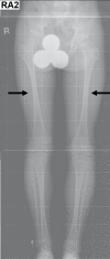
  • Skeletal radiographs show delayed anterior fontanelle closure and short long bones with cortical thickening and stenosis of the medullary cavity (see Figure 2).
  • Brain imaging shows calcification (typically of the basal ganglia but also reported in dentate nuclei and parts of the cerebrum and cerebellum).
  • Other imaging findings including wormian bones, prominent odontoid process, craniosynostosis (coronal and/or basal sutures), and coxa valga have also been reported.
Figure 2.

Figure 2.

Radiographs of the lower limbs in an individual with Kenny-Caffey syndrome (at age 13 years) showing slender femoral diaphyses with cortical thickening and medullary stenosis (black arrows) Reprinted with permission from Cheng et al [2021]

Laboratory findings. Primary hypoparathyroidism with hypocalcemia and hyperphosphatemia

Osteocraniostenosis

Clinical findings

  • Intrauterine growth deficiency with micromelia and disproportionately small hands and feet
  • Craniofacial features such as ear anomalies, short nose, and narrow mouth
  • Micropenis and small testes
  • Respiratory insufficiency secondary to pulmonary hypoplasia
  • Splenic aplasia or hypoplasia
  • Extramedullary hematopoiesis

Imaging findings

  • Cloverleaf-shaped skull (can be detected antenatally)
  • Decreased skull ossification (can be detected antenatally)
  • Slender long bones with cortical thickening, stenosis of the medullary cavity, and flared metaphyses
  • Thin ribs and thoracic hypoplasia
  • Bone fractures in some individuals, typically affecting long bones (e.g., femur, radius, ulna) and occasionally rib fractures

Laboratory findings. Primary hypoparathyroidism with hypocalcemia and hyperphosphatemia

Establishing the Diagnosis

The diagnosis of a FAM111A-related skeletal dysplasia is established in a proband with suggestive findings and a heterozygous pathogenic (or likely pathogenic) variant in FAM111A identified by molecular genetic testing (see Table 1).

Note: (1) Per ACMG/AMP variant interpretation guidelines, the terms "pathogenic variant" and "likely pathogenic variant" are synonymous in a clinical setting, meaning that both are considered diagnostic, and both can be used for clinical decision making [Richards et al 2015]. Reference to "pathogenic variant" in this section is understood to include any likely pathogenic variant. (2) Identification of a heterozygous FAM111A variant of uncertain significance does not establish or rule out the diagnosis.

Molecular genetic testing approaches can include a combination of gene-targeted testing (single-gene testing, multigene panel) and comprehensive genomic testing (exome sequencing, genome sequencing) depending on the phenotype.

Gene-targeted testing requires that the clinician determine which gene(s) are likely involved, whereas genomic testing does not. Individuals with the distinctive findings described in Suggestive Findings are likely to be diagnosed using gene-targeted testing (see Option 1), whereas those with a phenotype indistinguishable from many other skeletal dysplasias are more likely to be diagnosed using genomic testing (see Option 2).

Option 1

Single-gene testing. Sequence analysis of FAM111A is performed first to detect small intragenic deletions/insertions and missense, nonsense, and splice site variants. Typically, if no variant is detected by the sequencing method used, the next step is to perform gene-targeted deletion/duplication analysis to detect exon and whole-gene deletions or duplications; however, to date such variants have not been identified as a cause of this disorder.

A skeletal dysplasia multigene panel that includes FAM111A and other genes of interest (see Differential Diagnosis) may be considered to identify the genetic cause of the condition while limiting identification of variants of uncertain significance and pathogenic variants in genes that do not explain the underlying phenotype. Note: (1) The genes included in the panel and the diagnostic sensitivity of the testing used for each gene vary by laboratory and are likely to change over time. (2) Some multigene panels may include genes not associated with the condition discussed in this GeneReview. (3) In some laboratories, panel options may include a custom laboratory-designed panel and/or custom phenotype-focused exome analysis that includes genes specified by the clinician. (4) Methods used in a panel may include sequence analysis, deletion/duplication analysis, and/or other non-sequencing-based tests.

For an introduction to multigene panels click here. More detailed information for clinicians ordering genetic tests can be found here.

Option 2

Comprehensive genomic testing does not require the clinician to determine which gene is likely involved. Exome sequencing is most commonly used; genome sequencing is also possible.

For an introduction to comprehensive genomic testing click here. More detailed information for clinicians ordering genomic testing can be found here.

Table 1.

Molecular Genetic Testing Used in FAM111A-Related Skeletal Dysplasias

Gene 1MethodProportion of Probands with a Pathogenic Variant 2 Detectable by Method
FAM111A Sequence analysis 3100% 4
Gene-targeted deletion/duplication analysis 5None reported 6
1.
2.

See Molecular Genetics for information on variants detected in this gene.

3.

Sequence analysis detects variants that are benign, likely benign, of uncertain significance, likely pathogenic, or pathogenic. Variants may include small intragenic deletions/insertions and missense, nonsense, and splice site variants; typically, exon or whole-gene deletions/duplications are not detected. For issues to consider in interpretation of sequence analysis results, click here.

4.
5.

Gene-targeted deletion/duplication analysis detects intragenic deletions or duplications. Methods used may include a range of techniques such as quantitative PCR, long-range PCR, multiplex ligation-dependent probe amplification (MLPA), and a gene-targeted microarray designed to detect single-exon deletions or duplications.

6.

Data derived from the subscription-based professional view of Human Gene Mutation Database [Stenson et al 2020]

Clinical Characteristics

Clinical Description

FAM111A-related skeletal dysplasias include the milder phenotype of Kenny-Caffey syndrome (KCS) and a more severe lethal phenotype, osteocraniostenosis (OCS). To date, at least 35 individuals have been identified with a FAM111A-related skeletal dysplasia [Unger et al 2013, Guo et al 2014, Isojima et al 2014, Nikkel et al 2014, Kim et al 2015, Abraham et al 2017, Wang et al 2019, Cavole et al 2020, Deconte et al 2020, Pemberton et al 2020, Quaio et al 2020, Cheng et al 2021, Dempsey et al 2021, Lang et al 2021, Müller et al 2021, Stranneheim et al 2021, Yerawar et al 2021, Bowling et al 2022, Rosato et al 2022, Eren et al 2023]. The following description of the phenotypic features associated with these conditions are based on these reports.

Table 2.

FAM111A-Related Skeletal Dysplasias: Comparison of Phenotypes by Select Features

FeatureProportion of Persons w/Feature
Kenny-Caffey syndrome
(22 reported) 1
Osteocraniostenosis
(13 reported) 1
Anthropometric Intrauterine growth deficiency3/159/10
Short stature19/206/6
Microcephaly4/203/4
Craniofacial Large anterior fontanelle / delayed anterior fontanelle closure13/194/4
Craniosynostosis3/201/1
Frontal bossing or prominent forehead17/194/4
Triangular face10/152/3
Short palpebral fissures11/181/2
Midface retrusion5/161/1
Low-set ears4/154/5
Short nose &/or narrow nasal ridge5/154/4
Micrognathia or microretrognathia10/163/3
Skeletal Cloverleaf-shaped skull0/229/9
Decreased skull ossification3/1110/10
Slender long bones3/219/9
Cortical thickening of long bones21/217/7
Stenosis of medullary cavity of long bones21/217/7
Flared metaphyses0/169/9
Thin ribs1/214/6
Thoracic hypoplasia0/192/4
Acromicria2/211/1
Fractures0/213/10
Endocrine Primary hypoparathyroidism w/hypocalcemia & hyperphosphatemia20/226/6
Ophthalmologic Microphthalmia1/194/8
Refractive errors 215/18NA
Papilledema/pseudopapilledema2/19NA
Dental Defective dentition 38/12NA
Dental caries4/11NA
Loss of secondary dentition3/10NA

NA = not applicable or insufficient information due to osteocraniostenosis being a perinatally lethal condition

1.
2.

Refractive errors include hyperopia, myopia, and astigmatism.

3.

Defective dentition includes enamel hypoplasia, small teeth, hypodontia/oligodontia, and abnormal eruption pattern of dentition.

Kenny-Caffey Syndrome (KCS)

Growth deficiency. The majority of individuals with KCS were born at term; intrauterine growth deficiency is not common. Postnatal short stature (height ≥2 standard deviations [SD] below the mean) is present in all affected individuals. The reported heights range from 2.6 SD below the mean to 8.2 SD below the mean [Unger et al 2013, Guo et al 2014, Isojima et al 2014, Nikkel et al 2014, Abraham et al 2017, Cavole et al 2020, Deconte et al 2020, Quaio et al 2020, Cheng et al 2021, Lang et al 2021, Yerawar et al 2021].

Relative macrocephaly is common due to the significant reduction in height with relatively preserved head circumference [Cheng et al 2021, Yerawar et al 2021].

Craniofacial features. Large anterior fontanelle and delayed anterior fontanelle closure are common in individuals with KCS. Craniosynostosis has been reported; it is predominantly basal type and leads to a V-shaped orbital roof [Unger et al 2013, Cheng et al 2021]. Characteristic facial features include frontal bossing or prominent forehead with relative macrocephaly, triangular face, short palpebral fissures, deeply set eyes, midface retrusion, short nose, narrow nasal ridge, and micrognathia or microretrognathia (see Figure 1).

Skeletal features. Some individuals with KCS have decreased skull ossification. Long bones have cortical thickening and stenosis of the medullary cavity. In some individuals the long bones are slender. However, fractures have not been reported. Thin ribs have been reported in one individual [Kim et al 2015].

Primary hypoparathyroidism and hypocalcemia were reported in most individuals. Most presented before age two months (usually during the neonatal period) with hypocalcemic seizures [Unger et al 2013, Isojima et al 2014, Nikkel et al 2014, Kim et al 2015, Abraham et al 2017, Wang et al 2019, Cavole et al 2020, Deconte et al 2020, Quaio et al 2020, Cheng et al 2021, Yerawar et al 2021, Eren et al 2023]. Severity of hypocalcemia is variable, and most affected individuals require lifelong vitamin D and calcium supplements.

Ectopic calcification. Primary hypoparathyroidism results in hyperphosphatemia; elevated serum phosphorous can cause ectopic soft tissue calcifications.

  • Calcification of basal ganglia is the most frequently detected abnormality on brain imaging in individuals with KCS (5/10 individuals). One individual with KCS showed multiple calcifications in the cerebral hemisphere and cerebellum [Cavole et al 2020].
  • Cataract was reported in two adults with KCS at ages 20 and 40 years, respectively [Unger et al 2013, Cheng et al 2021].
  • Nephrocalcinosis was reported in two adults with KCS [Cheng et al 2021].
  • Other organs can be affected as well as the skin and joints.

Other ophthalmologic features include myopia, hypermetropia, astigmatism, and pseudopapilledema. Refractive errors were present in more than 80% of individuals with KCS, requiring corrective lenses without affecting daily function. Microphthalmia was reported in one individual with KCS [Lang et al 2021].

Dental anomalies include oligodontia or hypodontia, thin enamel or enamel hypoplasia, increased dental caries (4 individuals), retention of primary dentition, and delayed eruption of secondary dentition or loss of secondary dentition prior to age 40 years (3 individuals). Dental manifestations were found in more than 60% of individuals with KCS, and many individuals required dental prostheses [Guo et al 2014, Nikkel et al 2014, Wang et al 2019, Cavole et al 2020, Cheng et al 2021].

Genitourinary anomalies. Small testes were reported in three males with KCS [Cavole et al 2020, Cheng et al 2021]. Micropenis was reported in one individual [Cavole et al 2020].

High-pitched voice was noted in four in individuals with KCS [Unger et al 2013, Cheng et al 2021].

Other

  • Developmental delay is rarely reported. The severity of developmental delay was reported as mild in one individual [Deconte et al 2020]. One individual with FAM111A-related skeletal dysplasia presented with microcephaly and intellectual disability. The clinical phenotype was described as intermediate between KCS and Sanjad-Sakati syndrome (OMIM 211410) [Cavole et al 2020].
  • Growth hormone deficiency was reported in three individuals with KCS [Isojima et al 2014, Kim et al 2015]; however, further investigation is required to establish a causal relationship.
  • Other musculoskeletal anomalies such as polysyndactyly, torticollis, rigid spine, scoliosis, coxa valga, knee and ankle valgus, and hypermobile joints have been rarely reported in individuals with KCS [Isojima et al 2014, Kim et al 2015, Cavole et al 2020, Cheng et al 2021].

Prognosis. Based on current evidence, individuals with KCS have a normal life span.

Osteocraniostenosis (OCS)

Growth deficiency. Intrauterine growth deficiency is almost universal. Postnatal short stature (height ≥2 SD below the mean) was present in all affected individuals.

Microcephaly (head circumference ≥2 SD below the mean) is common in individuals with OCS. Information regarding detailed structural brain imaging is limited in this group.

Craniofacial features. Cloverleaf-shaped skull with large anterior fontanel was reported in all individuals/fetuses with OCS [Unger et al 2013, Pemberton et al 2020, Rosato et al 2022, Eren et al 2023]. One fetus with OCS and microcephaly had craniosynostosis [Pemberton et al 2020]. Characteristic facial features included frontal bossing, triangular face, short palpebral fissures, midface retrusion, ear anomalies, low-set ears, short nose, narrow mouth, micrognathia, and retrognathia. Two fetuses with OCS had microretrognathia detected at 20 weeks' gestation by ultrasound [Müller et al 2021, Rosato et al 2022].

Skeletal features. Decreased skull ossification was reported in ten individuals with OCS and could be detected as early as 20 weeks' gestation [Unger et al 2013, Müller et al 2021, Rosato et al 2022. Eren et al 2023]. Hypomineralized skull is a distinguishing skeletal feature in OCS. The long bones are slender with cortical thickening, stenosis of the medullary cavity, and flared metaphyses in all individuals with OCS. Fractures of the long bones (e.g., femur, radius, ulna) and rib fractures have been reported in the antenatal and perinatal period in three individuals [Rosato et al 2022]. Additional skeletal features (e.g., camptodactyly, platyspondyly) were also noted [Rosato et al 2022].

Thin ribs and thoracic hypoplasia can be detected early in the antenatal period. Pulmonary hypoplasia often leads to respiratory distress in newborns and is the main cause of early mortality [Rosato et al 2022, Eren et al 2023]. Surviving neonates with OCS typically require aggressive respiratory support. One child with OCS survived until age 21 months [Unger et al 2013].

Primary hypoparathyroidism and hypocalcemia. Two of six individuals had documented hypocalcemic seizures that occurred early in the neonatal period [Unger et al 2013, Eren et al 2023]. Affected individuals required vitamin D and calcium supplementation.

Ophthalmologic features. Microphthalmia is reported in half of individuals. Other ocular features (e.g., papilledema, refractive errors, early-onset cataract) were not identified due to the perinatal lethality of OCS.

Genitourinary anomalies. Micropenis was reported in six individuals with OCS. [Unger et al 2013, Rosato et al 2022, Eren et al 2023]. Small testes were reported in one individual [Eren et al 2023].

Splenic aplasia or hypoplasia was reported in four individuals with OCS [Unger et al 2013, Müller et al 2021, Rosato et al 2022].

Genotype-Phenotype Correlations

Most FAM111A pathogenic variants are clustered near the protein C-terminus. To date, there is no established genotype-phenotype correlations, and FAM111A genotype cannot definitively predict KCS or OCS phenotype [Rosato et al 2022].

Penetrance

The penetrance is complete for FAM111A-related skeletal dysplasias.

Nomenclature

Kenny-Caffey syndrome has also been referred to as Kenny-Caffey syndrome type 2. "Type 2" is intended to distinguish Kenny-Caffey syndrome caused by heterozygous pathogenic variants in FAM111A from a clinically similar disorder caused by biallelic pathogenic variants in TBCE [Rosato et al 2022]. The TBCE-related phenotype is referred to as a "recessive variant of the Kenny Caffey" in OMIM (OMIM 244460) and designated "Kenny-Caffey syndrome type 1."

In the 2023 revision of the Nosology of Genetic Skeletal Disorders [Unger et al 2023], Kenny-Caffey syndrome caused by heterozygous FAM111A pathogenic variants is designated "Kenny-Caffey syndrome, dominant, FAM111A-related," while the autosomal recessive skeletal dysplasia associated with TBCE pathogenic variants is designated "Sanjad-Sakati syndrome, recessive, TBCE-related." While these two conditions have some phenotypic overlap, they represent distinct clinical and genotypic entities (see Table 3a).

Prevalence

The prevalence of FAM111A-related skeletal dysplasias is unknown. To date, 35 individuals with FAM111A-related skeletal dysplasias have been reported.

Differential Diagnosis

Genes of interest in the differential diagnosis of Kenny-Caffey syndrome (KCS) are summarized in Table 3a.

Table 3a.

Genes of Interest in the Differential Diagnosis of Kenny-Caffey Syndrome

GeneDisorderMOIFeatures of Differential Diagnosis Disorder
Overlapping w/KCSDistinguishing from KCS
LRP5 Osteosclerosis (OMIM 144750)AD
  • Sclerosis of long bones
  • Dental manifestations
  • Normal stature
  • Elongated mandible
  • ↑ calvarial density
SOST SOST-related sclerosing bone dysplasias (incl sclerosteosis & van Buchem disease)AR
  • Diaphyseal sclerosis
  • Frontal bossing
  • Tall stature
  • Progressive skeletal overgrowth
  • Thickened ribs
  • Facial bone hyperplasia (e.g., prominent mandible)
  • Hyperostosis of skull
  • Cranial nerve impingement
  • Variable syndactyly (usually fingers 2-3)
  • ↑ serum parathyroid hormone
TBXAS1 Hematodiaphyseal dysplasia Ghosal (OMIM 231095)ARCortical hyperostosis
  • Thick long bones of extremities
  • Wide diaphyseal medullary cavities
  • Marrow hypocellularity
  • More likely to have hematologic abnormalities (e.g., anemia, leukopenia, thrombocytopenia)
TGFB1 Camurati-Engelmann disease (diaphyseal dysplasia Camurati-Engelmann)AD
  • Frontal bossing
  • Sclerosis / cortical thickening of long bones
  • Medullary stenosis of long bones
  • Macrocephaly w/skull hyperostosis
  • Enlargement of mandible
  • Proptosis
  • Cranial nerve impingement resulting in facial palsy
TBCE Sanjad-Sakati syndrome (OMIM 241410)AR
  • Short stature
  • Delayed anterior fontanelle closure
  • Frontal bossing / prominent forehead
  • Micrognathia
  • Micropenis/cryptorchidism
  • Low parathyroid hormone
  • Hypocalcemia
  • Medullary stenosis of tubular bones
  • Thin long bones
  • Patchy osteosclerosis
  • Severe intrauterine growth restriction
  • Microcephaly more common than in KCS
  • Recurrent infection
  • Intellectual disability / developmental delay (which is very rarely reported in KCS)

AD = autosomal dominant; AR = autosomal recessive; KCS = Kenny-Caffey syndrome; MOI = mode of inheritance

Genes of interest in the differential diagnosis of osteocraniostenosis (OCS) are summarized in Table 3b. Of note, unlike OCS, none of the disorders listed in Table 3b are associated with cortical thickening and medullary stenosis of long bones.

Table 3b.

Genes of Interest in the Differential Diagnosis of Osteocraniostenosis

Gene(s)DisorderMOIFeatures of Differential Diagnosis Disorder
Overlapping w/OCSDistinguishing from OCS 1
CEP120
CFAP410
DYNC2H1
DYNC2I1
DYNC2I2
DYNC2LI1
IFT52
IFT80
IFT81
IFT122
IFT140
IFT172
KIAA0586
KIAA0753
NEK1
TCTEX1D2
TRAF3IP1
TTC21B
WDR19
WDR35
Skeletal ciliopathies, incl perinatal lethal short-rib polydactyly syndromes & Jeune asphyxiating thoracic dystrophy (See OMIM PS208500.)AR
Digenic 2
  • May be lethal in perinatal period or infancy
  • Short long bones
  • Narrow thorax & short ribs
  • Pulmonary hypoplasia
  • Short stature & short limbs in infancy
  • Absence of cloverleaf-shaped skull
  • Polydactyly & multisystem manifestations are common.
  • Survivors may manifest only mild-to-moderate short stature.
  • Not assoc w/hypoparathyroidism
COL1A1
COL1A2
Perinatally lethal osteogenesis imperfect (previously OI type II) (See COL1A1/2 OI.)AD
  • Typically lethal in perinatal period
  • Absent calvarial mineralization
  • Large fontanelles
  • Shortened long bones
  • Severe short stature
  • Fractures in utero
  • Presence of blue sclera & markedly bowed long bones
  • Absence of splenic hypoplasia & cloverleaf-shaped skull
COL2A1
SLC26A2
TRIP11
Achondrogenesis (ACG) type IA, type IB, & type II (OMIM PS200600)AR
AD
  • Typically lethal in perinatal period
  • Short stature
  • Micromelia
  • Short ribs
  • Poorly ossified skull in TRIP11-related ACG
  • Minimal or absent ossification of vertebral bodies, iliac & ischial bones, & limbs
  • Absence of cloverleaf-shaped skull
COL2A1 Platyspondylic dysplasia, Torrance type (OMIM 151210)AD
  • Typically lethal in perinatal period
  • Thin ribs
  • Short long bones
  • Extreme platyspondyly
  • Hypoplastic ilia
  • Macrocephaly
FGFR3 Homozygous achondroplasiaAD
  • Typically lethal in perinatal period
  • Short stature
  • Flared metaphyses
  • Frontal bossing
  • Midface retrusion
  • Family history of achondroplasia in both parents
  • Megalencephaly
  • Upper airway obstruction
  • Foramen magnum stenosis
  • Dysplastic ilium
  • Severe bowing of femurs
SADDAN (severe achondroplasia w/developmental delay & acanthosis nigricans) (OMIM 616482)AD
  • Large anterior fontanelle
  • Short stature
  • Frontal bossing
  • Midface retrusion
  • Small chest
  • Not typically lethal in perinatal period
  • Severe tibial & clavicular bowing
  • Seizures
  • Foramen magnum stenosis / hydrocephalus
  • Presence of acanthosis nigricans
Thanatophoric dysplasia AD
  • Usually lethal in perinatal period
  • Cloverleaf-shaped skull
  • Large anterior fontanelle
  • Frontal bossing
  • Narrow thorax
  • Platyspondyly
  • Well-ossified skull
  • Severely bowed femurs
GPX4 Spondylometaphyseal dysplasia, Sedaghatian type (OMIM 250220)AR
  • Typically lethal in perinatal period
  • Short ribs
  • Irregular metaphyses
  • Turricephaly
  • Platyspondyly
  • Cardiac manifestations (e.g., atrial septal defect & cardiac arrhythmia)
  • Irregular lacy iliac crest
HSPG2 Dyssegmental dysplasia, Silverman-Handmaker type (OMIM 224410)AR
  • Typically lethal
  • Narrow thorax
  • Short stature
  • Flat face
  • Micrognathia
  • Cryptorchidism
  • Anisospondyly
  • Cleft palate
  • Encephalocele
INPPL1 Opsismodysplasia (OMIM 258480)AR
  • May be lethal in perinatal period
  • Large fontanelles
  • Frontal bossing
  • Short nose
  • Small chest
  • Marked delay in epiphyseal appearance
  • Severe scoliosis
  • Severe platyspondyly
  • Hypophosphatemia in some persons
  • Square iliac bones
PAM16 MAGMAS-related skeletal dysplasia (spondylometaphyseal dysplasia, Megarbane-Dagher-Melike type) (OMIM 613320)AR
  • May be lethal in perinatal period
  • Short stature
  • Narrow chest w/short ribs
  • Frontal bossing / prominent forehead
  • Large fontanelles
  • Short nose
  • Wormian bones
  • Prominent abdomen
  • Round face
  • Severe platyspondyly
  • Absence of epiphyseal ossification of knees
  • Square iliac bones
  • Horizontal acetabulae w/medial & lateral spurs
  • Hypoplastic ischia
  • Bone abnormalities improve w/age
PEX7 Classic rhizomelic chondrodysplasia punctata type 1 (RCDP1)AR
  • May be lethal in neonatal period
  • Micrognathia
  • Flat face
  • Frontal bossing
  • Punctate calcifications in cartilage w/epiphyseal & metaphyseal abnormalities
  • Coronal cleft or notch of vertebral bodies
  • Joint contractures
  • Skin manifestations (e.g., ichthyosis)
  • Cleft palate
SLC35D1 Schneckenbecken dysplasia (OMIM 269250)AR
  • Lethal in perinatal period
  • Short stature
  • Narrow chest
  • Midface retrusion
  • Hypoplastic iliac bones w/characteristic appearance resembling a snail
  • Broad long bones
  • Precocious ossification of tarsus
SOX9 Campomelic dysplasia AD
  • Typically lethal in perinatal period
  • Short stature
  • Enlarged & elongated skull
  • Skin dimples often present
  • Profound hypoplasia of body of scapulae
  • Poorly developed & immaturely ossified tubular bones
  • Non-mineralized thoracic pedicles
  • ≤75% of persons w/46,XY karyotype have either female external genitalia or ambiguous genitalia.

AD = autosomal dominant; AR = autosomal recessive; MOI = mode of inheritance; OCS = osteocraniostenosis

1.

In addition to absence of cortical thickening and medullary stenosis of long bones

2.

Biallelic inheritance of pathogenic variants in DYNC2H1 and NEK1 has been reported (see OMIM 613091).

Management

No clinical practice guidelines for FAM111A-related skeletal dysplasias have been published.

Evaluations Following Initial Diagnosis

To establish the extent of disease and needs in an individual diagnosed with a FAM111A-related skeletal dysplasia, the evaluations summarized in Table 4 (if not performed as part of the evaluation that led to the diagnosis) are recommended.

Table 4.

Recommended Evaluations Following Initial Diagnosis in Individuals with a FAM111A-Related Skeletal Dysplasia

System/ConcernEvaluationComment
Constitutional Growth assessment incl height, weight, body mass index & growth velocity, limb proportions, & upper-to-lower body segment proportions
Respiratory
  • Chest x-ray
  • Assessment of respiratory rate, effort, skin color, & oxygen saturations
In those w/OCS
Musculoskeletal
  • Complete radiologic skeletal survey
  • Orthopedic consultation
  • Functional assessment
  • Assess extent of skeletal malformations.
  • Eval by orthopedic specialist experienced in skeletal dysplasia
  • Consider eval of functional limitations & ADL.
  • Referral to PT &/or OT as needed
Neurologic
  • Head CT to assess for hydrocephalus or other brain malformation &/or craniosynostosis
  • Consider brain MRI to assess for intracranial calcifications, predominantly of basal ganglia.
Endocrine
  • Serum calcium, phosphorous, 25-hydroxyvitamin D, magnesium, & parathyroid hormone
  • Assess for complications from acute or chronic hypocalcemia (e.g., tetany, soft tissue calcifications).
  • Referral to endocrinology
  • Exam for micropenis &/or small testes
Consider renal ultrasound for nephrocalcinosis in older children & adults.
Ophthalmologic Ophthalmology examEval for refractive errors, microphthalmia, astigmatism, pseudopapilledema, & cataract
Dental Dental assessmentWhen teeth erupt, preferably by age 2-3 yrs
Hematologic
  • Abdominal ultrasound to assess for splenic hypoplasia/aplasia
  • CBC
In those w/OCS
Developmental Developmental assessment to incl motor, adaptive, cognitive, & speech-language eval
  • In those w/OCS
  • As needed in those w/KCS; note almost all affected persons had normal development & cognition
Psychosocial
  • Assessment for adaptive needs due to short stature
  • Referral to support resources
Genetic counseling By genetics professionals 1To inform affected persons & their families re nature, MOI, & implications of FAM111A-related skeletal dysplasias to facilitate medical & personal decision making
Family support
& resources
Assess need for:

ADL = activities of daily living; CBC = complete blood count; KCS = Kenny-Caffey syndrome; MOI = mode of inheritance; OCS = osteocraniostenosis; OT = occupational therapy; PT = physical therapy

1.

Medical geneticist, certified genetic counselor, certified advanced genetic nurse

Treatment of Manifestations

Supportive care by a multidisciplinary team often includes pediatric endocrinologists, orthopedic surgeons, pulmonologists (for those with osteocraniostenosis) ophthalmologists, dental surgeons, hematologists, occupational therapists, physical therapists, and psychologists.

Table 5.

Treatment of Manifestations in Individuals with a FAM111A-Related Skeletal Dysplasia

Manifestation/ConcernTreatmentConsiderations/Other
Respiratory disease For surviving neonates w/OCS:
  • Aggressive respiratory support
  • Mgmt of restrictive lung disease w/respiratory specialist
Hypoparathyroidism/
Hypocalcemia
Supplemental calcium & activated forms of vitamin D (calcitriol or alfacalcidol) per endocrinologist
  • Hypocalcemic seizure can be present in early neonatal period.
  • Maintenance of normal serum calcium & phosphorus is recommended to minimize risk of developing cataracts & intracerebral calcification.
Ophthalmologic Mgmt of refractive errors & cataract per ophthalmologist
Dental manifestations Treatment per dentist &/or oral surgeon
Short stature
  • Environmental or occupational modifications as needed (e.g., lower desk)
  • Consider referral to OT
GH deficiency was identified in 3 individuals w/KCS; however, response to recombinant GH has not been evaluated. 1
Scoliosis Conservative or surgical treatment per orthopedist &/or neurosurgeonIn 2 adults w/KCS, severe torticollis & scoliosis were managed conservatively. 2
Development Individualized developmental support by allied health clinicians
Psychosocial
  • Referral to support resources
  • Referral to psychologist as needed

GH = growth hormone; KCS = Kenny-Caffey syndrome; OCS = osteocraniostenosis; OT = occupational therapy

1.
2.

Surveillance

Table 6.

Recommended Surveillance for Individuals with a FAM111A-Related Skeletal Dysplasia

System/ConcernEvaluationFrequency
Constitutional Measurement of linear growth, weight, & upper-to-lower body segment proportionsAt each visit &/or annually
Musculoskeletal Clinical exam for scoliosis
  • Referral for orthopedic assessment
  • Assess functional limitations & assessment w/PT & OT.
As needed
Hypoparathyroidism/
Hypocalcemia
  • Measurement of serum calcium, phosphate, & vitamin D
  • Clinical exam for manifestations of acute or chronic hypocalcemia
  • Every 3 mos until calcium level is normalized on treatment
  • Subsequently every 6 mos
  • Labs may be needed more frequently in growing children w/↑ calcium & vitamin D requirements.
Abdominal ultrasound to assess for nephrocalcinosis &/or nephrolithiasisAnnually while on treatment
Ophthalmologic Ophthalmology exam to assess for changes in refractive errors & cataractsAnnually or as indicated
Dental Dental examEvery 6 mos
Hematologic Assess for clinical manifestations of anemia.At each visit
CBCAs indicated
Development Monitor developmental progress & educational needs.At each visit throughout childhood
Psychosocial concerns Assess for changes in mood, affect, &/or psychosocial stressors.At each visit
Family/Community Care coordination & follow-up genetic counseling if questions arise (e.g., family planning)

CBC = complete blood count; OT = occupational therapist; PT = physical therapist

Evaluation of Relatives at Risk

See Genetic Counseling for issues related to testing of at-risk relatives for genetic counseling purposes.

Pregnancy Management

When prenatal ultrasound was performed, the most prominent clinical features of osteocraniostenosis (OCS) detected were cloverleaf-shaped skull, intrauterine growth deficiency, limb undergrowth, and occasionally intrauterine bone fractures. Most fetuses presented with these features at 20 weeks' gestation [Rosato et al 2022]. When OCS is suspected prenatally, referral should be made to a maternal-fetal medicine specialist for assessment and management advice. Management of an affected pregnancy is determined following discussion between the medical team and family regarding prognosis and the need for aggressive lifesaving measures in survivors. This is often center specific. Indicators of lethality on ultrasound can provide additional information. Consensus guidelines on perinatal management of skeletal dysplasias have been published [Savarirayan et al 2019].

See MotherToBaby for further information on medication use during pregnancy.

Therapies Under Investigation

Search ClinicalTrials.gov in the US and EU Clinical Trials Register in Europe for access to information on clinical studies for a wide range of diseases and conditions. Note: There may not be clinical trials for this disorder.

Genetic Counseling

Genetic counseling is the process of providing individuals and families with information on the nature, mode(s) of inheritance, and implications of genetic disorders to help them make informed medical and personal decisions. The following section deals with genetic risk assessment and the use of family history and genetic testing to clarify genetic status for family members; it is not meant to address all personal, cultural, or ethical issues that may arise or to substitute for consultation with a genetics professional. —ED.

Mode of Inheritance

Kenny-Caffey syndrome (KCS) and osteocraniostenosis (OCS) are autosomal dominant disorders.

Note: In one consanguineous family, apparent autosomal recessive inheritance was suggested by the identification of biallelic compound heterozygous FAM111A variants (inherited from unaffected parents) in an individual with features consistent with OCS [Eren et al 2023].

Kenny-Caffey Syndrome – Risk to Family Members

Parents of a proband

  • Most individuals diagnosed with KCS have the disorder as the result of a de novo FAM111A pathogenic variant.
  • Rarely, individuals diagnosed with KCS have an affected parent.
  • Molecular genetic testing is recommended for the parents of the proband to confirm their genetic status and to allow reliable recurrence risk counseling.
  • If the pathogenic variant identified in the proband is not identified in either parent and parental identity testing has confirmed biological maternity and paternity, the following possibilities should be considered:

Sibs of a proband. The risk to the sibs of the proband depends on the clinical/genetic status of the proband's parents:

Offspring of a proband. Each child of an individual with KCS has a 50% chance of inheriting the FAM111A pathogenic variant.

Other family members. The risk to other family members depends on the status of the proband's parents: if a parent has the FAM111A pathogenic variant, the parent's family members may be at risk.

Osteocraniostenosis – Risk to Family Members

Parents of a proband

Sibs of a proband. The risk to the sibs of the proband depends on the genetic status of the proband's parents: if the FAM111A pathogenic variant found in the proband cannot be detected in the leukocyte DNA of either parent and neither parent has signs of a skeletal dysplasia, the recurrence risk to sibs is estimated to be 1% because of the theoretic possibility of parental germline mosaicism [Rahbari et al 2016].

Offspring of a proband. Individuals with OCS do not reproduce.

Other family members. Given that probands with OCS typically have the disorder as the result of a de novo FAM111A pathogenic variant, the risk to other family members is presumed to be low.

Related Genetic Counseling Issues

Family planning

  • The optimal time for determination of genetic risk and discussion of the availability of prenatal/preimplantation genetic testing is before pregnancy.
  • It is appropriate to offer genetic counseling (including discussion of potential risks to offspring and reproductive options) to young adults with KCS.

Prenatal Testing and Preimplantation Genetic Testing

High-risk pregnancies. Once the FAM111A pathogenic variant has been identified in an affected family member, prenatal and preimplantation genetic testing are possible.

Low-risk pregnancies. Findings on routine prenatal ultrasound examination that suggest a possible diagnosis of OCS in a fetus not known to be at risk may include intrauterine growth restriction, cloverleaf-shaped skull, limb undergrowth, poorly ossified skull, and occasionally bone fractures. Consideration of molecular genetic testing for FAM111A pathogenic variants in these situations is appropriate.

Note: When a FAM111A-related skeletal dysplasia has been diagnosed prenatally, referral should be made to a maternal-fetal medicine specialist for assessment and management advice (see Pregnancy Management).

Differences in perspective may exist among medical professionals and within families regarding the use of prenatal testing. While most centers would consider use of prenatal testing to be a personal decision, discussion of these issues may be helpful.

Resources

GeneReviews staff has selected the following disease-specific and/or umbrella support organizations and/or registries for the benefit of individuals with this disorder and their families. GeneReviews is not responsible for the information provided by other organizations. For information on selection criteria, click here.

Molecular Genetics

Information in the Molecular Genetics and OMIM tables may differ from that elsewhere in the GeneReview: tables may contain more recent information. —ED.

Table A.

FAM111A-Related Skeletal Dysplasias: Genes and Databases

GeneChromosome LocusProteinHGMDClinVar
FAM111A 11q12​.1 Serine protease FAM111A FAM111A FAM111A

Data are compiled from the following standard references: gene from HGNC; chromosome locus from OMIM; protein from UniProt. For a description of databases (Locus Specific, HGMD, ClinVar) to which links are provided, click here.

Table B.

OMIM Entries for FAM111A-Related Skeletal Dysplasias (View All in OMIM)

127000KENNY-CAFFEY SYNDROME, TYPE 2; KCS2
602361GRACILE BONE DYSPLASIA; GCLEB
615292FAMILY WITH SEQUENCE SIMILARITY 111, MEMBER A; FAM111A

Molecular Pathogenesis

FAM111A encodes FAM111A, a 611-amino acid protein containing a C-terminal trypsin-like serine peptidase domain that includes the conserved catalytic triad of histidine, aspartate, and serine. It is expressed in bone and parathyroid glands and is believed to have a crucial role in intracellular pathways regulating skeletal development, linear growth, parathyroid gland development and regulation, and calcium homeostasis [Unger et al 2013, Isojima et al 2014, Abraham et al 2017]. Functional studies of FAM111A have shown it is a serine protease that is putatively involved in DNA replication and response to replication stress [Hoffmann et al 2020, Kojima et al 2020, Nie et al 2021]. In vitro experiments on FAM111A knockout cell lines indicate a minimal effect on cell viability but an increased sensitivity to specific agents inducing DNA replication stress [Hoffmann et al 2020, Kojima et al 2020].

To date, all FAM111A variants associated with osteocraniostenosis (OCS) or Kenny-Caffey syndrome (KCS) are single amino acid substitutions or deletions [Rosato et al 2022]. It has been proposed that the pathogenic mechanisms by which FAM111A defects cause OCS involve parathyroid hormone dysfunction or deficiency [Unger et al 2013]. However, the precise mechanisms are still unknown. Haploinsufficiency is believed to be unlikely, as null alleles are present in reference population databases and have not been identified in individuals with OCS or KCS. Previous studies examining 3D protein structure predicted that most variants are located within specific areas on the surface of the protein close to the protease domain [Unger et al 2013]. Recent studies have suggested that known disease-associated variants may increase an intra- and intermolecular autocleavage mechanism and display protease activity-dependent cytotoxicity [Hoffmann et al 2020, Kojima et al 2020, Nie et al 2021]. However, functional evidence for the pathogenicity of FAM111A variants is still lacking, especially with regards to molecular targets and subcellular localization of the respective variants. The molecular mechanisms by which variants also cause variable severity of OCS and KCS have not yet been elucidated.

Mechanism of disease causation. Unknown. A dominant gain-of-function mechanism has been proposed [Hoffmann et al 2020, Rosato et al 2022]. This is supported by the absence of deletion or truncating variants in reports of individuals with OCS or KCS.

Chapter Notes

Revision History

  • 6 April 2023 (sw) Review posted live
  • 9 December 2022 (sc) Original submission

References

Literature Cited

  • Abraham MB, Li D, Tang D, O'Connell SM, McKenzie F, Lim EM, Hakonarson H, Levine MA, Choong CS. Short stature and hypoparathyroidism in a child with Kenny-Caffey syndrome type 2 due to a novel mutation in FAM111A gene. Int J Pediatr Endocrinol. 2017;2017:1. [PMC free article: PMC5264330] [PubMed: 28138333]
  • Bowling KM, Thompson ML, Finnila CR, Hiatt SM, Latner DR, Amaral MD, Lawlor JMJ, East KM, Cochran ME, Greve V, Kelley WV, Gray DE, Felker SA, Meddaugh H, Cannon A, Luedecke A, Jackson KE, Hendon LG, Janani HM, Johnston M, Merin LA, Deans SL, Tuura C, Williams H, Laborde K, Neu MB, Patrick-Esteve J, Hurst ACE, Kandasamy J, Carlo W, Brothers KB, Kirmse BM, Savich R, Superneau D, Spedale SB, Knight SJ, Barsh GS, Korf BR, Cooper GM. Genome sequencing as a first-line diagnostic test for hospitalized infants. Genet Med. 2022;24:851–61. [PMC free article: PMC8995345] [PubMed: 34930662]
  • Cavole TR, Perrone E, de Faria Soares MF, Dias da Silva MR, Maeda SS, Lazaretti-Castro M, Alvarez Perez AB. Overlapping phenotype comprising Kenny-Caffey type 2 and Sanjad-Sakati syndromes: The first case report. Am J Med Genet A. 2020;182:3029–34. [PubMed: 33010201]
  • Cheng SSW, Chan PKJ, Luk HM, Mok MT, Lo IFM. Adult Chinese twins with Kenny-Caffey syndrome type 2: A potential age-dependent phenotype and review of literature. Am J Med Genet A. 2021;185:636–46. [PubMed: 33263187]
  • Deconte D, Kreusch TC, Salvaro BP, Perin WF, Ferreira MAT, Kopacek C, da Rosa EB, Heringer JI, Ligabue-Braun R, Zen PRG, Rosa RFM, Fiegenbaum M. Ophthalmologic Impairment and Intellectual Disability in a Girl Presenting Kenny-Caffey Syndrome Type 2. J Pediatr Genet. 2020;9:263–9. [PMC free article: PMC7396477] [PubMed: 32765931]
  • Dempsey E, Haworth A, Ive L, Dubis R, Savage H, Serra E, Kenny J, Elmslie F, Greco E, Thilaganathan B, Mansour S, Homfray T, Drury S. A report on the impact of rapid prenatal exome sequencing on the clinical management of 52 ongoing pregnancies: a retrospective review. BJOG. 2021;128:1012–9. [PubMed: 32981126]
  • Eren E, Tezcan Unlu H, Ceylaner S, Tarim O. Compound heterozygous variants in FAM111A cause autosomal recessive Kenny-Caffey syndrome type 2. J Clin Res Pediatr Endocrinol. 2023;15:97–102. [PMC free article: PMC9976165] [PubMed: 34382758]
  • Guo MH, Shen Y, Walvoord EC, Miller TC, Moon JE, Hirschhorn JN, Dauber A. Whole exome sequencing to identify genetic causes of short stature. Horm Res Paediatr. 2014;82:44–52. [PMC free article: PMC4130218] [PubMed: 24970356]
  • Hoffmann S, Pentakota S, Mund A, Haahr P, Coscia F, Gallo M, Mann M, Taylor NM, Mailand N. FAM111 protease activity undermines cellular fitness and is amplified by gain-of-function mutations in human disease. EMBO Rep. 2020;21:e50662. [PMC free article: PMC7534640] [PubMed: 32776417]
  • Isojima T, Doi K, Mitsui J, Oda Y, Tokuhiro E, Yasoda A, Yorifuji T, Horikawa R, Yoshimura J, Ishiura H, Morishita S, Tsuji S, Kitanaka S. A recurrent de novo FAM111A mutation causes Kenny-Caffey syndrome type 2. J Bone Miner Res. 2014;29:992–8. [PubMed: 23996431]
  • Kim JH, Shin YL, Yang S, Cheon CK, Cho JH, Lee BH, Kim GH, Lee JO, Seo EJ, Choi JH, Yoo HW. Diverse genetic aetiologies and clinical outcomes of paediatric hypoparathyroidism. Clin Endocrinol (Oxf). 2015;83:790–6. [PubMed: 26384470]
  • Kojima Y, Machida Y, Palani S, Caulfield TR, Radisky ES, Kaufmann SH, Machida YJ. FAM111A protects replication forks from protein obstacles via its trypsin-like domain. Nat Commun. 2020;11:1318. [PMC free article: PMC7067828] [PubMed: 32165630]
  • Lang E, Koller S, Atac D, Pfäffli OA, Hanson JVM, Feil S, Bähr L, Bahr A, Kottke R, Joset P, Fasler K, Barthelmes D, Steindl K, Konrad D, Wille DA, Berger W, Gerth-Kahlert C. Genotype-phenotype spectrum in isolated and syndromic nanophthalmos. Acta Ophthalmol. 2021;99:e594–e607. [PubMed: 32996714]
  • Müller R, Steffensen T, Krstić N, Cain MA. Report of a novel variant in the FAM111A gene in a fetus with multiple anomalies including gracile bones, hypoplastic spleen, and hypomineralized skull. Am J Med Genet A. 2021;185:1903–7. [PubMed: 33750016]
  • Nie M, Oravcová M, Jami-Alahmadi Y, Wohlschlegel JA, Lazzerini-Denchi E, Boddy MN. FAM111A induces nuclear dysfunction in disease and viral restriction. EMBO Rep. 2021;22:e50803. [PMC free article: PMC7857424] [PubMed: 33369867]
  • Nikkel SM, Ahmed A, Smith A, Marcadier J, Bulman DE, Boycott KM. Mother-to-daughter transmission of Kenny-Caffey syndrome associated with the recurrent, dominant FAM111A mutation p.Arg569His. Clin Genet. 2014;86:394–5. [PubMed: 24635597]
  • Pemberton L, Barker R, Cockell A, Ramachandran V, Haworth A, Homfray T. Case report: targeted whole exome sequencing enables the first prenatal diagnosis of the lethal skeletal dysplasia Osteocraniostenosis. BMC Med Genet. 2020;21:7. [PMC free article: PMC6947839] [PubMed: 31910817]
  • Quaio CRDC, Moreira CM, Novo-Filho GM, Sacramento-Bobotis PR, Groenner Penna M, Perazzio SF, Dutra AP, da Silva RA, Santos MNP, de Arruda VYN, Freitas VG, Pereira VC, Pintao MC, Fornari ARDS, Buzolin AL, Oku AY, Burger M, Ramalho RF, Marco Antonio DS. E Ferreira EN, Pereira OJE, Cantagalli VD, Trindade ACG, de Sousa RRF, Reys Furuzawa C, Verzini F, Matalhana SD, Romano N, Paixão D, Olivati C, Spolador GM, Maciel GAR, Rocha VZ, Miguelez J, de Carvalho MHB, de Souza AWS, Andrade LEC, Chauffaille ML, Perazzio ADSB, Catelani ALPM, Mitne-Neto M, Kim CA, Baratela WADR. Diagnostic power and clinical impact of exome sequencing in a cohort of 500 patients with rare diseases. Am J Med Genet C Semin Med Genet. 2020;184:955–64. [PubMed: 33258288]
  • Rahbari R, Wuster A, Lindsay SJ, Hardwick RJ, Alexandrov LB, Turki SA, Dominiczak A, Morris A, Porteous D, Smith B, Stratton MR, Hurles ME, et al. Timing, rates and spectra of human germline mutation. Nat Genet. 2016;48:126–33. [PMC free article: PMC4731925] [PubMed: 26656846]
  • Richards S, Aziz N, Bale S, Bick D, Das S, Gastier-Foster J, Grody WW, Hegde M, Lyon E, Spector E, Voelkerding K, Rehm HL, et al. Standards and guidelines for the interpretation of sequence variants: a joint consensus recommendation of the American College of Medical Genetics and Genomics and the Association for Molecular Pathology. Genet Med. 2015;17:405–24. [PMC free article: PMC4544753] [PubMed: 25741868]
  • Rosato S, Unger S, Campos-Xavier B, Caraffi SG, Beltrami L, Pollazzon M, Ivanovski I, Castori M, Bonasoni MP, Comitini G, Nikkels PGJ, Lindstrom K, Umandap C, Superti-Furga A, Garavelli L. Clinical and Molecular Diagnosis of Osteocraniostenosis in Fetuses and Newborns: Prenatal Ultrasound, Clinical, Radiological and Pathological Features. Genes (Basel). 2022;13:261. [PMC free article: PMC8871755] [PubMed: 35205306]
  • Savarirayan R., Irving M., Bacino C.A., Bostwick B., Charrow J., Cormier-Daire V., Le Quan Sang K.H., Dickson P., Harmatz P., Phillips J., Owen N., Cherukuri A., Jayaram K., Jeha G.S., Larimore K., Chan M.L., Huntsman Labed A., Day J., Hoover-Fong J. C-type natriuretic peptide analogue therapy in children with achondroplasia. N Engl J Med. 2019;381:25–35. [PubMed: 31269546]
  • Stenson PD, Mort M, Ball EV, Chapman M, Evans K, Azevedo L, Hayden M, Heywood S, Millar DS, Phillips AD, Cooper DN. The Human Gene Mutation Database (HGMD®): optimizing its use in a clinical diagnostic or research setting. Hum Genet. 2020;139:1197–207. [PMC free article: PMC7497289] [PubMed: 32596782]
  • Stranneheim H, Lagerstedt-Robinson K, Magnusson M, Kvarnung M, Nilsson D, Lesko N, Engvall M, Anderlid BM, Arnell H, Johansson CB, Barbaro M, Björck E, Bruhn H, Eisfeldt J, Freyer C, Grigelioniene G, Gustavsson P, Hammarsjö A, Hellström-Pigg M, Iwarsson E, Jemt A, Laaksonen M, Enoksson SL, Malmgren H, Naess K, Nordenskjöld M, Oscarson M, Pettersson M, Rasi C, Rosenbaum A, Sahlin E, Sardh E, Stödberg T, Tesi B, Tham E, Thonberg H, Töhönen V, von Döbeln U, Vassiliou D, Vonlanthen S, Wikström AC, Wincent J, Winqvist O, Wredenberg A, Ygberg S, Zetterström RH, Marits P, Soller MJ, Nordgren A, Wirta V, Lindstrand A, Wedell A. Integration of whole genome sequencing into a healthcare setting: high diagnostic rates across multiple clinical entities in 3219 rare disease patients. Genome Med. 2021;13:40. [PMC free article: PMC7968334] [PubMed: 33726816]
  • Unger S, Ferreira CR, Mortier GR, Ali H, Bertola DR, Calder A, Cohn DH, Cormier-Daire V, Girisha KM, Hall C, Krakow D, Makitie O, Mundlos S, Nishimura G, Robertson SP, Savarirayan R, Sillence D, Simon M, Sutton VR, Warman ML, Superti-Furga A. Nosology of genetic skeletal disorders: 2023 revision. Am J Med Genet A. 2023. Epub ahead of print.
  • Unger S, Górna MW, Le Béchec A, Do Vale-Pereira S, Bedeschi MF, Geiberger S, Grigelioniene G, Horemuzova E, Lalatta F, Lausch E, Magnani C, Nampoothiri S, Nishimura G, Petrella D, Rojas-Ringeling F, Utsunomiya A, Zabel B, Pradervand S, Harshman K, Campos-Xavier B, Bonafé L, Superti-Furga G, Stevenson B, Superti-Furga A. FAM111A mutations result in hypoparathyroidism and impaired skeletal development. Am J Hum Genet. 2013;92:990–5. [PMC free article: PMC3675238] [PubMed: 23684011]
  • Wang Y, Nie M, Wang O, Li Y, Jiang Y, Li M, Xia W, Xing X. Genetic Screening in a Large Chinese Cohort of Childhood Onset Hypoparathyroidism by Next-Generation Sequencing Combined with TBX1-MLPA. J Bone Miner Res. 2019;34:2254–63. [PubMed: 31433868]
  • Yerawar C, Kabde A, Deokar P. Kenny-Caffey syndrome type 2. QJM. 2021;114:267–69. [PubMed: 32428224]
Copyright © 1993-2024, University of Washington, Seattle. GeneReviews is a registered trademark of the University of Washington, Seattle. All rights reserved.

GeneReviews® chapters are owned by the University of Washington. Permission is hereby granted to reproduce, distribute, and translate copies of content materials for noncommercial research purposes only, provided that (i) credit for source (http://www.genereviews.org/) and copyright (© 1993-2024 University of Washington) are included with each copy; (ii) a link to the original material is provided whenever the material is published elsewhere on the Web; and (iii) reproducers, distributors, and/or translators comply with the GeneReviews® Copyright Notice and Usage Disclaimer. No further modifications are allowed. For clarity, excerpts of GeneReviews chapters for use in lab reports and clinic notes are a permitted use.

For more information, see the GeneReviews® Copyright Notice and Usage Disclaimer.

For questions regarding permissions or whether a specified use is allowed, contact: ude.wu@tssamda.

Bookshelf ID: NBK590151PMID: 37023242

Views

Tests in GTR by Gene

Related information

  • OMIM
    Related OMIM records
  • PMC
    PubMed Central citations
  • PubMed
    Links to PubMed
  • Gene
    Locus Links

Similar articles in PubMed

See reviews...See all...

Recent Activity

Your browsing activity is empty.

Activity recording is turned off.

Turn recording back on

See more...