|
Disorders of metal metabolism
|
|
SLC39A14
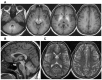
| SLC39A14 deficiency (HMNDYT2) | AR | Manganese transporter defect caused by impaired manganese uptake into liver. Affected persons present w/hypermanganesemia & rapidly progressive childhood-onset PD-DYT due to cerebral manganese deposition. Brain MRI appears the same as in HMNDYT1. Distinguishing features: absence of liver disease & polycythemia due to lack of hepatic manganese deposition (can be assessed by liver MRI) |
|
ATP7B
|
Wilson disease
| AR | Disorder of copper metabolism that can present w/hepatic, neurologic, or psychiatric disturbances (or a combination) from age 3 yrs to >50 yrs. Neurologic presentations incl mvmt disorders or rigid dystonia (mask-like facies, rigidity, gait disturbance, pseudobulbar involvement). |
|
Monogenic Parkinson disease 1 & hereditary disorders in the DiffDx of Parkinson disease
|
ATP13A2
DNAJC6
FBXO7
PODXL
SLC6A3
SYNJ1
| Juvenile-onset Parkinson disease (PD) 3 | AR | Age of onset of PD generally <20 yrs. Clinical presentation often incl addl signs incl dystonia, spasticity, & dementia. |
GBA1 (GBA)
LRRK2
PARK7
PINK1
PRKN
SNCA
VPS13C
VPS35 | Early-onset adult & late-onset adult Parkinson disease 2 | AD
AR | PD is characterized by rest tremor, muscle rigidity, slowed movement, & often postural instability. Onset is typically unilateral & may incl other abnormal movements (e.g., postural or action tremor & limb dystonia). Common assoc non-motor findings incl insomnia, depression, anxiety, REM sleep behavior disorder, fatigue, constipation, dysautonomia, & hyposmia. As part of a prodromal phase, non-motor features may predate formal diagnosis of PD by yrs. |
|
ATN1
|
DRPLA
| AD | Progressive disorder of ataxia, myoclonus, epilepsy, & progressive intellectual deterioration in children; ataxia, choreoathetosis, & dementia or character changes in adults. Clinical presentation varies w/age of onset. Cardinal features in adults: ataxia, choreoathetosis, dementia; in children: progressive intellectual deterioration, behavioral changes, myoclonus, epilepsy |
|
HTT
|
Huntington disease
| AD | Progressive disorder of motor, cognitive, & psychiatric disturbances. Mean age of onset 35-44 yrs. Affected persons may present w/neurologic manifestations or psychiatric changes. |
|
Hereditary dystonia
(selected examples of early-onset dystonia &/or dopa-responsive dystonia)
|
|
GCH1
|
GTP cyclohydrolase 1-deficient dopa-responsive dystonia
| AD
AR | Childhood-onset dystonia & dramatic & sustained response to low doses of oral administration of levodopa. Most common presenting findings: posturing or irregular tremor of a leg or arm (due to dystonic muscle contractions) |
|
KMT2B
|
KMT2B-related dystonia
| AD | Complex childhood-onset mvmt disorder. Typically presents w/progressive disease course evolving commonly from lower-limb focal dystonia into generalized dystonia w/prominent cervical, cranial, & laryngeal involvement |
|
SPR
|
Sepiapterin reductase deficiency
| AR | Rare cause of partially dopa-responsive childhood-onset dystonia characterized by axial hypotonia, motor & speech delay, weakness, & oculogyric crises; symptoms show diurnal fluctuation & sleep benefit. |
|
TH
| TH-deficient dopa-responsive dystonia (See Tyrosine Hydroxylase Deficiency.) | AR | Age of onset 12 mos-2 yrs. Initial manifestations: typically lower-limb dystonia &/or difficulty in walking. Diurnal fluctuation of symptoms may be present. |
|
THAP1
| DYT-THAP1 | AD | Although some phenotypic overlap w/DYT-TOR1A is observed, the onset of DYT-THAP1 is later (mean age 19 yrs) & cranial involvement is more prominent esp in muscles of the tongue, larynx, & face; dysphonia is a predominant feature. |
|
TOR1A
|
DYT1 early-onset isolated dystonia
| AD | Typically presents in childhood or adolescence & only on occasion in adulthood. Most common presenting findings: posturing or irregular tremor of a leg or arm (due to dystonic muscle contractions) |
|
Neurodegenerative diseases assoc w/dystonia
|
ATP13A2
C19orf12
COASY
CP
DCAF17
FA2H
FTL
PANK2
PLA2G6
WDR45
|
Neurodegeneration with brain iron accumulation disorders
| AR
AD
XL 3 | Characterized by abnormal accumulation of iron in basal ganglia. Generalized cerebral atrophy & cerebellar atrophy are frequently observed. Hallmark clinical manifestations: progressive dystonia & dysarthria, spasticity, parkinsonism, neuropsychiatric abnormalities, optic atrophy or retinal degeneration. Although cognitive decline occurs in some genetic types, more often cognition is relatively spared. Onset from infancy to adulthood. |
DLAT
DLD
PDHA1
PDHB
PDHX
PDP1
PDK3
| Primary pyruvate dehydrogenase complex deficiency (PDCD) | XL
AR 4 | Most commonly manifests as syndrome of neurologic signs (congenital microcephaly, hypotonia, epilepsy, &/or ataxia), abnormal brain imaging (dysgenesis of corpus callosum, Leigh syndrome), & metabolic abnormalities (↑ plasma pyruvate, lactic acidemia, &/or metabolic acidosis). DD is nearly universal. Mean age of diagnosis of primary PDCD typically~45 mos (median ~20 mos). |
| GBA1 (GBA) | Gaucher disease (GD) | AR | GD types 2 & 3 are characterized by presence of primary neurologic disease. Disease w/onset age <2 yrs, limited psychomotor development, & rapidly progressive course w/death by age 1-2 yrs is classified as GD type 2. Persons w/GD type 3 may have onset <2 yrs, but often more slowly progressive course, w/survival into 3rd-4th decade. |
NPC1
NPC2
|
Niemann-Pick disease type C
| AR | Principal manifestations are age dependent. Perinatal period & infancy: features are predominantly visceral, w/hepatosplenomegaly, jaundice, & (in some) pulmonary infiltrates. Late infancy onward: presentation dominated by neurologic manifestations. The youngest children may present w/hypotonia & DD, w/subsequent emergence of ataxia, dysarthria, dysphagia, &, in some, epileptic seizures, dystonia, & gelastic cataplexy. |
|
Hereditary spastic paraplegia
(selected examples of more commonly involved genes)
|
ATL1
KIF1A
REEP1
SPAST
| Autosomal dominant HSP 5 | AD | The predominant signs & symptoms of HSP are lower-extremity weakness & spasticity. When symptoms begin in very early childhood, they may be non-progressive & resemble spastic diplegic cerebral palsy. |
CYP7B1
SPG11
SPG7
| Autosomal recessive HSP 5 | AR |