U.S. flag

An official website of the United States government

NCBI Bookshelf. A service of the National Library of Medicine, National Institutes of Health.

Adam MP, Feldman J, Mirzaa GM, et al., editors. GeneReviews® [Internet]. Seattle (WA): University of Washington, Seattle; 1993-2024.

Cover of GeneReviews®

GeneReviews® [Internet].

Show details

Leukoencephalopathy with Brain Stem and Spinal Cord Involvement and Lactate Elevation

Synonyms: LBSL, Mitochondrial Aspartyl-tRNA Synthetase Deficiency

, MD, PhD, , PhD, , PhD, and , MD, PhD.

Author Information and Affiliations

Initial Posting: ; Last Update: February 18, 2021.

Estimated reading time: 23 minutes

Summary

Clinical characteristics.

Leukoencephalopathy with brain stem and spinal cord involvement and lactate elevation (LBSL) is characterized by slowly progressive cerebellar ataxia and spasticity with dorsal column dysfunction (decreased position and vibration sense) in most individuals. The neurologic dysfunction involves the legs more than the arms. The tendon reflexes are retained. Deterioration of motor skills usually starts in childhood or adolescence, but occasionally not until adulthood. Dysarthria develops over time. Occasional findings include epilepsy; learning problems; cognitive decline; and reduced consciousness, neurologic deterioration, and fever following minor head trauma.

Individuals with neonatal or early-infantile onset have a severe disease course often associated with early death. Those with childhood onset have slow progression with wheelchair dependence in the teens or twenties. Adult onset is associated with slow progression and mild impairment.

Diagnosis/testing.

The clinical diagnosis of LBSL can be established in a proband with characteristic abnormalities observed on brain and spinal cord MRI using MRI-based criteria. The molecular diagnosis can be established in a proband with suggestive findings and biallelic pathogenic variants in DARS2 identified by molecular genetic testing. If molecular results are inconclusive, a functional assay to identify reduced MtAspRS enzyme activity in lymphoblasts can confirm the diagnosis.

Management.

Treatment of manifestations: Supportive therapy includes: physical therapy and rehabilitation to improve motor function and prevent contractures and scoliosis; and anti-seizure medication, speech therapy, special education, and social work support as needed.

Surveillance: Assess for new neurologic manifestations at each visit; monitor those with seizures as needed; consider brain MRI every few years to monitor progression; monitor developmental progress and educational needs at each visit throughout childhood; assess for family support needs at each visit.

Genetic counseling.

LBSL is inherited in an autosomal recessive manner. If both parents are known to be heterozygous for a DARS2 pathogenic variant, each sib of an affected individual has at conception a 25% chance of being affected, a 50% chance of being an asymptomatic carrier, and a 25% chance of being unaffected and not a carrier. Once the DARS2 pathogenic variants have been identified in an affected family member, prenatal testing for a pregnancy at increased risk and preimplantation genetic testing are possible.

Diagnosis

Suggestive Findings

Diagnosis of leukoencephalopathy with brain stem and spinal cord involvement and lactate elevation (LBSL) should be suspected in individuals with characteristic abnormalities observed on brain and spinal cord MRI [van der Knaap et al 2003, Scheper et al 2007, Steenweg et al 2012]. However, atypical presentations with antenatal or early-infantile onset characterized by either profound cerebral atrophy or a leukoencephalopathy in the absence of typical involvement of tracts in the brain stem and spinal cord have been described; see * NOTE: (1) [Stellingwerff et al 2021].

MRI Criteria for LBSL

See Steenweg et al [2012], Stellingwerff et al [2021], Figure 1, Figure 2, and Figure 3.

Figure 1. . T2-weighted (A, C, D) and FLAIR (B) axial images of the brain in an adult with LBSL show extensive, patchy abnormalities with increased signal in the periventricular and deep cerebral white matter and posterior limb of the internal capsule.

Figure 1.

T2-weighted (A, C, D) and FLAIR (B) axial images of the brain in an adult with LBSL show extensive, patchy abnormalities with increased signal in the periventricular and deep cerebral white matter and posterior limb of the internal capsule. In the posterior (more...)

Figure 2. . Axial FLAIR images of the brain (A-C) in a young individual (toddler) with early presentation of LBSL show abnormal signal intensity (with some areas of rarefaction) of almost all cerebral white matter, only sparing the directly subcortical areas.

Figure 2.

Axial FLAIR images of the brain (A-C) in a young individual (toddler) with early presentation of LBSL show abnormal signal intensity (with some areas of rarefaction) of almost all cerebral white matter, only sparing the directly subcortical areas. The (more...)

Figure 3. . The MRI in a severe, early-onset form of LBSL at age two days (A, B, C) and age five months (D, E, F) shows striking progression of abnormalities.

Figure 3.

The MRI in a severe, early-onset form of LBSL at age two days (A, B, C) and age five months (D, E, F) shows striking progression of abnormalities. At two days, sagittal T1-weighted (A) and axial T2-weighted images (B) show hypoplasia of the cerebrum with (more...)

Major criteria. EITHER:

  • Signal abnormalities (abnormally low signal on T1-weighted images and abnormally high signal on T2-weighted images) in the following:
    • Cerebral white matter, which is either nonhomogeneous and spotty or homogeneous and confluent, with relative sparing of the directly subcortical white matter
    • Dorsal columns and lateral corticospinal tracts of the spinal cord (Visualization of such abnormalities in the cervical spinal cord is sufficient.)
    • Pyramids and/or decussation of the medial lemniscus in the medulla oblongata
    OR
  • Profound atrophy of the cerebral hemispheres in infancy (often with numerous tortuous blood vessels at the brain surface in early stages)

Supportive criteria. Signal abnormalities in the following:

  • Splenium of the corpus callosum
  • Posterior limb of the internal capsule
  • Superior cerebellar peduncles
  • Inferior cerebellar peduncles
  • Intraparenchymal part of the trigeminal nerve
  • Mesencephalic trigeminal tracts
  • Anterior spinocerebellar tracts in the medulla
  • Cerebellar white matter

* NOTE: (1) Individuals with antenatal and early-infantile onset have either a cerebral cortical degeneration followed by profound cerebral atrophy or a leukoencephalopathy. The brain stem abnormalities typical of LBSL may or may not be present [Steenweg et al 2011, Stellingwerff et al 2021]. Therefore, individuals with early onset may not meet MRI criteria. (2) Lactate is elevated within the abnormal cerebral white matter in most but not all affected individuals [van der Knaap et al 2003, Steenweg et al 2011]. Lactate elevation in proton magnetic resonance spectroscopy of abnormal white matter has been mentioned as a diagnostic criterion but has a low distinguishing value. If the MRI meets the criteria for LBSL, this diagnosis should be considered, whether lactate is elevated or not. If the MRI does not meet the criteria for LBSL, elevated lactate may be a general indicator of a mitochondrial leukoencephalopathy, but not specifically LBSL.

Establishing the Diagnosis

Clinical Diagnosis

The clinical diagnosis of LBSL can be established in a proband with MRI-based criteria; all major criteria and at least one supportive criterion should be fulfilled (see MRI Criteria for LBSL) [Steenweg et al 2012]. See also Figure 1, Figure 2, and Figure 3.

Molecular Diagnosis

The molecular diagnosis of LBSL can be established in proband with suggestive findings and biallelic pathogenic variants in DARS2 identified by molecular genetic testing (see Table 1).

If molecular results are inconclusive, a functional assay to identify reduced MtAspRS enzyme activity in lymphoblasts can confirm the diagnosis [van Berge et al 2014].

Molecular genetic testing approaches can include a combination of gene-targeted testing (single-gene testing, multigene panel) and comprehensive genomic testing (exome sequencing, genome sequencing, exome array) depending on the phenotype.

Gene-targeted testing requires that the clinician determine which gene(s) are likely involved, whereas genomic testing does not. Individuals with the distinctive findings described in Suggestive Findings are likely to be diagnosed using gene-targeted testing (see Option 1), whereas those with a phenotype indistinguishable from many other inherited disorders with leukoencephalopathy are more likely to be diagnosed using genomic testing (see Option 2).

Option 1

  • Single-gene testing. Sequence analysis of DARS2 is performed first to detect small intragenic deletions/insertions and missense, nonsense, and splice site variants. Note: Depending on the sequencing method used, single-exon, multiexon, or whole-gene deletions/duplications may not be detected. If only one or no variant is detected by the sequencing method used, the next step is to perform gene-targeted deletion/duplication analysis to detect exon and whole-gene deletions or duplications.
  • A multigene panel that includes DARS2 and other genes of interest (see Differential Diagnosis) may be considered to identify the genetic cause of the condition while limiting identification of variants of uncertain significance and pathogenic variants in genes that do not explain the underlying phenotype. Note: (1) The genes included in the panel and the diagnostic sensitivity of the testing used for each gene vary by laboratory and are likely to change over time. (2) Some multigene panels may include genes not associated with the condition discussed in this GeneReview. (3) In some laboratories, panel options may include a custom laboratory-designed panel and/or custom phenotype-focused exome analysis that includes genes specified by the clinician. (4) Methods used in a panel may include sequence analysis, deletion/duplication analysis, and/or other non-sequencing-based tests.
    For an introduction to multigene panels click here. More detailed information for clinicians ordering genetic tests can be found here.

Option 2. When the phenotype is indistinguishable from many other inherited disorders characterized by leukoencephalopathy, comprehensive genomic testing (which does not require the clinician to determine which gene is likely involved) is an option. Exome sequencing is most commonly used; genome sequencing is also possible. Importantly, the most common DARS2 pathogenic variant involves intron 2, which is easily missed by exome sequencing. If a heterozygous DARS2 variant is identified in an individual with leukoencephalopathy, analysis of intron 2 should be performed.

If exome sequencing is not diagnostic, exome array (when clinically available) may be considered to detect (multi)exon deletions or duplications that cannot be detected by sequence analysis [Lan et al 2017].

For an introduction to comprehensive genomic testing click here. More detailed information for clinicians ordering genomic testing can be found here.

Table 1.

Molecular Genetic Testing Used in Leukoencephalopathy with Brain Stem and Spinal Cord Involvement and Lactate Elevation

Gene 1MethodProportion of Pathogenic Variants 2 Detectable by This Method
DARS2 Sequence analysis 3~90% 4, 5
Gene-targeted deletion/duplication analysis 6Rare 7
UnknownNASee footnote 8.
1.
2.

See Molecular Genetics for information on allelic variants detected in this gene.

3.

Sequence analysis detects variants that are benign, likely benign, of unknown significance, likely pathogenic, or pathogenic. Variants may include small intragenic deletions/insertions and missense, nonsense, and splice site variants; typically, exon or whole-gene deletions/duplications are not detected. For issues to consider in interpretation of sequence analysis results, click here.

4.

Affected individuals are almost invariably compound heterozygous for two pathogenic variants in DARS2. A few individuals with homozygous pathogenic variants have been identified.

5.

In a few individuals it has not been possible to determine both pathogenic variants: in four of 43 families the second pathogenic variant could not be found in gDNA; in two of three the second pathogenic variant was detected using cDNA; cells of the fourth individual were not available for isolation of mRNA for cDNA synthesis.

6.

Gene-targeted deletion/duplication analysis detects intragenic deletions or duplications. Methods used may include quantitative PCR, long-range PCR, multiplex ligation-dependent probe amplification (MLPA), and a gene-targeted microarray designed to detect single-exon deletions or duplications.

7.

Intragenic deletion of exon 12 has been reported [Lan et al 2017].

8.

In one person fulfilling the MRI criteria for LBSL, no pathogenic variants in DARS2 were detected in either gDNA or cDNA [Scheper et al 2007; Scheper & van der Knaap, personal communication].

Clinical Characteristics

Clinical Description

The clinical picture of LBSL consists of slowly progressive cerebellar ataxia, spasticity, and dorsal column dysfunction, involving the legs more than the arms. Most affected individuals have decreased position and vibration sense of the legs more than the arms, leading to increased difficulty walking in the dark. Manual dexterity becomes impaired to a variable degree.

To date, more than 100 individuals have been identified with biallelic pathogenic variants in DARS2. Large series [Steenweg et al 2011, van Berge et al 2014, Stellingwerff et al 2021] as well as multiple case reports [Köhler et al 2015, Shimojima et al 2017, Yahia et al 2018, Yelam et al 2019, N'Gbo N'Gbo Ikazabo et al 2020, Yazici Gencdal et al 2020] have been described. Prospective natural history data are currently not available. The following description of the phenotypic features associated with this condition is based on the cited reports.

Table 2.

Leukoencephalopathy with Brain Stem and Spinal Cord Involvement and Lactate Elevation: Frequency of Select Features

Feature% of Persons w/FeatureComment
Normal early development 80%Infantile form was recently identified [Stellingwerff et al 2021]; relative incidence is not clear yet but probably low.
Loss of motor skills 100%Core feature of LBSL
Cerebellar ataxia Very common
Spasticity Very common
Axonal neuropathy Uncommon↓ or absent tendon reflexes, distal weakness, sensory loss
Learning disability 20%Most often learning disability; severe intellectual impairment is rare.
Cognitive decline 20%
Epilepsy UncommonUsually easily controlled w/ASM

ASM = anti-seizure medication

Variable severity. The range of disease spectrum includes:

  • Antenatal and early-infantile onset. Severe form characterized by microcephaly, severe developmental delay, epilepsy, and often early death [Steenweg et al 2012, Stellingwerff et al 2021]
  • Childhood onset. Most common form with slow progression. Most individuals with childhood onset become partially or fully wheelchair dependent in their teens, twenties, or later [van Berge et al 2014].
  • Adult onset. Slowly progressive form with only mild impairment [van Berge et al 2014]. Individuals with adult-onset disease are not known to become wheelchair dependent [van Berge et al 2014].

Motor involvement. In most affected individuals, initial motor development is normal. Deterioration of motor skills usually starts in childhood or adolescence [Uluc et al 2008, van Berge et al 2014] and occasionally in infancy [Steenweg et al 2012] or adulthood [van Berge et al 2014]. Slowly progressive cerebellar ataxia and spasticity occur in most individuals. Axonal peripheral neuropathy may add to the distal weakness. The motor dysfunction involves the legs more than the arms.

In the individuals with antenatal or early-infantile onset, motor involvement is severe and typically no or very few motor milestones are achieved [Stellingwerff et al 2021].

Sensory involvement. There is severe dorsal column dysfunction with decreased position and vibration sense on neurologic examination, causing sensory ataxia especially in the legs. Evidence of an axonal neuropathy is found in some (not all) affected individuals, leading to decreased or absent tendon reflexes and distal sensory loss affecting the legs more than the arms [van der Knaap et al 2003, Távora et al 2007, Uluc et al 2008, Isohanni et al 2010]. However, true peripheral neuropathy is probably not common as neurophysiologic examination is often normal [van der Knaap et al 2003].

Speech. Dysarthria develops over time. Swallowing is generally not affected in individuals with childhood or adult onset [van Berge et al 2014].

Cognitive skills. Some have learning problems from early on, but most have normal intellectual capacity. Cognitive decline may occur and is usually slow [van der Knaap et al 2003, Serkov et al 2004]. Individuals with antenatal or early-infantile presentation typically have severe cognitive impairment [Stellingwerff et al 2021].

Epilepsy. Some affected individuals develop epilepsy. Seizures are infrequent and easily controlled with medication [van der Knaap et al 2003]. In individuals with antenatal and early-infantile onset, epilepsy is almost invariably present and often severe [Stellingwerff et al 2021].

Microcephaly. In individuals with antenatal or early-infantile onset, microcephaly is almost invariably present and often severe [Stellingwerff et al 2021]. See also Figure 3.

Response to minor head trauma. Some affected individuals experience lowered consciousness, neurologic deterioration, and fever following minor head trauma [Serkov et al 2004]. Recovery is only partial.

Routine laboratory tests, including cerebrospinal fluid (CSF) analysis, are usually normal. In a few individuals mild and inconsistent mild elevation of lactate concentration has been noted in blood or CSF or both. No published information is available.

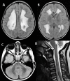
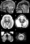
Neuropathologic findings have been described in two sibs with a severe variant of LBSL [Yamashita et al 2013]. Electron microscopy revealed vacuolar changes and myelin splitting in the affected white matter [Yamashita et al 2013]. Quantitative MR parameters are in line with these findings, with T2 hyperintense white matter showing increased fractional anisotropy and reduced apparent diffusion coefficient [Steenweg et al 2011].

Genotype-Phenotype Correlations

An overview study of 66 affected individuals revealed preliminary evidence in support of a genotype-phenotype correlation [van Berge et al 2014]. A recent study on antenatal and early-infantile presentation of LBSL substantiates genotype-phenotype correlation. Individuals with this presentation lack the common "leaky" splice site variants and instead have more severe loss-of-function variants [Stellingwerff et al 2021] (see Molecular Pathogenesis).

Prevalence

LBSL is rare. Carrier rate in the general population is low, with the exception of Finland, where a carrier rate of 1:95 has been reported [Isohanni et al 2010]. To date, only one family with parental consanguinity has been observed [Miyake et al 2011]. Almost all affected individuals are compound heterozygous for two pathogenic variants. Only four individuals from two families have been described with homozygous pathogenic variants [Miyake et al 2011, Synofzik et al 2011]. Strikingly, no instances of homozygosity have been seen among Finnish persons with LBSL [van Berge et al 2014].

Differential Diagnosis

Classic Leukoencephalopathy with Brain Stem and Spinal Cord Involvement and Lactate Elevation

The classic clinical picture of leukoencephalopathy with brain stem and spinal cord involvement and lactate elevation (LBSL) consists of slowly progressive cerebellar ataxia, spasticity, and dorsal column dysfunction, involving the legs more than the arms. The tendon reflexes are retained. Based on these findings alone, many disorders can be considered [Parodi et al 2018]; however, characteristic brain and spinal cord MRI findings (see Figure 1) distinguish LBSL from other disorders with overlapping clinical features.

Ataxia. MRI findings distinguish LBSL from other hereditary ataxias [Steenweg et al 2011, van der Knaap et al 2019]. Specifically, the spotty white matter abnormalities with focal diffusion restriction seen in CSF1R-related adult-onset leukoencephalopathy with axonal spheroids and pigmented glia (ALSP), an autosomal dominant disorder, can be confused with adult LBSL; however, in ALSP, the brain stem and spinal cord involvement typical of LBSL are lacking [Steenweg et al 2011, Mochel et al 2019].

Ataxia and spasticity in combination with MRI abnormalities

  • The clinical findings of ataxia and spasticity in combination with MRI abnormalities of the dorsal columns, lateral corticospinal tracts, and cerebral white matter are compatible with vitamin B12 deficiency (combined cord degeneration) [Locatelli et al 1999]. However, the brain stem abnormalities typically seen in LBSL do not occur in vitamin B12 deficiency. In vitamin B12 deficiency the cervical spinal cord is mainly affected [Locatelli et al 1999], whereas in LBSL the entire spinal cord is affected [van der Knaap et al 2003]. See also Figure 1.
  • Hypomyelination with brain stem and spinal cord involvement and leg spasticity (HBSL; OMIM 615281), caused by biallelic pathogenic variants in DARS1, shares part of the selective vulnerability of brain stem and spinal cord structures, but the signal abnormalities in HBSL are mild – indicative of hypomyelination – while they are prominent in LBSL [Taft et al 2013]. See also Figure 1 and Figure 2.

Elevated lactate in MRS or body fluids or both combined with clinical findings of a spinocerebellar ataxia or white matter abnormalities on MRI or both should lead to the consideration of mitochondrial disorders [Vernon & Bindoff 2018]. Although the brain stem and spinal cord are frequently affected in mitochondrial disorders, the selective involvement of specific brain stem and spinal cord tracts is unique for LBSL [van der Knaap & Valk 2005].

Variant LBSL: Antenatal and Early-Infantile Onset

A variant form of LBSL is characterized by antenatal or early infantile presentation and profoundly delayed or absent development and epilepsy [Stellingwerff et al 2021]. MRI reveals severe cerebral atrophy. If the brain stem and spinal cord abnormalities typical of LBSL are present, the correct diagnosis can still be suggested. However, in some individuals with early-onset LBSL, brain stem and spinal cord abnormalities are absent and in such cases the MRI shows a picture of an early onset severe neuronal degenerative disorder (see Figure 3). In these individuals, LBSL needs to be distinguished from infantile neuronal ceroid lipofuscinosis (OMIM PS256730), and other early-onset tRNA synthetase defects related to variants in AARS1 (OMIM 616339) and RARS1 (OMIM 616140) [Simons et al 2015, Mendes et al 2020].

Additionally, Stellingwerff et al [2021] reported an infantile-onset LBSL variant form with a leukoencephalopathy but lacking the typical brain stem or spinal cord abnormalities (see Figure 2). In such cases other mitochondrial leukodystrophies should be considered [Zhang et al 2019].

Management

Evaluations Following Initial Diagnosis

To establish the extent of disease and needs in an individual diagnosed with leukoencephalopathy with brain stem and spinal cord involvement and lactate elevation (LBSL), the evaluations summarized in Table 3 (if not performed as part of the evaluation that led to the diagnosis) are recommended.

Table 3.

Recommended Evaluations Following Initial Diagnosis in Individuals with Leukoencephalopathy with Brain Stem and Spinal Cord Involvement and Lactate Elevation

System/ConcernEvaluationComment
Neurologic/
Neurodevelopment
Neurologic examConsider:
  • Brain & spinal cord MRI & proton MRS of abnormal cerebral white matter;
  • EEG if seizures are suspected.
PT/OT/rehab medicine assessmentTo assess for weakness, spasticity, & gait issues
Speech therapy assessmentIn those w/dysarthria
Cognitive assessmentIn those w/suspected cognitive involvement
Genetic counseling By genetics professionals 1To inform patients & families re nature, MOI, & implications of LBSL to facilitate medical & personal decision making
Family support/
resources
For affected infants/children assess:

LBSL = leukoencephalopathy with brain stem and spinal cord involvement and lactate elevation; MOI = mode of inheritance; OT = occupational therapy; PT = physical therapy

1.

Medical geneticist, certified genetic counselor, or certified advanced genetic nurse

Treatment of Manifestations

Table 4.

Treatment of Manifestations in Individuals with Leukoencephalopathy with Brain Stem and Spinal Cord Involvement and Lactate Elevation

Manifestation/ConcernTreatmentConsiderations/Other
Spasticity PT & rehab medicine to improve motor function & prevent contractures & scoliosisAssess need for positioning & mobility devices, disability parking placard.
Seizures ASM if epileptic seizures are present
Dysarthria Speech therapy
Intellectual disability Early intervention & IEP as indicated
Family/Community
  • Ensure appropriate social work involvement to connect families w/local resources, respite, & support.
  • Care coordination to manage multiple subspecialty appointments, equipment, medications, & supplies
Ongoing assessment of need for palliative care involvement &/or home nursing

ASM = anti-seizure medication; IEP = individualized education plan; PT = physical therapy

Surveillance

Table 5.

Recommended Surveillance for Individuals with Leukoencephalopathy with Brain Stem and Spinal Cord Involvement and Lactate Elevation

System/ConcernEvaluationFrequency
Neurologic Assess for new manifestations (e.g., seizures, changes in tone, movement disorders).At each visit
Monitor those w/seizures.As clinically indicated
Brain MRICan be considered every few yrs to monitor progression
Development Monitor developmental progress & educational needs.At each visit throughout childhood
Miscellaneous/
Other
Assess family need for social work support (e.g., palliative/respite care, home nursing; other local resources) & care coordination.At each visit

Agents/Circumstances to Avoid

Case reports have described neurologic deterioration following head trauma (as has been reported for several other leukodystrophies including vanishing white matter and adrenoleukodystrophy). The exact risk is not clear. No specific guidelines exist for contact sports or other activities. Affected individuals should be counseled on the above-mentioned observations and can then make an individual decision based on personal preference [van der Knaap et al 2006, Budhram & Pandey 2017].

Evaluation of Relatives at Risk

See Genetic Counseling for issues related to testing of at-risk relatives for genetic counseling purposes.

Therapies Under Investigation

Search ClinicalTrials.gov in the US and EU Clinical Trials Register in Europe for access to information on clinical studies for a wide range of diseases and conditions. Note: There may not be clinical trials for this disorder.

Other

Studies of muscle biopsies, fibroblasts, and lymphoblasts show no evidence of mitochondrial dysfunction; therefore, there is no rationale for the "mitochondrial cocktail" of vitamins and cofactors, often given to persons with mitochondrial dysfunction.

Genetic Counseling

Genetic counseling is the process of providing individuals and families with information on the nature, mode(s) of inheritance, and implications of genetic disorders to help them make informed medical and personal decisions. The following section deals with genetic risk assessment and the use of family history and genetic testing to clarify genetic status for family members; it is not meant to address all personal, cultural, or ethical issues that may arise or to substitute for consultation with a genetics professional. —ED.

Mode of Inheritance

Leukoencephalopathy with brain stem and spinal cord involvement and lactate elevation (LBSL) is inherited in an autosomal recessive manner.

Risk to Family Members

Parents of a proband

  • The parents of an affected individual are obligate heterozygotes (i.e., presumed to be carriers of one DARS2 pathogenic variant based on family history).
  • Molecular genetic testing is recommended for the parents of a proband to confirm that both parents are heterozygous for a DARS2 pathogenic variant and to allow reliable recurrence risk assessment. If a pathogenic variant is detected in only one parent, the following possibilities should be considered:
  • Heterozygotes (carriers) are asymptomatic and are not at risk of developing the disorder.

Sibs of a proband

  • If both parents are known to be heterozygous for a DARS2 pathogenic variant, each sib of an affected individual has at conception a 25% chance of being affected, a 50% chance of being an asymptomatic carrier, and a 25% chance of being unaffected and not a carrier.
  • The clinical manifestations in sibs with biallelic DARS2 pathogenic variant are highly variable. Yahia et al [2018] described clinical presentations ranging "from an apparently healthy individual (with findings limited to brisk reflexes incidentally recognized at age 20 years) to disabled patients" within the same family.
  • Heterozygotes (carriers) are asymptomatic and are not at risk of developing the disorder.

Offspring of a proband. The offspring of an individual with LBSL are obligate heterozygotes (carriers) for a pathogenic variant in DARS2.

Other family members. Each sib of the proband's parents is at a 50% risk of being a carrier of a DARS2 pathogenic variant.

Carrier Detection

Carrier testing for at-risk family members requires prior identification of the DARS2 pathogenic variants in the family.

Related Genetic Counseling Issues

Family planning

  • The optimal time for determination of genetic risk and discussion of the availability of prenatal/preimplantation genetic testing is before pregnancy.
  • It is appropriate to offer genetic counseling (including discussion of potential risks to offspring and reproductive options) to young adults who are affected, are carriers, or are at risk of being carriers.

DNA banking. Because it is likely that testing methodology and our understanding of genes, allelic variants, and diseases will improve in the future, consideration should be given to banking DNA from probands in whom a molecular diagnosis has not been confirmed (i.e., the causative genetic alteration/s are unknown).

Prenatal Testing and Preimplantation Genetic Testing

Once the DARS2 pathogenic variants have been identified in an affected family member, prenatal testing for a pregnancy at increased risk and preimplantation genetic testing are possible.

Differences in perspective may exist among medical professionals and within families regarding the use of prenatal testing. While most centers would consider use of prenatal testing to be a personal decision, discussion of these issues may be helpful.

Resources

GeneReviews staff has selected the following disease-specific and/or umbrella support organizations and/or registries for the benefit of individuals with this disorder and their families. GeneReviews is not responsible for the information provided by other organizations. For information on selection criteria, click here.

Molecular Genetics

Information in the Molecular Genetics and OMIM tables may differ from that elsewhere in the GeneReview: tables may contain more recent information. —ED.

Table A.

Leukoencephalopathy with Brain Stem and Spinal Cord Involvement and Lactate Elevation: Genes and Databases

GeneChromosome LocusProteinLocus-Specific DatabasesHGMDClinVar
DARS2 1q25​.1 Aspartate--tRNA ligase, mitochondrial DARS2 database DARS2 DARS2

Data are compiled from the following standard references: gene from HGNC; chromosome locus from OMIM; protein from UniProt. For a description of databases (Locus Specific, HGMD, ClinVar) to which links are provided, click here.

Table B.

OMIM Entries for Leukoencephalopathy with Brain Stem and Spinal Cord Involvement and Lactate Elevation (View All in OMIM)

610956ASPARTYL-tRNA SYNTHETASE 2; DARS2
611105LEUKOENCEPHALOPATHY WITH BRAINSTEM AND SPINAL CORD INVOLVEMENT AND LACTATE ELEVATION; LBSL

Molecular Pathogenesis

DARS2 encodes a mitochondrial aspartyl-tRNA synthetase (mtAspRS), which charges tRNAAsp in mitochondria. Mitochondrial AspRS possesses strictly conserved residues found in all known AspRS sequences, including those for ATP binding and tRNA binding. Residues involved in amino acid binding include those typical for class II aaRSs and those specific for aspartic acid recognition [Bonnefond et al 2005]. Based on the homology to its bacterial counterparts, mtAspRS is thought to form homodimers [Delarue et al 1994].

Almost all affected individuals have a pathogenic variant that affects splicing of exon 3. Incorrect splicing of this exon results in a frameshift in the reading frame and nonsense-mediated decay of the wrongly spliced mRNA. It should be noted that these pathogenic variants upstream of exon 3 diminish but do not completely abolish correct splicing. As a result, a low amount of wild type protein is produced in the cells of an affected individual. A total lack of mtAspRS activity is thought to be incompatible with life.

Another common pathogenic variant, c.492+2T>C (p.Met134_Lys165del) leads to a deletion of part of the protein. Though not fully understood, this deletion is likely to have a severe effect on the function of the protein.

Mechanism of disease causation. Loss of function. Several pathogenic missense variants have been shown to severely reduce the amino acylation function in assays with purified bacterially expressed recombinant proteins [Scheper et al 2007].

DARS2-specific laboratory technical considerations. In almost all affected individuals one pathogenic variant is present upstream of exon 3. The variant c.228-21_228-20delTTinsC is most often observed. In other affected individuals nucleotide changes are seen in the same region, within a stretch of ten or 11 C-residues that lies ten nucleotides upstream of exon 3 [Scheper et al 2007].

Table 6.

Notable DARS2 Pathogenic Variants

Reference SequencesDNA Nucleotide ChangePredicted
Protein Change
Comment [Reference]
NM_108122​.5
NP_060592​.2
c.228-21_228-20delTTinsCp.Arg76SerfsTer5Most common pathogenic variant reported
c.492+2T>Cp.Met134_Lys165delSee footnote 1.
c.455G>Tp.Cys152Phe
NG_016138​.1 Exon 12 deletion 2See footnote 2. Lan et al [2017]

Variants listed in the table have been provided by the authors. GeneReviews staff have not independently verified the classification of variants.

GeneReviews follows the standard naming conventions of the Human Genome Variation Society (varnomen​.hgvs.org). See Quick Reference for an explanation of nomenclature.

1.

Several individuals share haplotypes involving five or six microsatellite markers on chromosome 1p25. The pathogenic variants c.492+2T>C and c.455G>T are correlated with two of these haplotypes and are often seen in affected individuals of northeastern European origin [Scheper et al 2007, Isohanni et al 2010].

2.

Variant designation that does not conform to current naming conventions; no breakpoints were delineated in the original report.

Chapter Notes

Author Notes

Center for Children with White Matter Disorders at Amsterdam University Medical Centers

Acknowledgments

We would like to thank Drs Ali Fatemi and Amena Smith for ongoing collaboration in LBSL research.

Author History

Truus EM Abbink, PhD (2021-present)
Marc Engelen, MD, PhD (2021-present)
Gajja S Salomons, PhD (2015-present)
Gert C Scheper, PhD; Vrije Universiteit Medical Center, Amsterdam (2010-2015)
Marjo S van der Knaap, MD, PhD (2010-present)

Revision History

  • 18 February 2021 (sw) Comprehensive update posted live
  • 12 February 2015 (me) Comprehensive update posted live
  • 25 May 2010 (me) Review posted live
  • 22 February 2010 (mvdk) Original submission

References

Literature Cited

  • Bonnefond L, Fender A, Rudinger-Thirion J, Giegé R, Florentz C, Sissler M. Toward the full set of human mitochondrial aminoacyl-tRNA synthetases: characterization of AspRS and TyrRS. Biochemistry. 2005;44:4805–16. [PubMed: 15779907]
  • Budhram A, Pandey SK. Activation of cerebral X-linked adrenoleukodystrophy after head trauma. Can J Neurol Sci. 2017;44:597–8. [PubMed: 28534457]
  • Delarue M, Poterszman A, Nikonov S, Garber M, Moras D, Thierry JC. Crystal structure of a prokaryotic aspartyl tRNA-synthetase. EMBO J. 1994;13:3219–29. [PMC free article: PMC395218] [PubMed: 8045252]
  • Isohanni P, Linnankivi T, Buzkova J, Lonnqvist T, Pihko H, Valanne L, Tienari PJ, Elovaara I, Pirttila T, Reunanen M, Koivisto K, Marjavaara S, Suomalainen A. DARS2 mutations in mitochondrial leukoencephalopathy and multiple sclerosis. J Med Genet. 2010;47:66–70. [PubMed: 19592391]
  • Jónsson H, Sulem P, Kehr B, Kristmundsdottir S, Zink F, Hjartarson E, Hardarson MT, Hjorleifsson KE, Eggertsson HP, Gudjonsson SA, Ward LD, Arnadottir GA, Helgason EA, Helgason H, Gylfason A, Jonasdottir A, Jonasdottir A, Rafnar T, Frigge M, Stacey SN, Th Magnusson O, Thorsteinsdottir U, Masson G, Kong A, Halldorsson BV, Helgason A, Gudbjartsson DF, Stefansson K. Parental influence on human germline de novo mutations in 1,548 trios from Iceland. Nature. 2017;549:519–22. [PubMed: 28959963]
  • Köhler C, Heyer C, Hoffjan S, Stemmler S, Lücke T, Thielsa C, Kohlschütter A, Lobele U, Horvath R, Kleinle S, Benet-Pages A, Abicht A. Early-onset leukoencephalopathy due to a homozygous missense mutation in the DARS2 gene. Mol Cell Probes. 2015;29:319–22. [PubMed: 26327357]
  • Lan MY, Chang YY, Yeh TH, Lin TK, Lu CS. Leukoencephalopathy with brainstem and spinal cord involvement and lactate elevation (LBSL) with a novel DARS2 mutation and isolated progressive spastic paraparesis. J Neurol Sci. 2017;372:229–31. [PubMed: 28017220]
  • Locatelli ER, Laureno R, Ballard P, Mark AS. MRI in vitamin B12 deficiency myelopathy. Can J Neurol Sci. 1999;26:60–3. [PubMed: 10068811]
  • Mendes MI, Green LMC, Bertini E, Tonduti D, Aiello C, Smith D, Salsano E, Beerepoot S, Hertecant J, von Spiczak S, Livingston JH, Emrick L, Fraser J, Russell L, Bernard G, Magri S, Di Bella D, Taroni F, Koenig MK, Moroni I, Cappuccio G, Brunetti-Pierri N, Rhee J, Mendelsohn BA, Helbig I, Helbig K, Muhle H, Ismayl O, Vanderver AL, Salomons GS, van der Knaap MS, Wolf NI. RARS1-related hypomyelinating leukodystrophy: expanding the spectrum. Ann Clin Transl Neurol. 2020;7:83–93. [PMC free article: PMC6952319] [PubMed: 31814314]
  • Miyake N, Yamashita S, Kurosawa K, Miyatake S, Tsurusaki Y, Doi H, Saitsu H, Matsumoto N. A novel homozygous mutation of DARS2 may cause a severe LBSL variant. Clin Genet. 2011;80:293–6. [PubMed: 21815884]
  • Mochel F, Delorme C, Czernecki V, Froger J, Cormier F, Ellie E, Fegueux N, Lehéricy S, Lumbroso S, Schiffmann R, Aubourg P, Roze E, Labauge P, Nguyen S. Haematopoietic stem cell transplantation in CSF1R-related adult-onset leukoencephalopathy with axonal spheroids and pigmented glia. J Neurol Neurosurg Psychiatry. 2019;90:1375–6. [PubMed: 31213485]
  • N'Gbo N'Gbo Ikazabo R, Mostosi C, Jissendi P, Labaisse M, Vandernoot I. A new DARS2 mutation discovered in an adult patient. Case Rep Neurol. 2020;12:107–13. [PMC free article: PMC7154256] [PubMed: 32308605]
  • Parodi L, Coarelli G, Stevanin G, Brice A, Durr A. Hereditary ataxias and paraparesias: clinical and genetic update. Curr Opin Neurol. 2018;31:462–71. [PubMed: 29847346]
  • Scheper GC, van der Klok T, van Andel RJ, van Berkel CG, Sissler M, Smet J, Muravina TI, Serkov SV, Uziel G, Bugiani M, Schiffmann R, Krageloh-Mann I, Smeitink JA, Florentz C, Van CR, Pronk JC, van der Knaap MS. Mitochondrial aspartyl-tRNA synthetase deficiency causes leukoencephalopathy with brain stem and spinal cord involvement and lactate elevation. Nat Genet. 2007;39:534–9. [PubMed: 17384640]
  • Serkov SV, Pronin IN, Bykova OV, Maslova OI, Arutyunov NV, Muravina TI, Kornienko VN, Fadeeva LM, Marks H, Bonnemann C, Schiffmann R, van der Knaap MS. Five patients with a recently described novel leukoencephalopathy with brainstem and spinal cord involvement and elevated lactate. Neuropediatrics. 2004;35:1–5. [PubMed: 15002045]
  • Shimojima K, Higashiguchi T, Kishimoto T, Miyatake S, Miyake N, Takanashi J, Matsumoto N, Yamamoto T. A novel DARS2 mutation in a Japanese patient with leukoencephalopathy with brainstem and spinal cord involvement but no lactate elevation. Hum Genome Var. 2017;4:17051. [PMC free article: PMC5678206] [PubMed: 29138691]
  • Simons C, Griffin LB, Helman G, Golas G, Pizzino A, Bloom M, Murphy JL, Crawford J, Evans SH, Topper S, Whitehead MT, Schreiber JM, Chapman KA, Tifft C, Lu KB, Gamper H, Shigematsu M, Taft RJ, Antonellis A, Hou YM, Vanderver A. Loss-of-function alanyl-tRNA synthetase mutations cause an autosomal-recessive early-onset epileptic encephalopathy with persistent myelination defect. Am J Hum Genet. 2015;96:675–81. [PMC free article: PMC4385183] [PubMed: 25817015]
  • Steenweg ME, Pouwels PJ, Wolf NI, van Wieringen WN, Barkhof F, van der Knaap MS. Leucoencephalopathy with brainstem and spinal cord involvement and high lactate: quantitative magnetic resonance imaging. Brain. 2011;134:3333–41. [PubMed: 22006980]
  • Steenweg ME, van Berge L, van Berkel CG, de Coo IF, Temple IK, Brockmann K, Mendonça CI, Vojta S, Kolk A, Peck D, Carr L, Uziel G, Feigenbaum A, Blaser S, Scheper GC, van der Knaap MS. Early-onset LBSL: how severe does it get? Neuropediatrics. 2012;43:332–8. [PubMed: 23065766]
  • Stellingwerff MD, Figuccia S, Bellacchio E, Alvarez EK, Castiglioni C, Topaloglu P, Stutterd CA, Erasmus CE, Sanchez-Valle A, Lebon S, Hughes S, Schmitt-Mechelke T, Vasco G, Chow G, Rahikkala E, Dallabona C, Okuma C, Aiello C, Goffrini P, Abbink TEM, Bertini ES, Van der Knaap MS. LBSL: case series and DARS2 variant analysis in early severe forms with unexpected presentations. Neurol Genet. 2021;7:e559. [PMC free article: PMC8105885] [PubMed: 33977142]
  • Synofzik M, Schicks J, Lindig T, Biskup S, Schmidt T, Hansel J, Lehmann-Horn F, Schöls L. Acetazolamide-responsive exercise-induced episodic ataxia associated with a novel homozygous DARS2 mutation. J Med Genet. 2011;48:713–5. [PubMed: 21749991]
  • Taft RJ, Vanderver A, Leventer RJ, Damiani SA, Simons C, Grimmond SM, Miller D, Schmidt J, Lockhart PJ, Pope K, Ru K, Crawford J, Rosser T, de Coo IF, Juneja M, Verma IC, Prabhakar P, Blaser S, Raiman J, Pouwels PJ, Bevova MR, Abbink TE, van der Knaap MS, Wolf NI. Mutations in DARS cause hypomyelination with brain stem and spinal cord involvement and leg spasticity. Am J Hum Genet. 2013;92:774–80. [PMC free article: PMC3644624] [PubMed: 23643384]
  • Távora DG, Nakayama M, Gama RL, Alvim TC, Portugal D, Comerlato EA. Leukoencephalopathy with brainstem and spinal cord involvement and high brain lactate: report of three Brazilian patients. Arq Neuropsiquiatr. 2007;65:506–11. [PubMed: 17665025]
  • Uluc K, Baskan O, Yildirim KA, Ozsahin S, Koseoglu M, Isak B, Scheper GC, Gunal DI, van der Knaap MS. Leukoencephalopathy with brain stem and spinal cord involvement and high lactate: a genetically proven case with distinct MRI findings. J Neurol Sci. 2008;273:118–22. [PubMed: 18619624]
  • van Berge L, Hamilton EM, Linnankivi T, Uziel G, Steenweg ME, Isohanni P, Wolf NI, Krägeloh-Mann I, Brautaset NJ, Andrews PI, de Jong BA, al Ghamdi M, van Wieringen WN, Tannous BA, Hulleman E, Würdinger T, van Berkel CG, Polder E, Abbink TE, Struys EA, Scheper GC, van der Knaap MS. Leukoencephalopathy with brainstem and spinal cord involvement and lactate elevation: clinical and genetic characterization and target for therapy. Brain. 2014;137:1019–29. [PubMed: 24566671]
  • van der Knaap MS, van der Voorn P, Barkhof F, van Coster R, Krageloh-Mann I, Feigenbaum A, Blaser S, Vles JS, Rieckmann P, Pouwels PJ. A new leukoencephalopathy with brainstem and spinal cord involvement and high lactate. Ann Neurol. 2003;53:252–8. [PubMed: 12557294]
  • van der Knaap MS, Valk J. Magnetic Resonance of Myelin Myelination and Myelin Disorders. 3 ed. Berlin: Springer; 2005.
  • van der Knaap MS, Pronk JC, Scheper GC. Vanishing white matter disease. Lancet Neurol. 2006;5:413–23. [PubMed: 16632312]
  • van der Knaap MS, Schiffman R, Mochel F, Wolf NI. Diagnosis, prognosis and treatment of leukodystrophies. Lancet Neurol. 2019;18:962–72. [PubMed: 31307818]
  • Vernon HJ, Bindoff LA. Mitochondrial ataxias. Handb Clin Neurol. 2018;155:129–41. [PubMed: 29891055]
  • Yahia A, Elsayed L, Babai A, Salih MA, Misbah El-Sadig S, Amin M, Koko M, Abubakr R, Idris R, Taha SOMA, Elmalik SA, Brice A, Eltahir Ahmed A, Stevanin G. Intra-familial phenotypic heterogeneity in a Sudanese family with DARS2-related leukoencephalopathy, brainstem and spinal cord involvement and lactate elevation: a case report. BMC Neurology. 2018;18:175. [PMC free article: PMC6198356] [PubMed: 30352563]
  • Yamashita S, Miyake N, Matsumoto N, Osaka H, Iai M, Aida N, Tanaka Y. Neuropathology of leukoencephalopathy with brainstem and spinal cord involvement and high lactate caused by a homozygous mutation of DARS2. Brain Dev. 2013;35:312–6. [PubMed: 22677571]
  • Yazici Gencdal I, Dincer A, Obuz O, Yapici Z. Leukoencephalopathy with brain stem and spinal cord involvement and lactate elevation (LBSL): a case with long-term follow-up. Neurologist. 2020;25:144–7. [PubMed: 32925487]
  • Yelam A, Nagarajan E, Chuquilin M, Govindarajan R. Leucoencephalopathy with brain stem and spinal cord involvement and lactate elevation: a novel mutation in the DARS2 gene. BMJ Case Rep. 2019;12:e227755. [PMC free article: PMC6340551] [PubMed: 30635318]
  • Zhang J, Liu M, Zhang Z, Zhou L, Kong W, Jiang Y, Wang J, Xiao J, Wu Y. Genotypic spectrum and natural history of cavitating leukoencephalopathies in childhood. Pediatr Neurol. 2019;94:38–47. [PubMed: 30770271]
Copyright © 1993-2024, University of Washington, Seattle. GeneReviews is a registered trademark of the University of Washington, Seattle. All rights reserved.

GeneReviews® chapters are owned by the University of Washington. Permission is hereby granted to reproduce, distribute, and translate copies of content materials for noncommercial research purposes only, provided that (i) credit for source (http://www.genereviews.org/) and copyright (© 1993-2024 University of Washington) are included with each copy; (ii) a link to the original material is provided whenever the material is published elsewhere on the Web; and (iii) reproducers, distributors, and/or translators comply with the GeneReviews® Copyright Notice and Usage Disclaimer. No further modifications are allowed. For clarity, excerpts of GeneReviews chapters for use in lab reports and clinic notes are a permitted use.

For more information, see the GeneReviews® Copyright Notice and Usage Disclaimer.

For questions regarding permissions or whether a specified use is allowed, contact: ude.wu@tssamda.

Bookshelf ID: NBK43417PMID: 20506600

Views

Tests in GTR by Gene

Related information

  • MedGen
    Related information in MedGen
  • OMIM
    Related OMIM records
  • PMC
    PubMed Central citations
  • PubMed
    Links to PubMed
  • Gene
    Locus Links

Similar articles in PubMed

See reviews...See all...

Recent Activity

Your browsing activity is empty.

Activity recording is turned off.

Turn recording back on

See more...