U.S. flag

An official website of the United States government

NCBI Bookshelf. A service of the National Library of Medicine, National Institutes of Health.

Adam MP, Feldman J, Mirzaa GM, et al., editors. GeneReviews® [Internet]. Seattle (WA): University of Washington, Seattle; 1993-2024.

Cover of GeneReviews®

GeneReviews® [Internet].

Show details

NOTCH3-Related Lateral Meningocele Syndrome

Synonym: Lehman Syndrome

, MD, FRCPC, , MD, MSc, and , MD.

Author Information and Affiliations

Initial Posting: ; Last Update: May 5, 2022.

Estimated reading time: 17 minutes

Summary

Clinical characteristics.

NOTCH3-related lateral meningocele syndrome (LMS) is characterized by multiple lateral spinal meningoceles (protrusions of the arachnoid and dura through spinal foramina), distinctive facial features, joint hyperextensibility, hypotonia, and skeletal, cardiac, and urogenital anomalies. Neurologic sequelæ of the meningoceles depend on size and location and can include neurogenic bladder, paresthesia, back pain, and/or paraparesis. Other neurologic findings can include Chiari I malformation, syringomyelia, and rarely, hydrocephalus. Additional findings of LMS include developmental delay, mixed or conductive hearing loss, and cleft palate. Skeletal abnormalities may include scoliosis, vertebral fusion, scalloping of vertebrae, and wormian bones. Infants may demonstrate feeding difficulties with poor weight gain.

Diagnosis/testing.

The diagnosis of NOTCH3-related LMS syndrome is established in a proband with consistent clinical findings and a heterozygous pathogenic variant in NOTCH3.

Management.

Treatment of manifestations: Surgical intervention of lateral spinal meningoceles is generally avoided, but may be necessary due to neurologic manifestations secondary to meningocele size and location. Symptomatic treatment of neurologic sequelae of lateral meningoceles (neurogenic bladder, paresthesias, back pain, and/or paraparesis) provided as needed. As needed: timely supportive interventions to optimize development; management by specialists in chronic pain management or rehabilitation medicine; physiotherapy to reduce the risk for joint subluxation and dislocation. Routine management of: cleft palate, cardiovascular issues, genitourinary abnormalities, ophthalmologic issues, hearing loss, feeding difficulties.

Surveillance: Close clinical and radiographic monitoring for progressive neurologic symptoms and increase in meningocele size; an initial yearly scan to monitor for stability and subsequent spacing to every two years if meningoceles are small in size. Ongoing monitoring by the appropriate subspecialists for developmental, musculoskeletal, cardiovascular, genitourinary, gastrointestinal, ophthalmologic, and/or hearing issues.

Genetic counseling.

All probands reported to date with NOTCH3-related LMS whose parents have undergone molecular genetic testing have the disorder as the result of a de novo NOTCH3 pathogenic variant. Each child of an individual with NOTCH3-related LMS has a 50% chance of inheriting the pathogenic variant. Once the NOTCH3 pathogenic variant has been identified in an affected family member, prenatal testing for a pregnancy at increased risk and preimplantation genetic testing for NOTCH3-related LMS are possible.

Diagnosis

Formal diagnostic clinical criteria for NOTCH3-related lateral meningocele syndrome (LMS) have not been established.

Suggestive Findings

NOTCH3-related LMS should be suspected in individuals with the following findings.

Multiple lateral spinal meningoceles (protrusion of the arachnoid and dura through the spinal foramina) are present in all affected individuals (Figure 1).

Figure 1. . Numerous lateral meningoceles (see arrows) protrude through the thoracic foramina in a sagittal view (a) and through the lumbar foramina in a sagittal (b) and axial (c) view.

Figure 1.

Numerous lateral meningoceles (see arrows) protrude through the thoracic foramina in a sagittal view (a) and through the lumbar foramina in a sagittal (b) and axial (c) view. The curved arrow in (a) shows a meningocele protruding from the middle cranial (more...)

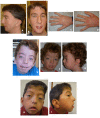
Characteristic craniofacial appearance [Gripp et al 2015, Ejaz et al 2016, Yamada et al 2022] includes widely spaced eyes, highly arched eyebrows, downslanted palpebral fissures, ptosis, malar flattening, long philtrum, thin vermilion of the upper lip, high and narrow palate, micrognathia, and coarse hair with a low posterior hairline (Figure 2).

Figure 2.

Figure 2.

Photographs of individuals with NOTCH3-related lateral meningocele syndrome A-D. Patient 1 at age 24 years:

Additional findings that may be present:

  • Neurologic. Chiari I malformation, hydrocephalus, syringomyelia, and neurogenic bladder.
  • Developmental delay or (rarely) intellectual disability
  • Musculoskeletal. Hypotonia and joint hypermobility
  • Dermatologic. Hyperextensibility of the skin
  • Cardiovascular malformations. Aortic abnormalities (bicuspid aortic valve, aortic dilatation, and coarctation of the aortic arch) and ventricular septal defects
  • Genitourinary. Cryptorchidism and (rarely) renal anomaly
  • Gastrointestinal. Feeding difficulties, dysphagia, and gastroesophageal reflux disease

Establishing the Diagnosis

The diagnosis of NOTCH3-related LMS is established in a proband with consistent clinical findings and a heterozygous pathogenic (or likely pathogenic) variant in NOTCH3 identified by molecular genetic testing (see Table 1).

Note: (1) Per ACMG/AMP variant interpretation guidelines, the terms "pathogenic variant" and "likely pathogenic variant" are synonymous in a clinical setting, meaning that both are considered diagnostic and can be used for clinical decision making [Richards et al 2015]. Reference to "pathogenic variants" in this section is understood to include likely pathogenic variants.. (2) Identification of a heterozygous NOTCH3 variant of uncertain significance does not establish or rule out the diagnosis.

Molecular genetic testing approaches can include a combination of gene-targeted testing (single-gene testing, multigene panel) and comprehensive genomic testing (exome sequencing, genome sequencing) depending on the phenotype.

Gene-targeted testing requires that the clinician determine which gene(s) are likely involved, whereas genomic testing does not. Individuals with the distinctive findings described in Suggestive Findings are likely to be diagnosed using gene-targeted testing (see Option 1), whereas those in whom the diagnosis of LMS has not been considered are more likely to be diagnosed using genomic testing (see Option 2).

Option 1

Single-gene testing. Sequence analysis of NOTCH3 is performed first to detect small intragenic deletions/insertions and missense, nonsense, and splice site variants. Note: Depending on the sequencing method used, single-exon, multiexon, or whole-gene deletions/duplications may not be detected.

Typically, if no variant is detected by the sequencing method used, the next step is to perform gene-targeted deletion/duplication analysis to detect exon and whole-gene deletions or duplications; however, to date, such variants have not been identified as a cause of this disorder.

A multigene panel that includes NOTCH3 and other genes of interest (see Differential Diagnosis) is most likely to identify the genetic cause of the condition while limiting identification of variants of uncertain significance and pathogenic variants in genes that do not explain the underlying phenotype. Note: (1) The genes included in the panel and the diagnostic sensitivity of the testing used for each gene vary by laboratory and are likely to change over time. (2) Some multigene panels may include genes not associated with the condition discussed in this GeneReview. (3) In some laboratories, panel options may include a custom laboratory-designed panel and/or custom phenotype-focused exome analysis that includes genes specified by the clinician. (4) Methods used in a panel may include sequence analysis, deletion/duplication analysis, and/or other non-sequencing-based tests.

For an introduction to multigene panels click here. More detailed information for clinicians ordering genetic tests can be found here.

Option 2

Comprehensive genomic testing does not require the clinician to determine which gene is likely involved. Exome sequencing is most commonly used; genome sequencing is also possible.

For an introduction to comprehensive genomic testing click here. More detailed information for clinicians ordering genomic testing can be found here.

Table 1.

Molecular Genetic Testing Used in NOTCH3-Related Lateral Meningocele Syndrome (LMS)

Gene 1MethodProportion of Probands with a Pathogenic Variant 2 Detectable by Method
NOTCH3 Sequence analysis 311/11 4
Gene-targeted deletion/duplication analysis 5Unknown 6
1.
2.

See Molecular Genetics for information on variants detected in this gene.

3.

Sequence analysis detects variants that are benign, likely benign, of uncertain significance, likely pathogenic, or pathogenic. Variants may include small intragenic deletions/insertions and missense, nonsense, and splice site variants; typically, exon or whole-gene deletions/duplications are not detected. For issues to consider in interpretation of sequence analysis results, click here.

4.
5.

Gene-targeted deletion/duplication analysis detects intragenic deletions or duplications. Methods used may include a range of techniques such as quantitative PCR, long-range PCR, multiplex ligation-dependent probe amplification (MLPA), and a gene-targeted microarray designed to detect single-exon deletions or duplications.

6.

No data on detection rate of gene-targeted deletion/duplication analysis are available.

Clinical Characteristics

Clinical Description

NOTCH3-related lateral meningocele syndrome (LMS) is characterized by multiple lateral spinal meningoceles, distinctive facial features, joint hyperextensibility, hypotonia, and skeletal, cardiac, and urogenital anomalies.

To date, 11 individuals have been identified with a pathogenic variant in NOTCH3 [Gripp et al 2015, Ejaz et al 2016, Brown et al 2017, Cappuccio et al 2020, Han et al 2022, Yamada et al 2022]. The following description of the phenotypic features associated with this condition is based on these reports.

Table 2.

NOTCH3-Related Lateral Meningocele Syndrome: Frequency of Select Features

Feature# of Persons with Feature
Multiple lateral spinal meningoceles11/11
Characteristic facial appearance11/11
Developmental delay10/11
Musculoskeletal findings suggestive of a connective tissue disorder11/11
Cardiovascular concerns10/11
Genitourinary issues4/8 (males w/cryptorchidism)
Gastrointestinal concerns10/11
Eye abnormality7/11
Hearing loss4/11
Cleft palate4/11

Multiple lateral spinal meningoceles (protrusions of the arachnoid and dura through the spinal foramina) are found in all affected individuals. Neurologic sequelæ of the meningoceles can include neurogenic bladder, paresthesia, back pain, and/or paraparesis depending on size and location. Other neurologic findings can include Chiari I malformation, syringomyelia, and rarely, hydrocephalus [Gripp et al 2015, Ejaz et al 2016, Cuoco et al 2020].

Craniofacial features include widely spaced eyes with downslanted palpebral fissures, highly arched eyebrows, ptosis, malar flattening, long philtrum, thin vermilion of the upper lip, high and narrow palate, micrognathia, and angulated ears (Figure 2). Cleft palate, dental crowding, epicanthal folds, dolichocephaly, and coarse hair with a low posterior hairline can also be seen.

A high nasal voice is noted in many individuals [Gripp et al 2015].

Developmental delay, particularly gross motor delay, is frequently seen in individuals with NOTCH3-related LMS, but cognition is typically preserved. Of ten individuals with NOTCH3-related LMS, all had developmental delay; only one also had intellectual disability [Gripp et al 2015, Ejaz et al 2016, Yamada et al 2022].

Musculoskeletal. Overlap with features of connective tissue disorders include neonatal hypotonia, abdominal hernias, ligamentous laxity, keloid scars, and back pain in later life.

Many individuals have skeletal changes including scoliosis, kyphosis, vertebral fusion, scalloping of vertebrae, and wormian bones [Gripp et al 2015, Cappuccio et al 2020].

Cardiovascular concerns have included atrial septal defect, ventricular septal defect, bicuspid aortic valve, dilatation of the aorta, and coarctation of the aortic arch [Gripp et al 2015, Ejaz et al 2016, Cappuccio et al 2020].

Genitourinary. Cryptorchidism is frequently seen. Prenatal hydronephrosis with postnatal left renal hypoplasia and bilateral renal cysts was reported in one individual [Cappuccio et al 2020].

Gastrointestinal. Infants with NOTCH3-related LMS may demonstrate feeding difficulties with poor weight gain. Feeding difficulties were severe enough to warrant gastrostomy tube feeding in two individuals [Ejaz et al 2016, Brown et al 2017].

Eye abnormalities can include proptosis and oculomotor restriction.

Hearing loss. Mixed or conductive hearing loss has been noted. Brain MRI of one individual showed an apical turn of the cochlea and modiolus, and dysmorphic vestibule [Cappuccio et al 2020]

Genotype-Phenotype Correlations

No genotype-phenotype correlations have been identified.

Penetrance

Penetrance appears to be complete but data are limited.

Nomenclature

Lehman et al [1977] first described a woman with dysmorphic facial features, skeletal sclerosis, and multiple meningoceles, and her mother with similar craniofacial dysmorphisms. Philip et al [1995], who published a second report, named the syndrome after Lehman. The authors prefer the term "lateral meningocele syndrome" as it emphasizes the hallmark feature of the condition.

The title of this GeneReview, NOTCH3-related lateral meningocele syndrome, is based on the dyadic naming approach proposed by Biesecker et al [2021] to delineate mendelian genetic disorders.

Prevalence

NOTCH3-related lateral meningocele syndrome is very rare, with 11 reported individuals. There does not appear to be increased prevalence in specific populations.

Differential Diagnosis

The differential diagnosis for NOTCH3-related lateral meningocele syndrome (LMS) is summarized in Table 4.

Table 4.

Genes and Disorders of Interest in the Differential Diagnosis of NOTCH3-Related Lateral Meningocele Syndrome (LMS)

Gene(s)DisorderMOIKey Features /Comment
BRAF, KRAS, LZTR1, MAP2K1, MRAS, NRAS, PTPN11, RAF1, RASA2, RIT1, RRAS2, SOS1, SOS2Noonan syndrome (NS)AD
(AR) 1
NOTCH3-related LMS & NS share similarities in their characteristic facial features (incl widely spaced eyes, ptosis, epicanthus, & low-set ears w/↑ posterior angulation) & a low posterior hairline. Prenatal signs of NS (e.g., nuchal edema & congenital cardiac defect) were also seen in 2 persons w/NOTCH3-related LMS. 2
FBN1 Marfan syndrome (MFS)ADNOTCH3-related LMS has significant overlap w/other connective tissue disorders. Spinal meningeal anomalies, specifically dural ectasias, are frequently seen in MFS. Persons w/MFS may also have joint laxity, scoliosis, cardiovascular anomalies, & some shared facial features (e.g., malar flattening & retrognathia).
NF1 Neurofibromatosis type 1 (NF1)ADLateral meningoceles & dural ectasia have been described in some persons w/NF1. 3 The distinctive facial features of NOTCH3-related LMS are not seen in persons w/NF1; other distinctive characteristics of NF1 incl café au lait spots, neurofibromas, & Lisch nodules. NF1 & NOTCH3-related LMS may also have similar skeletal & neurologic changes incl scoliosis, hydrocephalus, & developmental delays.
NOTCH2 Hadju-Cheney syndrome (OMIM 102500)ADSkeletal disorder characterized by dysmorphic facial features (e.g., malar flattening, thick eyebrows, micrognathia), osteoporosis w/acro-osteolysis, wormian bones, premature loss of dentition, & joint laxity. 1 person w/NOTCH3-related LMS was initially misdiagnosed w/Hadju-Cheney syndrome due to presence of acro-osteolysis. 4
FKBP14, PLOD1FKBP14- & PLOD1-related kyphoscoliotic Ehlers-Danlos syndrome (kEDS)ARLateral meningocele has been described in 1 person w/kEDS, & dural ectasia in at least 2 persons. 5 Persons w/kEDS also present w/joint hypermobility, congenital hypotonia, progressive scoliosis & kyphosis, motor delays, & hyperextensibility of the skin.
SMAD2, SMAD3, TGFB2, TGFB3, TGFBR1, TGFBR2Loeys-Dietz syndrome (LDS)ADOverlapping features in LDS & NOTCH3-related LMS incl spinal pathology (dural ectasia in LDS), some facial features (e.g. downslanted palpebral fissures, proptosis, high arched palate), congenital heart defects, joint hypermobility, & spine deformities.
1.

NS is most often inherited in an autosomal dominant manner. NS caused by pathogenic variants in LZTR1 can be inherited in either an autosomal dominant or an autosomal recessive manner.

2.
3.
4.
5.

Management

No clinical practice guidelines for NOTCH3-related lateral meningocele syndrome have been published.

Evaluations Following Initial Diagnosis

To establish the extent of disease and needs in an individual diagnosed with NOTCH3-related lateral meningocele syndrome (LMS), the evaluations summarized in Table 5 (if not performed as part of the evaluation that led to the diagnosis) are recommended.

Table 5.

Recommended Evaluations Following Initial Diagnosis in Individuals with NOTCH3-Related Lateral Meningocele Syndrome (LMS)

System/ConcernEvaluationComment
Lateral spinal
meningoceles
  • Spine MRI to assess for meningoceles
  • Neurosurgical assessment to evaluate effect of lateral spinal meningocele size & location on neurologic function
Associated
neurologic issues
Neurologic evalAssess for neuropathy, pain, neurogenic bladder &/or paraparesis.
Brain MRIAssess for Chiari I malformation or hydrocephalus.
Developmental
delay
Developmental assessment
  • To incl motor, adaptive, cognitive, & speech-language eval
  • Eval for early intervention / special education
Musculoskeletal Assessment for abdominal hernia, ligamentous laxity / joint concerns, scoliosis
Cardiovascular Eval w/cardiologist to incl echocardiogram
Genitourinary Assessment for cryptorchidism
Gastrointestinal Infant feeding evalAssess for GERD & any palate abnormalities.
Eyes Ophthalmologic evalAssess for ↓ vision, abnormal ocular mvmt, & structural eye abnormality.
Hearing Audiologic evalAssess for hearing loss.
Genetic
counseling
By genetics professionals 1To inform affected persons & their families re nature, MOI, & implications of NOTCH3-related LMS in order to facilitate medical & personal decision making
Family support
& resources
Assess need for:

GERD = gastroesophageal reflux disease; MOI = mode of inheritance

1.

Medical geneticist, certified genetic counselor, certified advanced genetic nurse

Treatment of Manifestations

The following medical issues are managed in a routine manner: cleft palate, cardiovascular issues, genitourinary abnormalities, ophthalmologic issues, hearing loss, feeding difficulties.

Table 6.

Treatment of Manifestations in Individuals with NOTCH3-Related Lateral Meningocele Syndrome (LMS)

Manifestation/
Concern
TreatmentConsiderations/Other
Lateral spinal
meningoceles
  • There is no standard operative mgmt of lateral spinal meningoceles [Cuoco et al 2020].
  • Symptomatic treatment of neurologic sequelæ (e.g., neurogenic bladder, paresthesias, back pain, &/or paraparesis) as needed
  • Surgical intervention is generally avoided but may be necessary due to neurologic manifestations secondary to meningocele size & location [Cuoco et al 2020].
  • 1 person benefited from CSF diversion w/a VP shunt for symptomatic relief of thoracolumbar meningocele [Brown et al 2017].
  • When required, surgical approach is individualized & can incl laminectomy for smaller meningoceles, costotransversectomy for larger meningoceles.
Developmental
delay
Timely supportive interventions as needed to optimize development through OT & education resources
Musculoskeletal
issues
  • PT to ↓ risk for joint subluxation & dislocation
  • Mgmt by specialists in chronic pain mgmt or rehab medicine as needed

OT = occupational therapy; PT = physical therapy

Surveillance

There are no established surveillance guidelines for lateral spinal meningoceles. Close clinical and radiographic monitoring is recommended for progressive neurologic symptoms and meningocele size enlargement [Cuoco et al 2020]. MRI of the spine can be considered at a one- to two-year interval; an initial yearly scan to monitor for stability and subsequent spacing to every two years if meningoceles are small in size has been suggested. Larger lesions may require closer follow up [JA Cuoco 2022, personal communication].

Ongoing monitoring by the appropriate subspecialists for developmental, musculoskeletal, cardiovascular, genitourinary, gastrointestinal, ophthalmologic, and/or hearing issues is indicated.

Evaluation of Relatives at Risk

See Genetic Counseling for issues related to testing of at-risk relatives for genetic counseling purposes.

Therapies Under Investigation

Search ClinicalTrials.gov in the US and EU Clinical Trials Register in Europe for access to information on clinical studies for a wide range of diseases and conditions. Note: There may not be clinical trials for this disorder.

Genetic Counseling

Genetic counseling is the process of providing individuals and families with information on the nature, mode(s) of inheritance, and implications of genetic disorders to help them make informed medical and personal decisions. The following section deals with genetic risk assessment and the use of family history and genetic testing to clarify genetic status for family members; it is not meant to address all personal, cultural, or ethical issues that may arise or to substitute for consultation with a genetics professional. —ED.

Mode of Inheritance

NOTCH3-related lateral meningocele syndrome (LMS) is inherited in an autosomal dominant manner.

Risk to Family Members

Parents of a proband

Sibs of a proband. The risk to the sibs of the proband depends on the genetic status of the proband’s parents:

  • If a parent of the proband has the NOTCH3 pathogenic variant identified in the proband, the risk to the sibs of inheriting the pathogenic variant is 50%.
  • If the NOTCH3 pathogenic variant identified in the proband cannot be detected in the leukocyte DNA of either parent, the recurrence risk to sibs is estimated to be 1% because of the theoretic possibility of parental germline mosaicism [Rahbari et al 2016].
  • If the parents are clinically unaffected but their genetic status is unknown, the risk to the sibs of a proband is presumed to be low but slightly greater than that of the general population because of the theoretic possibility of parental germline mosaicism.

Offspring of a proband. Each child of an individual with NOTCH3-related LMS has a 50% chance of inheriting the NOTCH3 pathogenic variant.

Other family members. The risk to other family members depends on the status of the proband's parents: if a parent has the NOTCH3 pathogenic variant, members of the parent's family may be at risk.

Related Genetic Counseling Issues

Family planning

  • The optimal time for determination of genetic risk and discussion of the availability of prenatal/preimplantation genetic testing is before pregnancy.
  • It is appropriate to offer genetic counseling (including discussion of potential risks to offspring and reproductive options) to the parents of affected individuals and young adults who are affected.

Prenatal Testing and Preimplantation Genetic Testing

Once the NOTCH3 pathogenic variant has been identified in an affected family member, prenatal and preimplantation genetic testing for NOTCH3-related LMS are possible.

Differences in perspective may exist among medical professionals and within families regarding the use of prenatal testing. While most centers would consider use of prenatal testing to be a personal decision, discussion of these issues may be helpful.

Resources

GeneReviews staff has selected the following disease-specific and/or umbrella support organizations and/or registries for the benefit of individuals with this disorder and their families. GeneReviews is not responsible for the information provided by other organizations. For information on selection criteria, click here.

Molecular Genetics

Information in the Molecular Genetics and OMIM tables may differ from that elsewhere in the GeneReview: tables may contain more recent information. —ED.

Table A.

NOTCH3-Related Lateral Meningocele Syndrome: Genes and Databases

Data are compiled from the following standard references: gene from HGNC; chromosome locus from OMIM; protein from UniProt. For a description of databases (Locus Specific, HGMD, ClinVar) to which links are provided, click here.

Table B.

OMIM Entries for NOTCH3-Related Lateral Meningocele Syndrome (View All in OMIM)

130720LATERAL MENINGOCELE SYNDROME; LMNS
600276NOTCH RECEPTOR 3; NOTCH3

Molecular Pathogenesis

NOTCH3 encodes the neurogenic locus notch homolog protein 3 (NOTCH3), a transmembrane receptor involved in cell signaling and activation of target genes. It appears to be universally expressed and is a part of a highly conserved signaling pathway that is responsible for cell differentiation, proliferation, and apoptosis, through regulating transcription of target genes.

In Notch3 variant mouse models, an osteopenic phenotype was thought to result from induction of RANKL by cells of osteoblast lineages with subsequent increased osteoclastogenesis [Canalis 2021].

In all individuals with NOTCH3-related LMS to date, the causative pathogenic variants (nonsense and frameshift) have been in exon 33, the last exon of NOTCH3 [Gripp et al 2015, Ejaz et al 2016, Han et al 2022, Yamada et al 2022]. It is predicted that the transcripts of such variants in this location escape nonsense-mediated decay and express a truncated NOTCH3 protein, which has been documented in the cells of one individual [Gripp et al 2015]. Through negative regulation of protein stability these truncated NOTCH3 proteins would lack the C-terminal PEST domain, which is responsible for degradation of the intracellular signaling portion of activated NOTCH3. The truncated protein is more likely to escape degradation, thereby prolonging its cellular half-life [Wang et al 2015, Canalis 2021].

Mechanism of disease causation. NOTCH3-related LMS is thought to be caused by a gain-of-function mechanism [Gripp et al 2015, Wang et al 2015]. The precise mechanism by which excess NOTCH3 results in NOTCH3-related LMS is not understood.

Chapter Notes

Author Notes

Web page: Resham Ejaz - McMaster Experts

Acknowledgments

We thank the individuals with NOTCH3-related LMS and their families for their participation in research related to the condition.

Revision History

  • 5 May 2022 (ha) Comprehensive update posted live
  • 23 June 2016 (bp) Review posted live
  • 19 February 2016 (re) Original submission

References

Literature Cited

  • Avela K, Valanne L, Helenius I, Mäkitie O. Hajdu-Cheney syndrome with severe dural ectasia. Am J Med Genet A. 2011;155A:595-8. [PubMed: 21337686]
  • Biesecker LG, Adam MP, Alkuraya FS, Amemiya AR, Bamshad MJ, Beck AE, Bennett JT, Bird LM, Carey JC, Chung B, Clark RD, Cox TC, Curry C, Dinulos MBP, Dobyns WB, Giampietro PF, Girisha KM, Glass IA, Graham JM Jr, Gripp KW, Haldeman-Englert CR, Hall BD, Innes AM, Kalish JM, Keppler-Noreuil KM, Kosaki K, Kozel BA, Mirzaa GM, Mulvihill JJ, Nowaczyk MJM, Pagon RA, Retterer K, Rope AF, Sanchez-Lara PA, Seaver LH, Shieh JT, Slavotinek AM, Sobering AK, Stevens CA, Stevenson DA, Tan TY, Tan WH, Tsai AC, Weaver DD, Williams MS, Zackai E, Zarate YA. A dyadic approach to the delineation of diagnostic entities in clinical genomics. Am J Hum Genet. 2021;108:8-15. [PMC free article: PMC7820621] [PubMed: 33417889]
  • Brown EC, Gupta K, Cantab M, Sayama C. Neurosurgical management in lateral meningocele syndrome: case report. J Neurosurg Pediatr. 2017;19:232-8. [PubMed: 27911244]
  • Canalis E. The skeleton of lateral meningocele syndrome. Front Genet. 2021;11:620334. [PMC free article: PMC7841456] [PubMed: 33519922]
  • Cappuccio G, Apuzzo D, Alagia M, Torella A, Pinelli M, Franco B, Corrado B, Del Giudice E, D'Amico A, Nigro V, Brunetti-Pierri N, et al. Expansion of the phenotype of lateral meningocele syndrome. Am J Med Genet A. 2020;182:1259-62. [PMC free article: PMC7217177] [PubMed: 32141180]
  • Cuoco JA, Klein BJ, Busch CM, Gosnell HL, Kar A, Marvin EA, Apfel LS. Neurosurgical management of lateral meningocele syndrome: a clinical update for the pediatric neurosurgeon. Pediatr Neurosurg. 2020;55:2-11. [PubMed: 31838470]
  • Ejaz R, Qin W, Huang L, Blaser S, Tetreault M, Hartley T, Boycott KM, Carter MT, et al. Lateral meningocele (Lehman) syndrome: A child with a novel NOTCH3 mutation. Am J Med Genet A. 2016;170A:1070-5. [PubMed: 26754023]
  • Gripp KW. Lateral meningocele syndrome and Hajdu-Cheney syndrome: different disorders with overlapping phenotypes. Am J Med Genet. 2011;155A:1773-4. [PubMed: 21671395]
  • Gripp KW, Robbins KM, Sobreira NL, Witmer PD, Bird LM, Avela K, Makitie O, Alves D, Hogue JS, Zackai EH, Doheny KF, Stabley DL, Sol-Church K. Truncating mutations in the last exon of NOTCH3 cause lateral meningocele syndrome. Am J Med Genet Part A. 2015;167A:271-81. [PMC free article: PMC5589071] [PubMed: 25394726]
  • Han Y, Chen M, Wang H. Management of lateral meningocele syndrome in a child without neurological symptoms and literature review. Childs Nerv Syst. 2022. Epub ahead of print. [PubMed: 35128567]
  • Lehman RA, Stears JC, Wesenberg RL, Nusbaum ED. Familial osteosclerosis with abnormalities of the nervous system and meninges. J Pediatr. 1977;90:49-54 [PubMed: 830893]
  • Philip N, Andrac L, Moncla A, Sigaudy S, Zanon N, Lena G, Choux M. Multiple lateral meningoceles, distinctive facies and skeletal anomalies: a new case of Lehman syndrome. Clin Dysmorphol 1995;4:347-51. [PubMed: 8574426]
  • Rahbari R, Wuster A, Lindsay SJ, Hardwick RJ, Alexandrov LB, Turki SA, Dominiczak A, Morris A, Porteous D, Smith B, Stratton MR, Hurles ME, et al. Timing, rates and spectra of human germline mutation. Nat Genet. 2016;48:126-33. [PMC free article: PMC4731925] [PubMed: 26656846]
  • Richards S, Aziz N, Bale S, Bick D, Das S, Gastier-Foster J, Grody WW, Hegde M, Lyon E, Spector E, Voelkerding K, Rehm HL, et al. Standards and guidelines for the interpretation of sequence variants: a joint consensus recommendation of the American College of Medical Genetics and Genomics and the Association for Molecular Pathology. Genet Med. 2015;17:405-24. [PMC free article: PMC4544753] [PubMed: 25741868]
  • Ueda K, Honda O, Satoh Y, Kawai M, Gyobu T, Kanazawa T, Hidaka S, Yanagawa M, Sumikawa H, Tomiyama N. Computed tomography (CT) findings in 88 neurofibromatosis 1 (NF1) patients: Prevalence rates and correlations of thoracic findings. Eur J Radiol. 2015;84:1191-5. [PubMed: 25802206]
  • Wang K, Zhang Q, Li D, Ching K, Zhang C, Zheng X, Ozeck M, Shi S, Li X, Wang H, Reito P, Christensen J, Olson P. PEST domain mutations in Notch receptors comprise an oncogenic driver segment in triple-negative breast cancer sensitive to γ-secretase inhibitor. Clin Cancer Res. 2015;21:1487-96. [PubMed: 25564152]
  • Yamada M, Arimitsu T, Suzuki H, Miwa T, Kosaki K. Early diagnosis of lateral meningocele syndrome in an infant without neurological symptoms based on genomic analysis. Childs Nerv Syst. 2022;38:659-63. [PubMed: 34121137]
Copyright © 1993-2024, University of Washington, Seattle. GeneReviews is a registered trademark of the University of Washington, Seattle. All rights reserved.

GeneReviews® chapters are owned by the University of Washington. Permission is hereby granted to reproduce, distribute, and translate copies of content materials for noncommercial research purposes only, provided that (i) credit for source (http://www.genereviews.org/) and copyright (© 1993-2024 University of Washington) are included with each copy; (ii) a link to the original material is provided whenever the material is published elsewhere on the Web; and (iii) reproducers, distributors, and/or translators comply with the GeneReviews® Copyright Notice and Usage Disclaimer. No further modifications are allowed. For clarity, excerpts of GeneReviews chapters for use in lab reports and clinic notes are a permitted use.

For more information, see the GeneReviews® Copyright Notice and Usage Disclaimer.

For questions regarding permissions or whether a specified use is allowed, contact: ude.wu@tssamda.

Bookshelf ID: NBK368476PMID: 27336130

Views

Tests in GTR by Gene

Related information

  • OMIM
    Related OMIM records
  • PMC
    PubMed Central citations
  • PubMed
    Links to PubMed
  • Gene
    Locus Links

Similar articles in PubMed

See reviews...See all...

Recent Activity

Your browsing activity is empty.

Activity recording is turned off.

Turn recording back on

See more...