U.S. flag

An official website of the United States government

NCBI Bookshelf. A service of the National Library of Medicine, National Institutes of Health.

Adam MP, Feldman J, Mirzaa GM, et al., editors. GeneReviews® [Internet]. Seattle (WA): University of Washington, Seattle; 1993-2024.

Cover of GeneReviews®

GeneReviews® [Internet].

Show details

C3 Glomerulopathy

Synonyms: C3G, Glomerulonephritis with Dominant C3

, PhD and , MD.

Author Information and Affiliations

Initial Posting: ; Last Update: April 5, 2018.

Estimated reading time: 44 minutes

Summary

Clinical characteristics.

C3 glomerulopathy (C3G) is a complex ultra-rare complement-mediated renal disease caused by uncontrolled activation of the complement alternative pathway (AP) in the fluid phase (as opposed to cell surface) that is rarely inherited in a simple mendelian fashion. C3G affects individuals of all ages, with a median age at diagnosis of 23 years. Individuals with C3G typically present with hematuria, proteinuria, hematuria and proteinuria, acute nephritic syndrome or nephrotic syndrome, and low levels of the complement component C3. Spontaneous remission of C3G is uncommon, and about half of affected individuals develop end-stage renal disease (ESRD) within ten years of diagnosis, occasionally developing the late comorbidity of impaired visual acuity.

Diagnosis/testing.

The definitive diagnosis of C3G requires a renal biopsy with specialized immunofluorescence and electron microscopy studies both for diagnosis and to distinguish between the two major subtypes of C3G: C3 glomerulonephritis (C3GN) and dense deposit disease (DDD). Some individuals will have biallelic or heterozygous pathogenic variants identified by molecular genetic testing in one or more of the genes that have been implicated in the pathogenesis of C3G (i.e., C3, CD46, CFB, CFH, CFHR1, CFHR5, CFI, and DGKE).

Management.

Treatment of manifestations: Nonspecific therapies used to treat numerous chronic glomerular diseases, including angiotensin-converting enzyme inhibitors, angiotensin II type-1 receptor blockers, and lipid-lowering agents (in particular hydroxymethylglutaryl coenzyme A reductase inhibitors). Complement inhibition with a terminal pathway blocker may alter disease course in some individuals. When ESRD develops, treatment options are limited to dialysis or transplantation. C3G recurs in nearly all grafts and is the predominant cause of graft failure in 50%-90% of transplant recipients.

Prevention of primary manifestations: Plasma replacement therapy in individuals with pathogenic variants in CFH may be effective in controlling complement activation and slowing progression of ESRD.

Surveillance: Close monitoring of renal function by a nephrologist with familiarity with the C3G disease spectrum, complete biannual assessment of the complement pathway, periodic eye examinations to evaluate the fundus.

Evaluation of relatives at risk: If the family history is positive for renal disease, evaluation of apparently asymptomatic at-risk relatives can include molecular genetic testing (if the pathogenic variants in the family are known), urinalysis, and comprehensive analysis of the complement system.

Genetic counseling.

C3G is a complex genetic disorder that is rarely inherited in a simple mendelian fashion. Multiple affected persons within a single nuclear family are reported only occasionally, with both dominant and recessive inheritance being described.

Diagnosis

C3 glomerulopathy (C3G) is a complex ultra-rare complement-mediated renal disease caused by uncontrolled activation of the complement alternative pathway (AP) in the fluid phase (as opposed to cell surface); it is rarely inherited in a simple mendelian fashion.

Suggestive Findings

C3G should be suspected in individuals of all ages who present with one of the following:

  • Hematuria
  • Proteinuria
  • Hematuria and proteinuria
  • Acute nephritic syndrome
  • Nephrotic syndrome
  • Persistent hypocomplementemia (low serum levels of complement component C3)

Establishing the Diagnosis

The diagnosis of C3G is established in a proband with typical findings on renal biopsy. Some individuals will have biallelic or heterozygous pathogenic variants identified by molecular genetic testing in one or more of the genes listed in Table 1.

Note: Identification of a pathogenic variant may help to direct treatment of the individual.

Renal biopsy. The definitive diagnosis of C3G requires a renal biopsy with specialized studies (see Figure 1) both for diagnosis and to distinguish between C3 glomerulonephritis (C3GN) and dense deposit disease (DDD).

Figure 1.

Figure 1.

Disease-specific characteristic IF, EM, and LM biopsy images in C3 glomerulopathy (C3G) A. Immunofluoresence (IF) shows bright staining for C3, which must be at least two orders of magnitude greater than any other immune reactant. Note the diffuse glomerular (more...)

  • Immunofluorescence (IF). The diagnosis of C3G can only be made with IF studies of a renal biopsy.
    • The predominant staining of C3 is key in delivering a C3G diagnosis.
    • IF should be predominantly positive for C3 with C3 intensity at least two orders of magnitude greater than any other immune reactant (i.e., IgA, IgG, IgM, and C1q) (Figure 1A).
  • Electron microscopy (EM) is used to distinguish between C3GN and DDD, a clinically relevant distinction (Figure 1). EM should demonstrate dense transformation of the glomerulus.
    • In C3GN there are light, hump-like and clustered deposits, which are found in the mesangium or in the subendothelial and/or subepithelial spaces.
    • In DDD, the deposits are darker, denser, segmental, discontinuous, ribbon-like, or diffuse and are most frequently located in the lamina densa of the glomerular basement membrane (GBM) (Figure 1B-C).
  • Light microscopy (LM) is necessary to quantitate changes associated with chronic kidney disease and risk for progression of ESRD.
    LM most commonly demonstrates mild mesangial cell hypercellularity (45% of cases), although membranoproliferative (25%), crescentic (18%), and acute proliferative and exudative (12%) patterns are also seen (Figure 1D).

Note: Timing of the biopsy is important. If the presentation suggests post-infectious glomerulonephritis (PIGN; see Figure 2), waiting for three months is typically recommended. During that interval, the hypocomplementemia, hematuria, and proteinuria that are characteristic of both PIGN and C3G should resolve in cases of PIGN [Walker et al 2007, Nester & Smith 2016, Goodship et al 2017].

Figure 2.

Figure 2.

Schematic representation of disease types Post-infectious glomerulonephritis (PIGN), C3 glomerulopathy (C3G), and other disease types fall under the classification "glomerular diseases with dominant C3" immunofluorescence (IF) staining, with the term (more...)

Molecular genetic testing approaches can include a multigene panel, more comprehensive genomic testing, and serial single-gene testing:

  • A multigene panel that includes C3, CD46, CFB, CFH, CFHR1, CFHR5, CFI, DGKE, and other genes of interest (see Differential Diagnosis) may be considered. Note: (1) The genes included in the panel and the diagnostic sensitivity of the testing used for each gene vary by laboratory and are likely to change over time. (2) Some multigene panels may include genes not associated with the condition discussed in this GeneReview; thus, clinicians need to determine which multigene panel is most likely to identify the genetic cause of the condition while limiting identification of variants of uncertain significance and pathogenic variants in genes that do not explain the underlying phenotype. (3) In some laboratories, panel options may include a custom laboratory-designed panel and/or custom phenotype-focused exome analysis that includes genes specified by the clinician. (4) Methods used in a panel may include sequence analysis, deletion/duplication analysis, and/or other non-sequencing-based tests.
    For an introduction to multigene panels click here. More detailed information for clinicians ordering genetic tests can be found here.
    Note: Analysis of CFH-related genes is complicated by the high degree of sequence identity between CFH and the downstream CFH-related genes (CFHR1-CFHR5). This similarity results in susceptibility to nonallelic homologous recombination (NAHR) events, large-scale deletions or duplications (copy number variants), and generation of hybrid CFH genes. Molecular assays must be specifically designed to detect the spectrum of changes that can occur in this region.
  • More comprehensive genomic testing (when available) including exome sequencing and genome sequencing may be considered. Such testing may provide or suggest a diagnosis not previously considered (e.g., mutation of a different gene or genes that results in a similar clinical presentation).
    For an introduction to comprehensive genomic testing click here. More detailed information for clinicians ordering genomic testing can be found here.
  • Serial single-gene testing. Sequence analysis can be performed on a gene-by-gene basis, although this approach is generally not recommended because there are no phenotypic clues to inform the order of genes to be tested and because rare/novel variants can be present in multiple genes [Bu et al 2016]. If single-gene testing is performed, gene-targeted deletion/duplication testing over the CFHR1-CFHR5 region should also be completed in all cases.

Table 1.

Molecular Genetic Testing Used in C3G

Gene 1, 2Proportion of C3G Attributed to Pathogenic Variants in GeneProportion of Pathogenic Variants 3 Detectable by Method
Sequence analysis 4Gene-targeted deletion/duplication analysis 5
C3 ~11% 6~100%Unknown
CD46 0%~2% 7100%Unknown
CFB <1% 8100%Unknown
CFH ~12% 9~98%~2%
CFHR1
duplication 10
2 individualsNA100% 11
CFHR1/CFHR5 hybrid allele 103 individualsNA100% 12
CFHR3/CFHR1 hybrid allele 105 individualsNA100% 13
CFHR5 See footnote 14.See footnote 15.Unknown (general population); 100% (Cyprus) 14
CFHR5/CFHR2 hybrid allele 102 individualsNA100% 16
CFI ~5% 17100%Unknown
DGKΕ 13 individuals 18~100%Unknown
1.

Genes are listed in alphabetic order.

2.
3.

See Molecular Genetics for information on allelic variants detected in this gene.

4.

Sequence analysis detects variants that are benign, likely benign, of uncertain significance, likely pathogenic, or pathogenic. Variants may include small intragenic deletions/insertions and missense, nonsense, and splice site variants; typically, exon or whole-gene deletions/duplications are not detected. For issues to consider in interpretation of sequence analysis results, click here.

5.

Gene-targeted deletion/duplication analysis detects intragenic deletions or duplications. Methods used may include a range of techniques such as quantitative PCR, long-range PCR, multiplex ligation-dependent probe amplification (MLPA), and a gene-targeted microarray designed to detect single-exon deletions or duplications.

6.
7.
8.
9.
10.

CFHR hybrid alleles are the gene fusion products of nonallelic homologous recombination between the highly homologous CFHR genes [Gale et al 2010, Malik et al 2012, Tortajada et al 2013, Chen et al 2014, Medjeral-Thomas et al 2014, Xiao et al 2016, Togarsimalemath et al 2017] (for details of exon arrangements see Figure 3).

11.
12.
13.
14.

Four individuals of non-Cypriot origin; however, hundreds of affected individuals with a duplication of exons 2 and 3, presumably due to a founder effect, have been identified in Cyprus [Gale et al 2010, Athanasiou et al 2011, Deltas et al 2013].

15.

Three patients of non-Cypriot origin have been reported with variants detectable by sequencing [Sethi et al 2012a, Vernon et al 2012, Besbas et al 2014].

16.
17.
18.

See Figure 3.

Figure 3.

Figure 3.

Complement factor H-related hybrid proteins and C3G Adapted from Togarsimalemath et al [2017] and references therein

Clinical Characteristics

Clinical Description

Age of onset. C3 glomerulopathy (C3G) affects individuals of all ages. Lu et al [2012] report a 1:1 female:male distribution and a median age at diagnosis of 23 years.

In comparing the two major subtypes, the median age at time of diagnosis in C3 glomerulonephritis (C3GN) is higher than in dense deposit disease (DDD). In childhood, DDD is more frequently diagnosed than C3GN [Nester & Smith 2016, Riedl et al 2017].

Renal disease. Individuals with C3G typically present with one of the following findings:

  • Hematuria
  • Proteinuria
  • Hematuria and proteinuria
  • Acute nephritic syndrome
  • Nephrotic syndrome

Hypocomplementemia. Individuals with C3G have low levels of complement component C3. Complement dysregulation can be mediated by autoantibodies (see Pathophysiology).

Autoantibodies that may be detected in individuals with C3G:

  • Serum C3 nephritic factor (C3NeFs). C3NeFs are present in up to ~50% of individuals with C3GN and ~80% of individuals with DDD [Salvadori & Bertoni 2016].
  • Factor H autoantibodies (FHAAs). Blanc et al [2015] reported the prevalence of FHAAs in C3G individuals to be 11%.
  • Factor B autoantibodies (FBAAs). FBAAs have been linked to C3G; their role in disease remains unclear [Pickering et al 2013].

Course and progression

Acquired partial lipodystrophy (APL). APL may develop as a direct aftermath of complement activation in 5%-17% of persons with C3G [Barbour et al 2013b, Goodship et al 2017]. The association between APL and C3G is related to the effects of AP dysregulation on both kidneys and adipose tissue [Goodship et al 2017]. The deposition of activated complement components in adipose tissue destroys adipocytes in areas where factor D (fD, also known as adipsin) is high; loss of subcutaneous fat in the upper half of the body typically precedes the onset of kidney disease by several years.

Eye findings. Individuals with C3G develop drusen as a result of complement activation, often in early adulthood [Barbour et al 2013b, Thomas et al 2014, Goodship et al 2017]. The whitish-yellow deposits, which lie within Bruch's membrane beneath the retinal pigment epithelium of the retina, are similar in composition and structure to the deposits observed in the kidney [D'Souza et al 2009, Lu et al 2012, Barbour et al 2013b]. The retinal distribution of drusen is variable [Thomas et al 2014, Goodship et al 2017] and initially has little impact on visual acuity or visual fields. However, vision loss can occur later in life [Cebeci et al 2016].

Recent investigations convey the importance of the complications that result from drusen [Cebeci et al 2016, Dalvin et al 2016, Savige et al 2016]. Tests of retinal function such as dark adaptation, electroretinography, and electrooculography can gradually become abnormal, and vision can deteriorate as subretinal neovascular membranes, macular detachment, and central serous retinopathy develop [Cebeci et al 2016, Dalvin et al 2016, Savige et al 2016].

The long-term risk for visual problems in individuals with C3G is approximately 10%. No correlation exists between disease severity in the kidney and in the eye.

Pathophysiology

See Figure 4. Fluid-phase dysregulation of the alternate pathway (AP) of the complement cascade is the triggering pathophysiologic event in C3G, and dysregulation of the C3 convertase alone is necessary and sufficient to result in C3G [Martínez-Barricarte et al 2010, Paixão-Cavalcante et al 2012, Zhang et al 2012].

Figure 4.

Figure 4.

Complement alternative pathway (AP) Left. Three phases of complement activity are illustrated:

During disease progression, activation of downstream complement proteins in the solid phase, in particular cleavage of C5 to C5a and C5b, can contribute to tissue injury in the micro-environment of the renal glomerulus [Appel et al 2005, Smith et al 2007]. Current consensus considers that in C3G, uncontrolled regulation of the AP may be due to both genetic and/or acquired drivers of disease [Servais et al 2012, Nester & Smith 2016, Goodship et al 2017].

Acquired drivers of disease include autoantibodies such as C3 nephritic factors (C3NeFs), C4 nephritic factors (C4NeFs), C5 nephritic factors (C5NeFs), factor H autoantibodies (FHAA), and factor B autoantibodies (FBAA).

C3NeFs and C5NeFs are most commonly detected and are autoantibodies that recognize neoantigenic epitopes on C3bBb, the C3 convertase of the AP, and on C3bBbC3b, the C5 convertase of the terminal pathway, respectively (see Figure 4) [Paixão-Cavalcante et al 2012, Zhang et al 2012, Nester & Smith 2013a, Nicolas et al 2014]. C3 convertases cleave C3 into C3b and C3a, while C5 convertases cleave C5 into C5a and C5b. In the presence of C3NeFs and C5NeFs, the half-lives of C3 convertase and C5 convertase are increased. Persistent cleavage of C3 drives down serum concentrations of C3 and increases serum concentrations of its cleavage products, C3c and C3d, while persistent cleavage of C5 increases serum concentrations of soluble C5b-9. C4NeFs are found in fewer than 5% of individuals with C3GN and stabilize the C3 convertase of the classic and lectin pathways (C4b2a) [Zhang et al 2017].

Nephritic factors may persist in serum throughout the disease course [Schwertz et al 2001, Paixão-Cavalcante et al 2012, Zhang et al 2012]. Serum concentrations of C3NeFs can vary over time [Appel et al 2005, Paixão-Cavalcante et al 2012, Zhang et al 2012, Servais et al 2013, Rabasco et al 2015]. Their presence is nearly always associated with evidence of complement activation such as decrease in serum concentration of C3 and increase in serum concentration of C3 cleavage products (e.g., C3c and C3d), but the relationship between nephritic factors, C3, and prognosis is not clear [Paixão-Cavalcante et al 2012, Zhang et al 2012, Rabasco et al 2015]. The observed differences may be reconciled by several observations relevant to C3NeFs, which have been most thoroughly studied. First, not all C3NeFs recognize the same epitope on C3bBb; second, the methods for their detection vary; third, many studies do not report titers; and fourth, there is good evidence that the triggering epitopes can change over time [Ohi et al 1992, Spitzer & Stitzel 1996, Paixão-Cavalcante et al 2012, Zhang et al 2012].

The consequence of AP dysregulation in C3G is kidney damage. As the degree of chronic damage increases, renal outcome ultimately becomes independent of the degree of complement dysregulation. With sufficient chronic damage, even if complement normalcy is restored, the likelihood of improving or stabilizing renal function becomes remote and ESRD ensues.

Factor H autoantibodies (FHAA) have been reported in individuals with C3G; epitope mapping shows that these autoantibodies bind the N-terminus of fH [Zhang et al 2012, Blanc et al 2015, Goodship et al 2017].

Factor B autoantibodies (FBAA) have been linked to C3G; however, their role in disease remains unclear [Pickering et al 2013]. FBAAs were identified in a person with DDD without serum C3NeFs. FBAAs bind to and stabilize C3 convertase, targeting both fB and C3b, enhancing the consumption of C3. C5 convertase formation from C3 convertase is prevented, thus interfering with activation of the terminal complement cascade [Strobel et al 2010]. Additional studies have identified FBAAs targeting fB and C3b in two individuals with DDD; C3 convertase activity was increased although no C3NeFs were identified [Chen et al 2011].

As a general rule, C3Nefs, FHAAs, and FBAAs extend the half-life and stabilize C3 convertase, which leads to persistent AP activation in the fluid phase [Noris & Remuzzi 2015].

Genotype-Phenotype Correlations

To date, the most striking genotype-phenotype correlation has been with CFHR fusion genes and the C3GN phenotype (as opposed to the DDD phenotype) (see Figure 3).

Nomenclature

C3 glomerulonephritis (C3GN) and dense deposit disease (DDD). Prior to adopting the C3G classification [Pickering et al 2013], dense deposit disease (DDD) was also described as membranoproliferative glomerulonephritis type 2 (MPGN2). C3 glomerulonephritis (C3GN) was recognized as atypical MPGN1 (Burkholder variant of MPGN1) and atypical MPGN3 (Strife and Anders variant of MPGN3) [D'Agati & Bomback 2012, Sethi et al 2016].

Prevalence

The rarity of C3G makes it difficult to estimate prevalence, although from epidemiologic studies, its prevalence in the USA is estimated at 2-3 per 1,000,000 [Smith et al 2007].

Differential Diagnosis

Table 3.

Disorders to Consider in the Differential Diagnosis of C3G

DisorderGene(s)MOIClinical Features of This Disorder
Overlapping w/C3GDistinguishing from C3G
Post-infectious glomerulonephritis 1NAAcquiredHematuria, proteinuria, nephritic syndrome, edema, ↓ serum C3, C3 glomerular deposition, subepithelial hump-like depositsPost-infection (throat or skin) often due to Group A hemolytic streptococcus bacterium; ↓ levels of C3 resolve w/in 3 mos; glomerular codeposition of C3 & IgG
Immune-complex MPGN 2NAAcquiredHematuria, proteinuria, C3 deposits, subendothelial & subepithelial deposits, progressive diseaseImmune complex-mediated, often low complement C4 levels; codeposition of C3 & IgG/IgM/C1q/C4 on IF
Juvenile acute non-proliferative glomerulonephritis 3NAAcquiredMesangial cell proliferation, subepithelial deposits on EMC3 levels typically remaining in lower limits of nl
Familial lecithin-cholesterol acyltransferase deficiency 4 LCAT ARESRD, glomerular pattern of IF similar to dense deposit diseaseAbnl lipoprotein (lipoprotein X); corneal opacities; normochromic anemia; capillary endothelial damage; cross-striated & vacuole structures
Partial lipodystrophy 5Several genesAD/ARLoss of subcutaneous fat in upper half of the bodyNo renal disease; no dysregulation of complement alternative pathway
Age-related macular degeneration 6Many genesADDrusenNo renal disease
Malattia Leventinese & Doyne honeycomb retinal dystrophy 7 EFEMP1 ADDrusenNo renal disease

Management

Evaluations Following Initial Diagnosis

To establish the extent of disease and needs in an individual diagnosed with C3G, the following evaluations are recommended if they have not already been completed:

  • Evaluate the complement system by measuring serum/plasma concentrations of C3, C3c, C3d, C4, C5, fB, Ba, Bb, fH, fI, properdin, and s(C5b-9).
  • Quantitate the degree of complement function by measuring CH50 and APH50.
  • Measure autoantibodies including C3NeFs, C4NeFs, C5NeFs, FHAA, and FBAA.
  • Establish the extent of renal disease by measuring serum creatinine concentration, and monitor creatinine clearance, proteinuria, and hematuria.
  • Quantitate the degree of chronic renal damage by renal biopsy.
  • Obtain a baseline ophthalmologic examination.
  • Consult with a clinical geneticist and/or genetic counselor.

Treatment of Manifestations

Currently, there are no therapeutic agents specifically designed to target the underlying complement dysregulation that occurs in individuals with C3G. Nonspecific therapies are most commonly used.

Nonspecific therapies have been shown to be effective in numerous chronic glomerular diseases. The judicious use of these agents along with optimal blood pressure control is of benefit in individuals with C3G.

  • Angiotensin-converting enzyme inhibitors and angiotensin II type-1 receptor blockers decrease proteinuria in many glomerular diseases and slow the progression to renal failure [Licht et al 2006, Lu et al 2012, Nester & Smith 2016, Riedl et al 2017]. A retrospective study found that the combination of angiotensin blockers and immunosuppressants (steroids) is more effective than each therapy alone in preventing the development of renal failure [Nasr et al 2009, Nester & Smith 2013b, Thomas et al 2014, Cook 2017].
  • Lipid-lowering agents, and in particular hydroxymethylglutaryl coenzyme A reductase inhibitors, may delay progression of renal disease as well as correct endothelial cell dysfunction and alter long-term atherosclerotic risks in the presence of hyperlipidemia [Nester & Smith 2013b, Thomas et al 2014]. These agents are not widely used in children.
  • Complement inhibition with a terminal pathway blocker may alter disease course. Eculizumab is a recombinant humanized monoclonal antibody that targets C5. It blocks cleavage of C5 by C5 convertase, thereby having two effects: (1) an anti-inflammatory effect caused by preventing the release of C5a, a potent anaphylatoxin; and (2) an anticomplement effect caused by preventing formation of the terminal complement complex (see Figure 4). Reports of its use in individuals with C3G have demonstrated limited success [ClinicalTrials.gov NCT00838513, Bomback et al 2012].
    • Individuals with C3G treated with eculizumab have not produced a uniform response [Noris & Remuzzi 2015].
    • Early administration of eculizumab prior to sclerotic tissue formation provides better results, reduces proteinuria, and improves kidney health [Vivarelli & Emma 2014]; however, success of this therapeutic is limited in C3G because proximal complement control is not restored (see Figure 4).

Renal allografts. When end-stage renal disease (ESRD) develops, treatment options are limited to dialysis or transplantation. When an individual with C3G elects to undergo a renal transplant, it is important to recognize that C3G recurs in nearly all grafts and is the predominant cause of graft failure in 50%-90% of transplant recipients [Appel et al 2005, Angelo et al 2011, Lu et al 2012, Servais et al 2012, Zand et al 2014, Salvadori & Bertoni 2016, Goodship et al 2017]. Data suggesting that any therapeutic interventions reverse this course are limited, although isolated reports have described the use of plasmapheresis, which appears to be of equivocal benefit [Fremeaux-Bacchi et al 1994, Kurtz & Schlueter 2002]. A thorough complement and genetic evaluation of the transplant recipient is recommended pre-transplantation as results may inform post-transplant care. In addition, a genetic assessment is recommended for relatives being considered as kidney donors.

Prevention of Primary Manifestations

Most treatments for C3G are ineffective; however, plasma replacement therapy in individuals with pathogenic variants in CFH has been reported by some authors to be effective in controlling complement activation and slowing progression of ESRD [Licht et al 2006]. Other authors report that the benefit of plasma exchange is inconsistent in reducing progression to ESRD [Kurtz & Schlueter 2002, McCaughan et al 2012, Servais et al 2013, Thomas et al 2014].

Surveillance

The following are appropriate:

  • Close monitoring of renal function by a nephrologist with familiarity with the C3G disease spectrum
    Note: Frequency of follow up and testing required is determined by the degree of renal dysfunction.
  • Complete biannual assessment of the complement pathway
  • Periodic eye examinations to evaluate the fundus

Evaluation of Relatives at Risk

There are very few familial cases of C3G. However, if the family history is positive for renal disease, it is appropriate to evaluate apparently asymptomatic sibs of a proband and at-risk relatives to identify those who would benefit from periodic observation and continued follow up for management of renal disease.

Evaluations can include:

  • Molecular genetic testing if the pathogenic variants in the family are known. Penetrance rates, however, are not known.
  • Urinalysis
  • Comprehensive analysis of the complement system if the pathogenic variants in the family are not known.

See Genetic Counseling for issues related to testing of at-risk relatives for genetic counseling purposes.

Pregnancy Management

Chronic kidney disease does not preclude pregnancy, but any pregnancy in a woman with C3G should be followed by a nephrologist and obstetrician with expertise in caring for pregnant women with chronic kidney disease [Nava et al 2017, Piccoli et al 2018].

See MotherToBaby for further information on medication use during pregnancy.

Therapies Under Investigation

Numerous anti-complement therapies are entering clinical trials for individuals with C3G. These trials are registered under ClinicalTrials.gov.

Search ClinicalTrials.gov in the US and EU Clinical Trials Register in Europe for access to information on clinical studies for a wide range of diseases and conditions.

Genetic Counseling

Genetic counseling is the process of providing individuals and families with information on the nature, mode(s) of inheritance, and implications of genetic disorders to help them make informed medical and personal decisions. The following section deals with genetic risk assessment and the use of family history and genetic testing to clarify genetic status for family members; it is not meant to address all personal, cultural, or ethical issues that may arise or to substitute for consultation with a genetics professional. —ED.

Mode of Inheritance

C3G is a complex genetic disorder that is rarely inherited in a simple mendelian fashion. In most persons with C3G, inheritance is complex and incompletely understood. For these reasons, recurrence risk to family members is not known but likely very low.

  • In persons with C3G in whom two pathogenic variants can be identified, inheritance is autosomal recessive.
  • Multiple affected persons within a single nuclear family are only reported occasionally; in these instances, parental consanguinity is common [Licht et al 2006].
  • Autosomal dominant cases of C3G are reported in association with CFHR hybrid fusion proteins (see Figure 3).

Autosomal Recessive C3G – Risk to Family Members

Parents of a proband

  • The parents of an affected child are obligate heterozygotes (i.e., carriers of one pathogenic variant).
  • Heterozygotes (carriers) are asymptomatic and not at risk of developing the disorder.

Sibs of a proband

  • At conception, each sib of an affected individual has a 25% chance of being affected, a 50% chance of being an asymptomatic carrier, and a 25% chance of being unaffected and not a carrier.
  • Heterozygotes (carriers) are asymptomatic and are not at risk of developing the disorder.

Offspring of a proband. The offspring of an individual with autosomal recessive C3G are obligate heterozygotes (carriers) for a pathogenic variant.

Other family members. Each sib of the proband's parents is at a 50% risk of being a carrier of a pathogenic variant.

Carrier Detection

Carrier testing for at-risk relatives requires prior identification of the pathogenic variants in the family.

Autosomal Dominant C3G – Risk to Family Members

Parents of a proband

  • Few individuals (<1%) diagnosed with C3G have an affected parent.
  • Because simplex cases (i.e., a single occurrence in a family) have not been evaluated sufficiently to determine if the pathogenic variant occurred de novo, the proportion of C3G caused by a de novo pathogenic variant is unknown.
  • The family history of some individuals diagnosed with C3G may appear to be negative because of failure to recognize the disorder in family members, reduced penetrance, early death of the parent before the onset of symptoms, or late onset of the disease in the affected parent. Therefore, an apparently negative family history cannot be confirmed unless appropriate clinical evaluation and/or molecular genetic testing has been performed on the parents of the proband.

Sibs of a proband

  • The risk to the sibs of the proband depends on the genetic status of the proband's parents.
  • If a parent of the proband is affected, the risk to the sibs of inheriting the genetic variant is 50%. However, there is reduced penetrance in families [Xiao et al 2014].
  • If the parents have been tested for the pathogenic variant identified in the proband and:
    • A parent of the proband has the pathogenic variant, the risk to the sibs of inheriting the variant is 50%.
    • If the pathogenic variant found in the proband cannot be detected in the leukocyte DNA of either parent, the risk to sibs is presumed to be slightly greater than that of the general population (though still <1%) because of the theoretic possibility of parental germline mosaicism.
  • If the parents have not been tested for the pathogenic variant but are clinically unaffected, the risk to the sibs of a proband is extremely low. The sibs of a proband with clinically unaffected parents are still at increased risk for C3G because of the possibility of reduced penetrance in a parent or the theoretic possibility of parental germline mosaicism.

Offspring of a proband. Each child of an individual with autosomal dominant C3G has a 50% chance of inheriting the pathogenic variant; however, penetrance is extremely variable [Xiao et al 2014].

Other family members. The risk to other family members depends on the status of the proband's parents: if a parent has the pathogenic variant, his or her family members may be at risk.

Other Etiologies – Risk to Family Members

Parents, sibs, and offspring of a proband. The risk to the family members of a proband who does not have identified pathogenic variants or a family history consistent with autosomal recessive or dominant inheritance is low.

Related Genetic Counseling Issues

See Management, Evaluation of Relatives at Risk for information on evaluating at-risk relatives for the purpose of early diagnosis and treatment.

Other autoimmune diseases, in particular diabetes mellitus type 1 and celiac disease, are diagnosed more frequently in families with C3G (16% of families) than would be expected based on estimates in the general population [Smith et al 2007, Lu et al 2012, Barbour et al 2013a, Thomas et al 2014].

Family planning

  • The optimal time for determination of genetic risk, clarification of carrier status, and discussion of the availability of prenatal/preimplantation genetic testing is before pregnancy.
  • It is appropriate to offer genetic counseling (including discussion of potential risks to offspring and reproductive options) to young adults who are affected, are carriers, or are at risk of being carriers.

DNA banking. Because it is likely that testing methodology and our understanding of genes, pathogenic mechanisms, and diseases will improve in the future, consideration should be given to banking DNA from probands in whom a molecular diagnosis has not been confirmed (i.e., the causative pathogenic mechanism is unknown).

Prenatal Testing and Preimplantation Genetic Testing

Once the pathogenic variants have been identified in an affected family member, prenatal testing for a pregnancy at increased risk and preimplantation genetic testing are possible.

Differences in perspective may exist among medical professionals and within families regarding the use of prenatal testing. While most centers would consider use of prenatal testing to be a personal decision, discussion of these issues may be helpful.

Resources

GeneReviews staff has selected the following disease-specific and/or umbrella support organizations and/or registries for the benefit of individuals with this disorder and their families. GeneReviews is not responsible for the information provided by other organizations. For information on selection criteria, click here.

  • Kidneeds
    Dedicated to the study of C3 glomerulopathy
    Cedar Rapids IA
    Email: kidneedsMPGN@yahoo.com
  • Kidney Foundation of Canada
    Canada
    Phone: 514-369-4806; 800-361-7494
    Email: info@kidney.ca
  • Medline Plus
  • National Kidney Foundation (NFK)
    Phone: 855-NKF-CARES; 855-653-2273
    Email: nkfcares@kidney.org

Molecular Genetics

Information in the Molecular Genetics and OMIM tables may differ from that elsewhere in the GeneReview: tables may contain more recent information. —ED.

Table B.

OMIM Entries for C3 Glomerulopathy (View All in OMIM)

120700COMPLEMENT COMPONENT 3; C3
120920CD46 ANTIGEN; CD46
134370COMPLEMENT FACTOR H; CFH
134371COMPLEMENT FACTOR H-RELATED 1; CFHR1
138470COMPLEMENT FACTOR B; CFB
217030COMPLEMENT FACTOR I; CFI
601440DIACYLGLYCEROL KINASE, EPSILON, 64-KD; DGKE
608593COMPLEMENT FACTOR H-RELATED 5; CFHR5
609814COMPLEMENT FACTOR H DEFICIENCY; CFHD
610984COMPLEMENT FACTOR I DEFICIENCY; CFID
613779COMPLEMENT COMPONENT 3 DEFICIENCY, AUTOSOMAL RECESSIVE; C3D
615008NEPHROTIC SYNDROME, TYPE 7; NPHS7

Molecular Pathogenesis

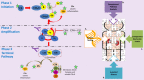
The complement system, composed of the classic pathway and the alternate pathway, is a component of the immune system that enhances the function of antibodies and phagocytes. C3 glomerulopathy (C3G) is caused by uncontrolled activation of the complement alternative pathway.

With the exception of DGKE, all the genes discussed in association with C3G encode proteins in the complement system. C3 and CFB are integral to complement activation and together form C3bBb, a C3 convertase that amplifies the initial complement response, and C3bBbC3b, a C5 convertase that cleaves C5 into C5a and C5b to trigger the terminal pathway. CD46, CFH, CFHR1, CFHR5, and CFI are complement regulators. The role of DGKE in complement activation, although minimal, is believed to be essential in normal podocyte function.

Familial cases of C3G are uncommon and when identified are most often highly penetrant heterozygous copy number variants involving the CFHR1-5 genes (see Figure 3), homozygous variants that lead to CFH deficiency, or heterozygous gain-of-function variants in C3. Common to all of these variants is an impact on the regulation of the AP in the fluid phase [Noris & Remuzzi 2017].

Since C3G is rarely inherited in a simple mendelian fashion, the study of rare variants and haplotypes associated with disease is important.

Several studies have shown that some common variants in complement genes are also associated with C3G and increase the odds ratio of developing disease [Abrera-Abeleda et al 2011, Kobayashi et al 2017]. While the identification of common variants that are C3G "risk alleles" cannot be used to direct clinical care, the identification of rare variants in complement genes does affect patient care, especially in the context of a comprehensive assessment of complement function, which includes plasma levels of complement proteins and their split products, assays for autoantibodies, and tests of overall complement activity. For more detailed information, see Osborne et al [2018].

C3

Gene structure. C3 comprises 41 exons that encode complement C3, which has a molecular weight of 176 kd. The mature protein forms a beta chain and an alpha chain. For a detailed summary of gene and protein information, see Table A.

Benign variants. Several C3 variants (some of which are common in the population) are associated with C3G and define an at-risk haplotype [Hageman et al 2005, Smith et al 2007, Fremeaux-Bacchi et al 2008, Abrera-Abeleda et al 2011, Iatropoulos et al 2016, Riedl et al 2017, Osborne et al 2018].

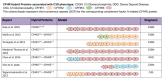
Pathogenic variants. Pathogenic variants and their location are shown in Table 4.

Table 4.

C3 Pathogenic Variants Discussed in This GeneReview

DNA Nucleotide ChangePredicted Protein ChangeAffected DomainReference Sequences
c.443G>Ap.Arg148GlnC3β chain NM_000064​.3
NP_000055​.2
c.1855G>Ap.Val619MetLinker
c.3125G>Ap.Arg1042GlnTED
c.3908G>Ap.Arg1303HisCUBf
c.3959G>Ap.Arg1320GlnCUBf
c.4552T>Cp.Cys1518ArgC345C
c.4873T>Cp.Asp1625HisC345C

Variants listed in the table have been provided by the authors. GeneReviews staff have not independently verified the classification of variants.

GeneReviews follows the standard naming conventions of the Human Genome Variation Society (varnomen​.hgvs.org). See Quick Reference for an explanation of nomenclature.

Normal gene product. 
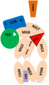
C3 encodes complement component C3, the central activating protein of the AP. This 1,663-amino-acid protein has 15 domains, including the anaphylatoxin (ANA), αNT, CUB (C1r/C1s, Uegf, Bmp1), C345C, thioester (TED), linker (LNK), and anchor domains and eight macroglobulin (MG) domains (see Figure 5). C3 is mainly synthesized in the liver, and it is the central protein of the complement system. Spontaneous or proteolytic cleavage of C3 generates the anaphylatoxin C3a (inflammatory effector cells) and the C3b fragment that deposits on cell surfaces triggering the complement cascade activation.

Figure 5.

Figure 5.

Schematic representation of complement component C3 The structural organization of C3 contains eight macroglobulins, anaphylatoxin (ANA), αNT, CUB (C1r/C1s, Uegf, Bmp1), C345C, linker, and thioester (TED) domains.

Abnormal gene product. Most pathogenic variants listed in Table 4 affect proper cleavage of C3 protein by affecting recognition sites for the binding of CFH and CFI, two regulators of complement activation. Pathogenic variants may also produce reduced quantities of C3 protein (i.e., truncating variants or frameshifts that lead to premature stop variants) [Martínez-Barricarte et al 2010].

CD46

Gene structure. CD46 (cluster differentiation 46) has 14 exons that encode the 43.7-kd membrane cofactor protein (MCP).

Benign variants. Several variants in CD46 have been associated with C3G and define an at-risk haplotype [Servais et al 2007, Fang et al 2008, Servais et al 2012, Osborne et al 2018].

Normal gene product. CD46 encodes MCP, a complement regulatory protein of 392 amino acids that is highly expressed in the kidney. It is a transmembrane protein and a member of the regulators of complement activation (RCA), and has eight domains: four short consensus repeat (SCRs 1-4-sushi) domains, which contain ligand-binding domains for decay and cofactor activity; an O-linked-glycosylation site, rich in serine, threonine, and proline (STP) domain, which can be alternately spliced; a helical transmembrane domain; and two cytoplasmic topological domains. Its major role in controlling complement activity is to inactivate C3b and C4b by functioning as a cofactor for factor I [Servais et al 2012, Liszewski & Atkinson 2015].

Abnormal gene product. Pathogenic variants in CD46 typically lead to reduced surface expression of MCP, which contributes to defective surface regulation of complement [Servais et al 2012].

CFB

Gene structure. CFB comprises 18 exons that encode complement factor B, which has a molecular weight of 93 kd. For a detailed summary of gene and protein information, see Table A.

Benign variants. Benign variants in CFB associated with complement regulation have been described [Gold et al 2006].

Pathogenic variants. The p.Ser367Arg pathogenic variant is in the von Willebrand factor A (VWFa) domain, the catalytic unit of Bb, and is a gain-of-function variant that contributes to AP dysregulation [Imamura et al 2015].

Table 5.

CFB Pathogenic Variants Discussed in This GeneReview

DNA Nucleotide ChangePredicted Protein ChangeReference Sequences
c.1099A>Cp.Ser367Arg NM_001710​.5
NP_001701​.2

Variants listed in the table have been provided by the authors. GeneReviews staff has not independently verified the classification of variants.

GeneReviews follows the standard naming conventions of the Human Genome Variation Society (varnomen​.hgvs.org). See Quick Reference for an explanation of nomenclature.

Normal gene product. CFB is composed of 764 amino acids and has five domains: three complement component protein (sushi) domains; a VWFa domain; and a peptidase S1 (serine protease) domain. Factor B (fB) is a component of the AP. It binds to C3b and is then cleaved by fD into Ba, a non-catalytic fragment that is released, and Bb, a catalytic subunit that remains bound to C3b to form C3 convertase (C3bBb). The fB cleavage site is near the N-terminus of the VWFa domain.

Abnormal gene product. Pathogenic variants in CFB may lead to gain-of-function properties that contribute to the dysregulation of the complement cascade [Alberts et al 2002, Imamura et al 2015].

CFH

Gene structure. CFH has 23 exons that encode complement factor H, a protein of 1,231 amino acids. For a detailed summary of gene and protein information, see Table A, Gene.

Benign variants. Several variants of CFH (some of which are common in the population) define an at-risk haplotype that has been associated with C3G [Hageman et al 2005, Servais et al 2007, Smith et al 2007, Zhang et al 2012, Johnson et al 2014, Xiao et al 2014, Merinero et al 2018, Riedl et al 2017, Osborne et al 2018].

Pathogenic variants. CFH pathogenic variants in C3G occur in heterozygous, homozygous, and compound heterozygous states. CFH has been implicated in C3G by the following:

  • A report of two sisters with DDD who were homozygous for the c.670_672delAAG (p.Lys224del) pathogenic variant in CFH [Licht et al 2006, Zipfel et al 2006]. Factor H (fH) carrying this pathogenic variant is present in the serum at normal concentrations but is nonfunctional [Zipfel et al 2006]. The normal N-terminal activities of fH (C3b binding and complement regulation) are defective, indicating that dysfunctional fH is associated with the development of DDD [Licht et al 2006].
  • Studies of skin fibroblasts from a child with fH deficiency and chronic hypocomplementemic renal disease and abnormal fH localization. One copy of a p.Cys536Arg missense variant and one copy of a p.Cys959Tyr missense variant were detected in CFH. Both pathogenic variants affect conserved cysteine residues characteristic of the short consensus-repeat (SCR) modules of fH and therefore predict profound changes in the higher-order structure of the 155-kd protein [Ault et al 1997].
  • Two brothers with MPGN were reportedly homozygous for a p.Arg127Leu amino acid change in CFH [Dragon-Durey et al 2004]. Homozygosity for this missense variant is associated with absence of fH in the serum, suggesting that this variant results in sequestration of the protein in the endoplasmic reticulum.

Table 6.

CFH Pathogenic Variants Discussed in This GeneReview

DNA Nucleotide Change
(Alias 1)
Predicted Protein Change
(Alias 1)
Reference Sequences
c.380G>Tp.Arg127Leu NM_000186​.3
NP_000177​.2
c.670_672delAAGp.Lys224del
(ΔLys224)
c.1606T>C
(1679T>C)
p.Cys536Arg
(Cys518Arg)
c.2655delp.Arg885SerfsTer13
c.2876G>A
(2949G>A)
p.Cys959Tyr
(Cys991Tyr)

Variants listed in the table have been provided by the authors. GeneReviews staff have not independently verified the classification of variants.

GeneReviews follows the standard naming conventions of the Human Genome Variation Society (varnomen​.hgvs.org). See Quick Reference for an explanation of nomenclature.

1.

Variant designation that does not conform to current naming conventions

Normal gene product. Complement factor H (fH) protein is the key regulator of the alternative pathway of complement and is composed of 1,231 amino acids. Its structural organization is based on 20 homologous repeat domains (short consensus repeats [SCRs] or sushi domains). Each SCR has 60 amino acids. The first four SCRs (1-4) at the N-terminus are essential for fluid-phase complement regulation, binding of fH to C3b, decay acceleration activity of C3bBb, and fI cofactor activity in mediating the cleavage of C3b to iCb3. The last two SCRs (19-20) at the C terminus bind to cell surfaces to regulate C3 convertase in that microenvironment. See Figure 6.

Figure 6.

Figure 6.

Schematic representation of factor H The 20 bead-like homologous short consensus repeats (SCRs) each have 60 amino acids. The first four SCRs (1-4) (red beads) regulate activation in the fluid phase, binding to C3b, decay acceleration activity, and fI (more...)

Abnormal gene product. CFH pathogenic variants associated with C3G are more frequently found in the N-terminal short consensus repeats (SCRs 1-4), a region essential for fluid-phase complement control. In contrast, in atypical hemolytic uremic syndrome (aHUS), pathogenic variants are more frequently located in the carboxy terminus (SCRs 18-20), a region involved in cell surface regulation.

CFHR1

Gene structure. CFHR1 comprises six exons that encode complement factor H-related protein 1. For a detailed summary of gene and protein information, see Table A, Gene.

Pathogenic variants. A duplication of exons 2 through 5 results in an abnormal fusion protein (Figure 3). The duplication alters the complex oligomerization of the FHR proteins, leading to increased FHR deregulation likely due to increased binding to C3b, iC3b, and C3dg, with decreased fH binding and decreased AP control (see Figure 3) [Tortajada et al 2013].

Table 7.

CFHR1 Pathogenic Variants Discussed in This GeneReview

DNA Nucleotide ChangePredicted Protein ChangeReference Sequences
Dup exons 1-4Duplication of all amino acids in SCR 1-4 of protein NM_002113​.2
NP_002104​.2

Variants listed in the table have been provided by the authors. GeneReviews staff have not independently verified the classification of variants.

GeneReviews follows the standard naming conventions of the Human Genome Variation Society (varnomen​.hgvs.org). See Quick Reference for an explanation of nomenclature.

Normal gene product. CFHR1 encodes complement factor H-related 1 (FHR1), a protein of 330 amino acids and a member of the regulators of complement activation (RCA) family. Its structural organization is highly homologous to fH, although FHR1 has only five SCRs. The last two SCRs (SCRs 4 and 5) share 96%-100% homology to SCRs 19 and 20 of fH. FHR1 homodimerizes through its first two SCRs and also forms heterodimers with FHR2 and FHR5.

Abnormal gene product. Fusion proteins of FHR1 lead to the formation of complex multimers with FHR1, FHR2, and FHR5 that alter complement regulation by outcompeting fH, which results in reduced convertase regulation (see Figure 3) [Xiao et al 2016].

CFHR5

Gene structure. CFHR5 has ten exons that encode complement factor H-related 5 (FHR5), a protein of 551 amino acids organized into nine SCRs. For a detailed summary of gene and protein information, see Table A, Gene.

Benign variants. Several CFHR5 variants (some of which are common in the population) are associated with C3G and define an at-risk haplotype [Abrera-Abeleda et al 2006, Zipfel et al 2015, Osborne et al 2018].

Pathogenic variants. Pathogenic variants are more frequently associated with C3GN than with DDD (CFHR5 nephropathy is a type of C3GN) [Goicoechea de Jorge et al 2009].

Normal gene product. The normal gene product encoded by CFHR5 is complement factor H-related protein 5 (FHR5), a plasma protein organized like fH in repetitive SCRs. FHR5 has nine SCRs and possesses fI-dependent cofactor activity that leads to inactivation of C3b [McRae et al 2001, Rodríguez de Córdoba et al 2004, McRae et al 2005]. In 92 renal biopsies from patients with different glomerular diseases, FHR5 was present in all complement-containing glomerular immune deposits [Murphy et al 2002], suggesting that FHR5 plays an important role in protecting the glomerulus from complement activation. The precise role of FHR5 in the physiopathology of C3G remains to be determined.

Abnormal gene product. Several abnormal gene products of CFHR5 have been reported in association with C3G. These products usually arise as a consequence of nonallelic homologous recombination, which results in hybrid gene formation (see Figure 3).

The presence of CFHR5 pathogenic variants in C3G is consistent with the hypothesis that FHRs play an important role in the complement regulation and disease pathogenesis [Abrera-Abeleda et al 2006, Zhang et al 2013].

CFI

Gene structure. CFI comprises 13 exons that encode complement factor I (fI), which has a molecular weight of 33 kd. For a detailed summary of gene and protein information, see Table A.

Benign variants. Several CFI variants (some of which are common in the population) are associated with C3G and define an at-risk haplotype [Fremeaux-Bacchi et al 2013, Imamura et al 2015, Chauvet et al 2017, Osborne et al 2018].

Pathogenic variants

  • c-4C>T – in the Kozac sequence. This noncoding variant is associated with DDD [Iatropoulos et al 2016].
  • p.Gly57Asp – in FIMAC domain, a region for fI-C3b degradation [Iatropoulos et al 2016]
  • c.Ala240Gly – in LDL receptor class A 1 and associated with C3G, aHUS, and AMD. The variant results in partial reduction in secretion [Nilsson et al 2010].

Table 8.

CFI Pathogenic Variants Discussed in This GeneReview

DNA Nucleotide ChangePredicted Protein Change
(Alias 1)
Reference Sequences
c.719C>Gp.Ala240Gly
(Ala222Gly)
NM_000204​.3
NP_000195​.2
c.-4C>T
c.170G>Ap.Gly57Asp

Variants listed in the table have been provided by the authors. GeneReviews staff have not independently verified the classification of variants.

GeneReviews follows the standard naming conventions of the Human Genome Variation Society (varnomen​.hgvs.org). See Quick Reference for an explanation of nomenclature.

1.

Variant designation that does not conform to current naming conventions

Normal gene product. CFI encodes complement fI, a protein of 583 amino acids synthesized in the liver. The fI protein cleaves fluid-phase and cell-bound C3b and C4b, inhibiting their activity in the complement cascade. Factor H and several other RCA proteins are obligatory cofactors for fI activity.

Abnormal gene product. Abnormal fI may lead to a partial reduction in secreted fI, thus compromising its cofactor activity with fH [Servais et al 2012]. Deficiency in fI can lead to complement dysregulation and consequently low serum levels of other complement proteins.

DGKE

Gene structure. DGKE comprises 11 exons that encode diacylglycerol kinase epsilon, which has a molecular weight of 64 kd. For a detailed summary of gene and protein information, see Table A.

Benign variants. Several DGKE variants (some of which are common in the population) are associated with C3G and define an at-risk haplotype [Osborne et al 2018].

Pathogenic variants. Several loss-of-function variants have been associated with C3G including the following:

Table 9.

DGKE Pathogenic Variants Discussed in This GeneReview

DNA Nucleotide ChangePredicted Protein ChangeReference Sequences
c.127C>Tp.Gln43Ter NM_003647​.2
NP_003638​.1
c.301A>Tp.Lys101Ter
c.610delAp.Thr204GlnfsTer6
c.966G>Ap.Trp322Ter
c.1050G>Ap.Trp350Ter

Note on variant classification: Variants listed in the table have been provided by the authors. GeneReviews staff have not independently verified the classification of variants.

Note on nomenclature: GeneReviews follows the standard naming conventions of the Human Genome Variation Society (varnomen​.hgvs.org). See Quick Reference for an explanation of nomenclature.

Normal gene product. DGKE is unrelated to the complement pathway. DGKE encodes diacylglycerol kinase epsilon (DGKE), a lipid kinase of 567 amino acids expressed in podocytes, glomerular capillary endothelial cells, and platelets. DGKE plays a key role in signal transduction by affecting the balance between diacylglycerol kinase and phosphatidic acid, thus controlling DAG levels within the cell [Lemaire et al 2013]. The role of DGKE in complement activation is minimal; however, it is believed to play an essential role in normal podocyte function [Ozaltin et al 2013].

In the kidney, DGKE is ubiquitously expressed in podocytes and endothelial cells. Although Dgke null mice do not have spontaneous clinical signs of kidney disease and have normal serum creatinine and urinary albumin, they develop subclinical microscopic anomalies of the glomerular endothelium that worsen with age, as well as glomerular capillary occlusion when exposed to nephrotoxic serum [Zhu et al 2016].

Abnormal gene product. Abnormal DGKE may lead to prothrombosis through sustained signaling by arachidonic acid-containing diacylglycerol (AA-DAG) [Lemaire et al 2013].

References

Literature Cited

  • Abrera-Abeleda MA, Nishimura C, Frees K, Jones M, Maga T, Katz LM, Zhang Y, Smith RJH. Allele variants of complement genes associated with dense deposit disease. J Am Soc Nephrol. 2011;22:1551–9. [PMC free article: PMC3148710] [PubMed: 21784901]
  • Abrera-Abeleda MA, Nishimura C, Smith JL, Sethi S, McRae JL, Murphy BF, Silvestri G, Skerka C, Józsi M, Zipfel PF, Hageman GS, Smith RJ. Variations in the complement regulatory genes factor H (CFH) and factor H related 5 (CFHR5) are associated with membranoproliferative glomerulonephritis type II (dense deposit disease). J Med Genet. 2006;43:582–9. [PMC free article: PMC2564553] [PubMed: 16299065]
  • Alberts B, Johnson A, Lewis J, et al. Studying gene expression and function. In: Molecular Biology of the Cell. 4 ed. New York: Garland Science. Available online. 2002. Accessed 2-23-22.
  • Angelo JR, Bell CS, Braun MC. Allograft failure in kidney transplant recipients with membranoproliferative glomerulonephritis. Am J Kidney Dis. 2011;57:291–9. [PubMed: 21215503]
  • Appel GB, Cook HT, Hageman G, Jennette JC, Kashgarian M, Kirschfink M, Lambris JD, Lanning L, Lutz HU, Meri S, Rose NR, Salant DJ, Sethi S, Smith RJ, Smoyer W, Tully HF, Tully SP, Walker P, Welsh M, Wurzner R, Zipfel PF. Membranoproliferative glomerulonephritis type II (dense deposit disease): an update. J Am Soc Nephrol. 2005;16:1392–403. [PubMed: 15800116]
  • Athanasiou Y, Voskarides K, Gale DP, Damianou L, Patsias C, Zavros M, Maxwell PH, Cook HT, Demosthenous P, Hadjisavvas A, Kyriacou K, Zouvani I, Pierides A, Deltas C. Familial C3 glomerulopathy associated with CFHR5 mutations: clinical characteristics of 91 patients in 16 pedigrees. Clin J Am Soc Nephrol. 2011;6:1436–46. [PMC free article: PMC3109942] [PubMed: 21566112]
  • Ault BH, Schmidt BZ, Fowler NL, Kashtan CE, Ahmed AE, Vogt BA, Colten HR. Human factor H deficiency. Mutations in framework cysteine residues and block in H protein secretion and intracellular catabolism. J Biol Chem. 1997;272:25168–75. [PubMed: 9312129]
  • Azukaitis K, Simkova E, Majid MA, Galiano M, Benz K, Amann K, Bockmeyer C, Gajjar R, Meyers KE, Cheong HI, Lange-Sperandio B, Jungraithmayr T, Fremeaux-Bacchi V, Bergmann C, Bereczki C, Miklaszewska M, Csuka D, Prohászka Z, Gipson P, Sampson MG, Lemaire M, Schaefer F. The phenotypic spectrum of nephropathies associated with mutations in diacylglycerol kinase ε. J Am Soc Nephrol. 2017;28:3066–75. [PMC free article: PMC5619969] [PubMed: 28526779]
  • Barbour TD, Pickering MC, Cook HT. Recent insights into C3 glomerulopathy. Nephrol Dial Transplant. 2013a;28:1685–93. [PMC free article: PMC3707523] [PubMed: 23479095]
  • Barbour TD, Pickering MC, Terence Cook H. Dense deposit disease and C3 glomerulopathy. Semin Nephrol. 2013b;33:493–507. [PMC free article: PMC3820036] [PubMed: 24161036]
  • Besbas N, Gulhan B, Gucer S, Korkmaz E, Ozaltin F. A novel CFHR5 mutation associated with C3 glomerulonephritis in a Turkish girl. J Nephrol. 2014;27:457–60. [PubMed: 24536001]
  • Blanc C, Togarsimalemath SK, Chauvet S, Le Quintrec M, Moulin B, Buchler M, Jokiranta TS, Roumenina LT, Fremeaux-Bacchi V, Dragon-Durey MA. Anti-factor H autoantibodies in C3 glomerulopathies and in atypical hemolytic uremic syndrome: one target, two diseases. J Immunol. 2015;194:5129–38. [PubMed: 25917093]
  • Bomback AS, Smith RJ, Barile GR, Zhang Y, Heher EC, Herlitz L, Stokes MB, Markowitz GS, D'Agati VD, Canetta PA, Radhakrishnan J, Appel GB. Eculizumab for dense deposit disease and C3 glomerulonephritis. Clin J Am Soc Nephrol. 2012;7:748–56. [PMC free article: PMC3338285] [PubMed: 22403278]
  • Bu F, Borsa NG, Jones MB, Takanami E, Nishimura C, Hauer JJ, Azaiez H, Black-Ziegelbein EA, Meyer NC, Kolbe DL, Li Y, Frees K, Schnieders MJ, Thomas C, Nester C, Smith RJ. High-throughput genetic testing for thrombotic microangiopathies and C3 glomerulopathies. J Am Soc Nephrol. 2016;27:1245–53. [PMC free article: PMC4814193] [PubMed: 26283675]
  • Cameron JS, Turner DR, Heaton J, Williams DG, Ogg CS, Chantler C, Haycock GB, Hicks J. Idiopathic mesangiocapillary glomerulonephritis. Comparison of types I and II in children and adults and long-term prognosis. Am J Med. 1983;74:175–92. [PubMed: 6337487]
  • Cebeci Z, Bayraktar S, Oray M, Kir N. Multimodal imaging of membranoproliferative glomerulonephritis type II. Saudi J Ophthalmol. 2016;30:260–3. [PMC free article: PMC5161817] [PubMed: 28003788]
  • Chauvet S, Fremeaux-Bacchi V, Petitprez F, Karras A, Daniel L, Burtey S, Choukroun G, Delmas Y, Guerrot D, François A, Le Quintrec M, Javaugue V, Ribes D, Vrigneaud L, Arnulf B, Goujon JM, Ronco P, Touchard G, Bridoux F. Treatment of B-cell disorder improves renal outcome of patients with monoclonal gammopathy-associated C3 glomerulopathy. Blood. 2017;129:1437–47. [PubMed: 28069603]
  • Chen Q, Muller D, Rudolph B, Hartmann A, Kuwertz-Broking E, Wu K, Kirschfink M, Skerka C, Zipfel PF. Combined C3b and factor B autoantibodies and MPGN type II. N Engl J Med. 2011;365:2340–2. [PubMed: 22168663]
  • Chen Q, Wiesener M, Eberhardt HU, Hartmann A, Uzonyi B, Kirschfink M, Amann K, Buettner M, Goodship T, Hugo C, Skerka C, Zipfel PF. Complement factor H-related hybrid protein deregulates complement in dense deposit disease. J Clin Invest. 2014;124:145–55. [PMC free article: PMC3871254] [PubMed: 24334459]
  • Cook HT. C3 glomerulopathy. F1000Res. 2017;6:248. [PMC free article: PMC5357035] [PubMed: 28357053]
  • D'Agati VD, Bomback AS. C3 glomerulopathy: what's in a name? Kidney Int. 2012;82:379–81. [PubMed: 22846813]
  • Daina E, Noris M, Remuzzi G. Eculizumab in a patient with dense-deposit disease. N Engl J Med. 2012;366:1161–3. [PubMed: 22435382]
  • Dalvin LA, Fervenza FC, Sethi S, Pulido JS. Shedding light on fundus drusen associated with membranoproliferative glomerulonephritis: breaking stereotypes of types I, II, and III. Retin Cases Brief Rep. 2016;10:72–8. [PubMed: 26110522]
  • Deltas C, Gale D, Cook T, Voskarides K, Athanasiou Y, Pierides A. C3 glomerulonephritis/CFHR5 nephropathy is an endemic disease in Cyprus: clinical and molecular findings in 21 families. Adv Exp Med Biol. 2013;735:189–96. [PubMed: 23402027]
  • Dragon-Durey MA, Fremeaux-Bacchi V, Loirat C, Blouin J, Niaudet P, Deschenes G, Coppo P, Herman Fridman W, Weiss L. Heterozygous and homozygous factor h deficiencies associated with hemolytic uremic syndrome or membranoproliferative glomerulonephritis: report and genetic analysis of 16 cases. J Am Soc Nephrol. 2004;15:787–95. [PubMed: 14978182]
  • D'Souza YB, Jones CJ, Short CD, Roberts IS, Bonshek RE. Oligosaccharide composition is similar in drusen and dense deposits in membranoproliferative glomerulonephritis type II. Kidney Int. 2009;75:824–7. [PubMed: 19177159]
  • Eisinger AJ, Shortland JR, Moorhead PJ. Renal disease in partial lipodystrophy. Q J Med. 1972;41:343–54. [PubMed: 4561162]
  • Fang CJ, Fremeaux-Bacchi V, Liszewski MK, Pianetti G, Noris M, Goodship TH, Atkinson JP. Membrane cofactor protein mutations in atypical hemolytic uremic syndrome (aHUS), fatal Stx-HUS, C3 glomerulonephritis, and the HELLP syndrome. Blood. 2008;111:624–32. [PMC free article: PMC2200836] [PubMed: 17914026]
  • Fremeaux-Bacchi V, Fakhouri F, Garnier A, Bienaimé F, Dragon-Durey MA, Ngo S, Moulin B, Servais A, Provot F, Rostaing L, Burtey S, Niaudet P, Deschênes G, Lebranchu Y, Zuber J, Loirat C. Genetics and outcome of atypical hemolytic uremic syndrome: a nationwide French series comparing children and adults. Clin J Am Soc Nephrol. 2013;8:554–62. [PMC free article: PMC3613948] [PubMed: 23307876]
  • Fremeaux-Bacchi V, Fakhouri F, Loirat C. Hemolytic-uremic syndrome: what is the mechanism? Rev Prat. 2008;58:2093–6. [PubMed: 19213534]
  • Fremeaux-Bacchi V, Weiss L, Brun P, Kazatchkine MD. Selective disappearance of C3NeF IgG autoantibody in the plasma of a patient with membranoproliferative glomerulonephritis following renal transplantation. Nephrol Dial Transplant. 1994;9:811–4. [PubMed: 7970124]
  • Fujita T, Nozu K, Iijima K, Kamioka I, Kaito H, Tanaka R, Nakanishi K, Matsuo M, Yoshikawa N. Long-term follow-up of juvenile acute nonproliferative glomerulitis (JANG). Pediatr Nephrol. 2007;22:1957–61. [PubMed: 17674054]
  • Gale DP, de Jorge EG, Cook HT, Martínez-Barricarte R, Hadjisavvas A, McLean AG, Pusey CD, Pierides A, Kyriacou K, Athanasiou Y, Voskarides K, Deltas C, Palmer A, Fremeaux-Bacchi V, de Cordoba SR, Maxwell PH, Pickering MC. Identification of a mutation in complement factor H-related protein 5 in patients of Cypriot origin with glomerulonephritis. Lancet. 2010;376:794–801. [PMC free article: PMC2935536] [PubMed: 20800271]
  • Gale DP, Owen-Casey M. Membranoproliferative glomerulonephritis and C3 glomerulopathy. In: Harber M, ed. Practical Nephrology. London: Springer; 2014:189-202.
  • Goicoechea de Jorge E, Gale DP, Cook HT, Martínez-Barricate A, Hadjisavvas A, Pusey CD, Palmer A, Fremeaux-Bacchi V, Rodriguez de Cordoba S, Maxwell PH, Pickering MC. A mutant complement factor H-related 5 protein is associated with familial C3 glomerulonephritis. Mol Immunol. 2009;46:2822.
  • Gold B, Merriam JE, Zernant J, Hancox LS, Taiber AJ, Gehrs K, Cramer K, Neel J, Bergeron J, Barile GR, Smith RT, Hageman GS, Dean M, Allikmets R, et al. Variation in factor B (BF) and complement component 2 (C2) genes is associated with age-related macular degeneration. Nat Genet. 2006;38:458–62. [PMC free article: PMC2921703] [PubMed: 16518403]
  • Goodship TH, Cook HT, Fakhouri F, Fervenza FC, Fremeaux-Bacchi V, Kavanagh D, Nester CM, Noris M, Pickering MC, Rodríguez de Córdoba S, Roumenina LT, Sethi S, Smith RJ, et al. Atypical hemolytic uremic syndrome and C3 glomerulopathy: conclusions from a "Kidney Disease: Improving Global Outcomes" (KDIGO) Controversies Conference. Kidney Int. 2017;91:539–51. [PubMed: 27989322]
  • Habib R, Gubler MC, Loirat C, Maiz HB, Levy M. Dense deposit disease: a variant of membranoproliferative glomerulonephritis. Kidney Int. 1975;7:204–15. [PubMed: 1095806]
  • Hageman GS, Anderson DH, Johnson LV, Hancox LS, Taiber AJ, Hardisty LI, Hageman JL, Stockman HA, Borchardt JD, Gehrs KM, Smith RJ, Silvestri G, Russell SR, Klaver CC, Barbazetto I, Chang S, Yannuzzi LA, Barile GR, Merriam JC, Smith RT, Olsh AK, Bergeron J, Zernant J, Merriam JE, Gold B, Dean M, Allikmets R. A common haplotype in the complement regulatory gene factor H (HF1/CFH) predisposes individuals to age-related macular degeneration. Proc Natl Acad Sci U S A. 2005;102:7227–32. [PMC free article: PMC1088171] [PubMed: 15870199]
  • Hulleman JD. Malattia Leventinese/Doyne honeycomb retinal dystrophy: similarities to age-related macular degeneration and potential therapies. Adv Exp Med Biol. 2016;854:153–8. [PubMed: 26427406]
  • Iatropoulos P, Noris M, Mele C, Piras R, Valoti E, Bresin E, Curreri M, Mondo E, Zito A, Gamba S, Bettoni S, Murer L, Fremeaux-Bacchi V, Vivarelli M, Emma F, Daina E, Remuzzi G. Complement gene variants determine the risk of immunoglobulin-associated MPGN and C3 glomerulopathy and predict long-term renal outcome. Mol Immunol. 2016;71:131–42. [PubMed: 26895476]
  • Imamura H, Konomoto T, Tanaka E, Hisano S, Yoshida Y, Fujimura Y, Miyata T, Nunoi H. Familial C3 glomerulonephritis associated with mutations in the gene for complement factor B. Nephrol Dial Transplant. 2015;30:862–4. [PubMed: 25758434]
  • Johnson SA, Wong EK, Taylor CM. Making sense of the spectrum of glomerular disease associated with complement dysregulation. Pediatr Nephrol. 2014;29:1883–94. [PubMed: 23852337]
  • Kambham N. Postinfectious glomerulonephritis. Adv Anat Pathol. 2012;19:338–47. [PubMed: 22885383]
  • Khalighi MA, Wang S, Henriksen KJ, Bock M, Keswani M, Meehan SM, Chang A. Revisiting post-infectious glomerulonephritis in the emerging era of C3 glomerulopathy. Clin Kidney J. 2016;9:397–402. [PMC free article: PMC4886922] [PubMed: 27274823]
  • Kobayashi Y, Yang S, Nykamp K, Garcia J, Lincoln SE, Topper SE. Pathogenic variant burden in the ExAC database: an empirical approach to evaluating population data for clinical variant interpretation. Genome Med. 2017;9:13. [PMC free article: PMC5295186] [PubMed: 28166811]
  • Kurtz KA, Schlueter AJ. Management of membranoproliferative glomerulonephritis type II with plasmapheresis. J Clin Apher. 2002;17:135–7. [PubMed: 12378549]
  • Lemaire M, Fremeaux-Bacchi V, Schaefer F, Choi M, Tang WH, Le Quintrec M, Fakhouri F, Taque S, Nobili F, Martínez F, Ji W, Overton JD, Mane SM, Nürnberg G, Altmüller J, Thiele H, Morin D, Deschenes G, Baudouin V, Llanas B, Collard L, Majid MA, Simkova E, Nürnberg P, Rioux-Leclerc N, Moeckel GW, Gubler MC, Hwa J, Loirat C, Lifton RP. Recessive mutations in DGKE cause atypical hemolytic-uremic syndrome. Nat Genet. 2013;45:531–6. [PMC free article: PMC3719402] [PubMed: 23542698]
  • Licht C, Fremeaux-Bacchi V. Hereditary and acquired complement dysregulation in membranoproliferative glomerulonephritis. Thromb Haemost. 2009;101:271–8. [PubMed: 19190809]
  • Licht C, Heinen S, Jozsi M, Loschmann I, Saunders RE, Perkins SJ, Skerka C, Kirschfink M, Hoppe B, Zipfel PF. Deletion of Lys224 in regulatory domain 4 of factor H reveals a novel pathomechanism for dense deposit disease (MPGNII). Kidney Int. 2006;70:42–50. [PubMed: 16612335]
  • Licht C, Mengel M. Membranoproliferative glomerulonephritis. In: Geary DF, Schaefer F, eds. Comprehensive Pediatric Nephrology. 1 ed. Philadelphia, PA: Mosby; 2008:269-89.
  • Liszewski MK, Atkinson JP. Complement regulator CD46: genetic variants and disease associations. Hum Genomics. 2015;9:7. [PMC free article: PMC4469999] [PubMed: 26054645]
  • Lu DF, McCarthy AM, Lanning LD, Delaney C, Porter C. A descriptive study of individuals with membranoproliferative glomerulonephritis. Nephrol Nurs J. 2007;34:295–302. [PubMed: 17644874]
  • Lu DF, Moon M, Lanning LD, McCarthy AM, Smith RJ. Clinical features and outcomes of 98 children and adults with dense deposit disease. Pediatr Nephrol. 2012;27:773–81. [PMC free article: PMC4423603] [PubMed: 22105967]
  • Malik TH, Lavin PJ, Goicoechea de Jorge E, Vernon KA, Rose KL, Patel MP, de Leeuw M, Neary JJ, Conlon PJ, Winn MP, Pickering MC. A hybrid CFHR3-1 gene causes familial C3 glomerulopathy. J Am Soc Nephrol. 2012;23:1155–60. [PMC free article: PMC3380655] [PubMed: 22626820]
  • Marks SD, Rees L. Spontaneous clinical improvement in dense deposit disease. Pediatr Nephrol. 2000;14:322–4. [PubMed: 10775078]
  • Martínez-Barricarte R, Heurich M, Valdes-Cañedo F, Vazquez-Martul E, Torreira E, Montes T, Tortajada A, Pinto S, Lopez-Trascasa M, Morgan BP, Llorca O, Harris CL, Rodríguez de Córdoba S. Human C3 mutation reveals a mechanism of dense deposit disease pathogenesis and provides insights into complement activation and regulation. J Clin Invest. 2010;120:3702–12. [PMC free article: PMC2947238] [PubMed: 20852386]
  • Mathieson PW, Peters DK. Lipodystrophy in MCGN type II: the clue to links between the adipocyte and the complement system. Nephrol Dial Transplant. 1997;12:1804–6. [PubMed: 9306323]
  • McCaughan JA, O'Rourke DM, Courtney AE. Recurrent dense deposit disease after renal transplantation: an emerging role for complementary therapies. Am J Transplant. 2012;12:1046–51. [PubMed: 22233157]
  • McRae JL, Cowan PJ, Power DA, Mitchelhill KI, Kemp BE, Morgan BP, Murphy BF. Human factor H-related protein 5 (FHR-5). A new complement-associated protein. J Biol Chem. 2001;276:6747–54. [PubMed: 11058592]
  • McRae JL, Duthy TG, Griggs KM, Ormsby RJ, Cowan PJ, Cromer BA, McKinstry WJ, Parker MW, Murphy BF, Gordon DL. Human factor H-related protein 5 has cofactor activity, inhibits C3 convertase activity, binds heparin and C-reactive protein, and associates with lipoprotein. J Immunol. 2005;174:6250–6. [PubMed: 15879123]
  • Medjeral-Thomas N, Malik TH, Patel MP, Toth T, Cook HT, Tomson C, Pickering MC. A novel CFHR5 fusion protein causes C3 glomerulopathy in a family without Cypriot ancestry. Kidney Int. 2014;85:933–7. [PMC free article: PMC3789233] [PubMed: 24067434]
  • Merinero HM, García SP, García-Fernández J, Arjona E, Tortajada A, Rodríguez de Córdoba S. Complete functional characterization of disease-associated genetic variants in the complement factor H gene. Kidney Int. 2018;93:470–81. [PubMed: 28941939]
  • Mullins RF, Aptsiauri N, Hageman GS. Structure and composition of drusen associated with glomerulonephritis: implications for the role of complement activation in drusen biogenesis. Eye (Lond). 2001;15:390–5. [PubMed: 11450763]
  • Murphy B, Georgiou T, Machet D, Hill P, McRae J. Factor H-related protein-5: a novel component of human glomerular immune deposits. Am J Kidney Dis. 2002;39:24–7. [PubMed: 11774097]
  • Nasr SH, Valeri AM, Appel GB, Sherwinter J, Stokes MB, Said SM, Markowitz GS, D'Agati VD. Dense deposit disease: clinicopathologic study of 32 pediatric and adult patients. Clin J Am Soc Nephrol. 2009;4:22–32. [PMC free article: PMC2615696] [PubMed: 18971369]
  • Nava J, Moran S, Figueroa V, Salinas A, Lopez M, Urbina R, Gutierrez A, Lujan JL, Orozco A, Montufar R, Piccoli GB. Successful pregnancy in a CKD patient on a low-protein, supplemented diet: an opportunity to reflect on CKD and pregnancy in Mexico, an emerging country. J Nephrol. 2017;30:877–82. [PubMed: 28918595]
  • Nester CM, Smith RJ. Complement inhibition in C3 glomerulopathy. Semin Immunol. 2016;28:241–9. [PubMed: 27402056]
  • Nester CM, Smith RJ. Diagnosis and treatment of C3 glomerulopathy. Clin Nephrol. 2013a;80:395–403. [PubMed: 23993166]
  • Nester CM, Smith RJ. Treatment options for C3 glomerulopathy. Curr Opin Nephrol Hypertens. 2013b;22:231–7. [PMC free article: PMC4437761] [PubMed: 23318699]
  • Nicolas C, Vuiblet V, Baudouin V, Macher MA, Vrillon I, Biebuyck-Gouge N, Dehennault M, Gié S, Morin D, Nivet H, Nobili F, Ulinski T, Ranchin B, Marinozzi MC, Ngo S, Fremeaux-Bacchi V, Pietrement C. C3 nephritic factor associated with C3 glomerulopathy in children. Pediatr Nephrol. 2014;29:85–94. [PubMed: 24068526]
  • Nilsson SC, Kalchishkova N, Trouw LA, Fremeaux-Bacchi V, Villoutreix BO, Blom AM. Mutations in complement factor I as found in atypical hemolytic uremic syndrome lead to either altered secretion or altered function of factor I. Eur J Immunol. 2010;40:172–85. [PubMed: 19877009]
  • Noris M, Remuzzi G. Genetics of immune-mediated glomerular diseases: focus on complement. Semin Nephrol. 2017;37:447–63. [PubMed: 28863792]
  • Noris M, Remuzzi G. Glomerular diseases dependent on complement activation, including atypical hemolytic uremic syndrome, membranoproliferative glomerulonephritis, and C3 glomerulopathy: core curriculum 2015. Am J Kidney Dis. 2015;66:359–75. [PMC free article: PMC4528072] [PubMed: 26032627]
  • Ohi H, Watanabe S, Fujita T, Yasugi T. Significance of C3 nephritic factor (C3NeF) in non-hypocomplementaemic serum with membranoproliferative glomerulonephritis (MPGN). Clin Exp Immunol. 1992;89:479–84. [PMC free article: PMC1554488] [PubMed: 1516262]
  • Osborne AJ, Breno M, Borsa NG, Bu F, Fremeaux-Bacchi V, Gale DP, van den Heuvel LP, Kavanagh D, Noris M, Pinto S, Rallapalli PM, Remuzzi G, Rodríguez de Cordoba S, Ruiz A, Smith RJH, Vieira-Martins P, Volokhina E, Wilson V, Goodship THJ, Perkins SJ. Statistical validation of rare complement variants provides insights into the molecular basis of atypical hemolytic uremic syndrome and C3 glomerulopathy. J Immunol. 2018;2018;200:2464–78. [PMC free article: PMC6324840] [PubMed: 29500241]
  • Ossoli A, Lucca F, Boscutti G, Remaley AT, Calabresi L. Familial LCAT deficiency: from pathology to enzyme replacement therapy. Clinical Lipidology. 2015;10:405–13.
  • Ozaltin F, Li B, Rauhauser A, An SW, Soylemezoglu O, Gonul II, Taskiran EZ, Ibsirlioglu T, Korkmaz E, Bilginer Y, Duzova A, Ozen S, Topaloglu R, Besbas N, Ashraf S, Du Y, Liang C, Chen P, Lu D, Vadnagara K, Arbuckle S, Lewis D, Wakeland B, Quigg RJ, Ransom RF, Wakeland EK, Topham MK, Bazan NG, Mohan C, Hildebrandt F, Bakkaloglu A, Huang CL, Attanasio M. DGKE variants cause a glomerular microangiopathy that mimics membranoproliferative GN. J Am Soc Nephrol. 2013;24:377–84. [PMC free article: PMC3582208] [PubMed: 23274426]
  • Paixão-Cavalcante D, López-Trascasa M, Skattum L, Giclas PC, Goodship TH, de Córdoba SR, Truedsson L, Morgan BP, Harris CL. Sensitive and specific assays for C3 nephritic factors clarify mechanisms underlying complement dysregulation. Kidney Int. 2012;82:1084–92. [PMC free article: PMC3608896] [PubMed: 22854646]
  • Piccoli GB, Alrukhaimi M, Liu ZH, Zakharova E, Levin A, et al. What we do and do not know about women and kidney diseases; questions unanswered and answers unquestioned: reflection on World Kidney Day and International Woman's Day. BMC Nephrol. 2018;19:66. [PMC free article: PMC5856379] [PubMed: 29544451]
  • Pickering MC, D'Agati VD, Nester CM, Smith RJ, Haas M, Appel GB, Alpers CE, Bajema IM, Bedrosian C, Braun M, Doyle M, Fakhouri F, Fervenza FC, Fogo AB, Fremeaux-Bacchi V, Gale DP, Goicoechea de Jorge E, Griffin G, Harris CL, Holers VM, Johnson S, Lavin PJ, Medjeral-Thomas N, Paul Morgan B, Nast CC, Noel LH, Peters DK, Rodríguez de Córdoba S, Servais A, Sethi S, Song WC, Tamburini P, Thurman JM, Zavros M, Cook HT. C3 glomerulopathy: consensus report. Kidney Int. 2013;84:1079–89. [PMC free article: PMC3842953] [PubMed: 24172683]
  • Rabasco C, Cavero T, Román E, Rojas-Rivera J, Olea T, Espinosa M, Cabello V, Fernández-Juarez G, González F, Ávila A, Baltar JM, Díaz M, Alegre R, Elías S, Antón M, Frutos MA, Pobes A, Blasco M, Martín F, Bernis C, Macías M, Barroso S, de Lorenzo A, Ariceta G, López-Mendoza M, Rivas B, López-Revuelta K, Campistol JM, Mendizábal S, de Córdoba SR, Praga M, et al. Effectiveness of mycophenolate mofetil in C3 glomerulonephritis. Kidney Int. 2015;88:1153–60. [PubMed: 26221755]
  • Recalde S, Tortajada A, Subias M, Anter J, Blasco M, Maranta R, Coco R, Pinto S, Noris M, García-Layana A, Rodríguez de Córdoba S. Molecular basis of factor H R1210C association with ocular and renal diseases. J Am Soc Nephrol. 2016;27:1305–11. [PMC free article: PMC4849834] [PubMed: 26376859]
  • Riedl M, Thorner P, Licht C. C3 glomerulopathy. Pediatr Nephrol. 2017;32:43–57. [PubMed: 27056062]
  • Rodríguez de Córdoba S, Esparza-Gordillo J, Goicoechea de Jorge E, Lopez-Trascasa M, Sanchez-Corral P. The human complement factor H: functional roles, genetic variations and disease associations. Mol Immunol. 2004;41:355–67. [PubMed: 15163532]
  • Salvadori M, Bertoni E. Complement related kidney diseases: recurrence after transplantation. World J Transplant. 2016;6:632–45. [PMC free article: PMC5175220] [PubMed: 28058212]
  • Savige J, Amos L, Ierino F, Mack HG, Symons RC, Hughes P, Nicholls K, Colville D. Retinal disease in the C3 glomerulopathies and the risk of impaired vision. Ophthalmic Genet. 2016;37:369–76. [PubMed: 26915021]
  • Schwertz R, Rother U, Anders D, Gretz N, Scharer K, Kirschfink M. Complement analysis in children with idiopathic membranoproliferative glomerulonephritis: a long-term follow-up. Pediatr Allergy Immunol. 2001;12:166–72. [PubMed: 11473682]
  • Servais A, Fremeaux-Bacchi V, Lequintrec M, Salomon R, Blouin J, Knebelmann B, Grünfeld JP, Lesavre P, Noël LH, Fakhouri F. Primary glomerulonephritis with isolated C3 deposits: a new entity which shares common genetic risk factors with haemolytic uraemic syndrome. J Med Genet. 2007;44:193–9. [PMC free article: PMC2598029] [PubMed: 17018561]
  • Servais A, Noël LH, Fremeaux-Bacchi V, Lesavre P. C3 glomerulopathy. Contrib Nephrol. 2013;181:185–93. [PubMed: 23689580]
  • Servais A, Noel LH, Roumenina LT, Le Quintrec M, Ngo S, Dragon-Durey MA, Macher MA, Zuber J, Karras A, Provot F, Moulin B, Grünfeld JP, Niaudet P, Lesavre P, Fremeaux-Bacchi V. Acquired and genetic complement abnormalities play a critical role in dense deposit disease and other C3 glomerulopathies. Kidney Int. 2012;82:454–64. [PubMed: 22456601]
  • Sessa A, Battini G, Meroni M, Daidone G, Carnera I, Brambilla PL, Vigano G, Giordano F, Pallotti F, Torri Tarelli L, Calabresi L, Rolleri M, Bertolini S. Hypocomplementemic type II membranoproliferative glomerulonephritis in a male patient with familial lecithin-cholesterol acyltransferase deficiency due to two different allelic mutations. Nephron. 2001;88:268–72. [PubMed: 11423760]
  • Sethi S, Fervenza FC. Membranoproliferative glomerulonephritis: pathogenetic heterogeneity and proposal for a new classification. Semin Nephrol. 2011;31:341–8. [PubMed: 21839367]
  • Sethi S, Fervenza FC, Zhang Y, Zand L, Meyer NC, Borsa N, Nasr SH, Smith RJ. Atypical post-infectious glomerulonephritis is associated with abnormalities in the alternative pathway of complement. Kidney Int. 2013;83:293–9. [PMC free article: PMC3561505] [PubMed: 23235567]
  • Sethi S, Fervenza FC, Zhang Y, Zand L, Vrana JA, Nasr SH, Theis JD, Dogan A, Smith RJ. C3 glomerulonephritis: clinicopathological findings, complement abnormalities, glomerular proteomic profile, treatment, and follow-up. Kidney Int. 2012a;82:465–73. [PMC free article: PMC4438675] [PubMed: 22673887]
  • Sethi S, Haas M, Markowitz GS, D'Agati VD, Rennke HG, Jennette JC, Bajema IM, Alpers CE, Chang A, Cornell LD, Cosio FG, Fogo AB, Glassock RJ, Hariharan S, Kambham N, Lager DJ, Leung N, Mengel M, Nath KA, Roberts IS, Rovin BH, Seshan SV, Smith RJ, Walker PD, Winearls CG, Appel GB, Alexander MP, Cattran DC, Casado CA, Cook HT, De Vriese AS, Radhakrishnan J, Racusen LC, Ronco P, Fervenza FC. Mayo Clinic/Renal Pathology Society Consensus Report on Pathologic Classification, Diagnosis, and Reporting of GN. J Am Soc Nephrol. 2016;27:1278–87. [PMC free article: PMC4849835] [PubMed: 26567243]
  • Sethi S, Nester CM, Smith RJ. Membranoproliferative glomerulonephritis and C3 glomerulopathy: resolving the confusion. Kidney Int. 2012b;81:434–41. [PMC free article: PMC4428602] [PubMed: 22157657]
  • Smith RJ, Alexander J, Barlow PN, Botto M, Cassavant TL, Cook HT, de Cordoba SR, Hageman GS, Jokiranta TS, Kimberling WJ, Lambris JD, Lanning LD, Levidiotis V, Licht C, Lutz HU, Meri S, Pickering MC, Quigg RJ, Rops AL, Salant DJ, Sethi S, Thurman JM, Tully HF, Tully SP, van der Vlag J, Walker PD, Wurzner R, Zipfel PF. New approaches to the treatment of dense deposit disease. J Am Soc Nephrol. 2007;18:2447–56. [PMC free article: PMC4853920] [PubMed: 17675665]
  • Sohn EH, Wang K, Thompson S, Riker MJ, Hoffmann JM, Stone EM, Mullins RF. Comparison of drusen and modifying genes in autosomal dominant radial drusen and age-related macular degeneration. Retina. 2015;35:48–57. [PMC free article: PMC5513174] [PubMed: 25077532]
  • Sotsiou F. Postinfectious glomerulonephritis. Nephrol Dial Transplant. 2001;16 Suppl 6:68–70. [PubMed: 11568248]
  • Spitzer RE, Stitzel AE. Loss of autoantibody activity by alteration in autoantigen. Clin Immunol Immunopathol. 1996;80:211–3. [PubMed: 8764567]
  • Stone EM, Lotery AJ, Munier FL, Heon E, Piguet B, Guymer RH, Vandenburgh K, Cousin P, Nishimura D, Swiderski RE, Silvestri G, Mackey DA, Hageman GS, Bird AC, Sheffield VC, Schorderet DF. A single EFEMP1 mutation associated with both Malattia Leventinese and Doyne honeycomb retinal dystrophy. Nat Genet. 1999;22:199–202. [PubMed: 10369267]
  • Strobel S, Zimmering M, Papp K, Prechl J, Józsi M. Anti-factor B autoantibody in dense deposit disease. Mol Immunol. 2010;47:1476–83. [PubMed: 20193965]
  • Thomas S, Ranganathan D, Francis L, Madhan K, John GT. Current concepts in C3 glomerulopathy. Indian J Nephrol. 2014;24:339–48. [PMC free article: PMC4244712] [PubMed: 25484526]
  • Togarsimalemath SK, Sethi SK, Duggal R, Quintrec ML, Jha P, Daniel R, Gonnet F, Bansal S, Roumenina LT, Fremeaux-Bacchi V, Kher V, Dragon-Durey MA. A novel CFHR1-CFHR5 hybrid leads to a familial dominant C3 glomerulopathy. Kidney Int. 2017;92:876–87. [PubMed: 28729035]
  • Tortajada A, Yébenes H, Abarrategui-Garrido C, Anter J, García-Fernández JM, Martínez-Barricarte R, Alba-Domínguez M, Malik TH, Bedoya R, Cabrera Pérez R, López Trascasa M, Pickering MC, Harris CL, Sánchez-Corral P, Llorca O, Rodríguez de Córdoba S. C3 glomerulopathy-associated CFHR1 mutation alters FHR oligomerization and complement regulation. J Clin Invest. 2013;123:2434–46. [PMC free article: PMC3668852] [PubMed: 23728178]
  • Vaclavik V, Munier FL. Malattia Leventinese (autosomal dominant drusen). In: Querques G, Souied E, eds. Macular Dystrophies. Cham, Switzerland: Springer; 2016:39-51.
  • Vernon KA, Goicoechea de Jorge E, Hall AE, Fremeaux-Bacchi V, Aitman TJ, Cook HT, Hangartner R, Koziell A, Pickering MC. Acute presentation and persistent glomerulonephritis following streptococcal infection in a patient with heterozygous complement factor H-related protein 5 deficiency. Am J Kidney Dis. 2012;60:121–5. [PMC free article: PMC3382710] [PubMed: 22503529]
  • Vivarelli M, Emma F. Treatment of C3 glomerulopathy with complement blockers. Semin Thromb Hemost. 2014;40:472–7. [PubMed: 24799307]
  • Walker PD, Ferrario F, Joh K, Bonsib SM. Dense deposit disease is not a membranoproliferative glomerulonephritis. Mod Pathol. 2007;20:605–16. [PubMed: 17396142]
  • West CD, McAdams AJ, Witte DP. Acute non-proliferative glomerulitis: a cause of renal failure unique to children. Pediatr Nephrol. 2000;14:786–93. [PubMed: 10955928]
  • Westland R, Bodria M, Carrea A, Lata S, Scolari F, Fremeaux-Bacchi V, D'Agati VD, Lifton RP, Gharavi AG, Ghiggeri GM, Sanna-Cherchi S. Phenotypic expansion of DGKE-associated diseases. J Am Soc Nephrol. 2014;25:1408–14. [PMC free article: PMC4073436] [PubMed: 24511134]
  • Xiao X, Ghossein C, Tortajada A, Zhang Y, Meyer N, Jones M, Borsa NG, Nester CM, Thomas CP, de Córdoba SR, Smith RJ. Familial C3 glomerulonephritis caused by a novel CFHR5-CFHR2 fusion gene. Mol Immunol. 2016;77:89–96. [PubMed: 27490940]
  • Xiao X, Pickering MC, Smith RJH. C3 glomerulopathy: the genetic and clinical findings in dense deposit disease and C3 glomerulonephritis. Semin Thromb Hemost. 2014;40:465–71. [PubMed: 24799308]
  • Zand L, Lorenz EC, Cosio FG, Fervenza FC, Nasr SH, Gandhi MJ, Smith RJ, Sethi S. Clinical findings, pathology, and outcomes of C3GN after kidney transplantation. J Am Soc Nephrol. 2014;25:1110–7. [PMC free article: PMC4005307] [PubMed: 24357668]
  • Zhang Y, Meyer NC, Fervenza FC, Lau W, Keenan A, Cara-Fuentes G, Shao D, Akber A, Fremeaux-Bacchi V, Sethi S, Nester CM, Smith RJ. C4 nephritic factors in C3 glomerulopathy: a case series. Am J Kidney Dis. 2017;70:834–43. [PMC free article: PMC5701858] [PubMed: 28838767]
  • Zhang Y, Meyer NC, Wang K, Nishimura C, Frees K, Jones M, Katz LM, Sethi S, Smith RJ. Causes of alternative pathway dysregulation in dense deposit disease. Clin J Am Soc Nephrol. 2012;7:265–74. [PMC free article: PMC3280037] [PubMed: 22223606]
  • Zhang Y, Nester CM, Holanda DG, Marsh HC, Hammond RA, Thomas LJ, Meyer NC, Hunsicker LG, Sethi S, Smith RJ. Soluble CR1 therapy improves complement regulation in C3 glomerulopathy. J Am Soc Nephrol. 2013;24:1820–9. [PMC free article: PMC3810083] [PubMed: 23907509]
  • Zhu J, Chaki M, Lu D, Ren C, Wang SS, Rauhauser A, Li B, Zimmerman S, Jun B, Du Y, Vadnagara K, Wang H, Elhadi S, Quigg RJ, Topham MK, Mohan C, Ozaltin F, Zhou XJ, Marciano DK, Bazan NG, Attanasio M. Loss of diacylglycerol kinase epsilon in mice causes endothelial distress and impairs glomerular Cox-2 and PGE2 production. Am J Physiol Renal Physiol. 2016;310:F895–908. [PMC free article: PMC4867310] [PubMed: 26887830]
  • Zipfel PF, Skerka C, Chen Q, Wiech T, Goodship T, Johnson S, Fremeaux-Bacchi V, Nester C, de Córdoba SR, Noris M, Pickering M, Smith R. The role of complement in C3 glomerulopathy. Mol Immunol. 2015;67:21–30. [PubMed: 25929733]
  • Zipfel PF, Smith RJH, Heinen ST. The role of complement in membranoproliferative glomerulonephritis. In: Zipfel PF, ed. Complement and Kidney Disease. Berlin, Germany: Springer; 2006:199-221.

Chapter Notes

Author Notes

Contact Information

Richard JH Smith
Division of Nephrology
University of Iowa
200 Hawkins Drive
Iowa City, IA 52242
Telephone: 319-356-3612
Fax: 319-356-4108
Email: richard-smith@uiowa.edu

Acknowledgments

Supported in part by grant RO1-DK110023 from the NIDDK (RJHS)

Author History

Johnny Cruz Corchado; University of Iowa (2011-2018)
Bertha Martín, PhD (2018-present)
Sanjeev Sethi, MD, PhD; Mayo Clinic (2007-2011)
Richard JH Smith, MD (2007-present)
Peter F Zipfel, PhD habil Prof; Hans Knöll Institute (2007-2011)

Revision History

  • 5 April 2018 (ha) Comprehensive update posted live
  • 19 May 2011 (me) Comprehensive update posted live
  • 2 January 2008 (rjhs/cd) Revision: clinical testing (sequence analysis) for CFHR5 mutations
  • 20 July 2007 (me) Review posted live
  • 17 August 2005 (rjhs) Original submission
Copyright © 1993-2024, University of Washington, Seattle. GeneReviews is a registered trademark of the University of Washington, Seattle. All rights reserved.

GeneReviews® chapters are owned by the University of Washington. Permission is hereby granted to reproduce, distribute, and translate copies of content materials for noncommercial research purposes only, provided that (i) credit for source (http://www.genereviews.org/) and copyright (© 1993-2024 University of Washington) are included with each copy; (ii) a link to the original material is provided whenever the material is published elsewhere on the Web; and (iii) reproducers, distributors, and/or translators comply with the GeneReviews® Copyright Notice and Usage Disclaimer. No further modifications are allowed. For clarity, excerpts of GeneReviews chapters for use in lab reports and clinic notes are a permitted use.

For more information, see the GeneReviews® Copyright Notice and Usage Disclaimer.

For questions regarding permissions or whether a specified use is allowed, contact: ude.wu@tssamda.

Bookshelf ID: NBK1425PMID: 20301598

Views

Tests in GTR by Gene

Related information

  • MedGen
    Related information in MedGen
  • OMIM
    Related OMIM records
  • PMC
    PubMed Central citations
  • PubMed
    Links to PubMed
  • Gene
    Locus Links

Similar articles in PubMed

See reviews...See all...

Recent Activity

Your browsing activity is empty.

Activity recording is turned off.

Turn recording back on

See more...