U.S. flag

An official website of the United States government

NCBI Bookshelf. A service of the National Library of Medicine, National Institutes of Health.

Adam MP, Feldman J, Mirzaa GM, et al., editors. GeneReviews® [Internet]. Seattle (WA): University of Washington, Seattle; 1993-2024.

Cover of GeneReviews®

GeneReviews® [Internet].

Show details

Mitochondrial Membrane Protein-Associated Neurodegeneration

Synonym: Neurodegeneration with Brain Iron Accumulation 4 (NBIA4)

, MS, CGC, , MD, , MD, , MD, and , MD.

Author Information and Affiliations

Initial Posting: ; Last Update: March 4, 2021.

Estimated reading time: 23 minutes

Summary

Clinical characteristics.

Mitochondrial membrane protein-associated neurodegeneration (MPAN) is characterized initially by gait changes followed by progressive spastic paresis, progressive dystonia (which may be limited to the hands and feet or more generalized), neuropsychiatric abnormalities (emotional lability, depression, anxiety, impulsivity, compulsions, hallucinations, perseveration, inattention, and hyperactivity), and cognitive decline. Additional early findings can include dysphagia, dysarthria, optic atrophy, axonal neuropathy, parkinsonism, and bowel/bladder incontinence. Survival is usually well into adulthood. End-stage disease is characterized by severe dementia, spasticity, dystonia, and parkinsonism.

Diagnosis/testing.

The diagnosis of MPAN is typically established in a proband with suggestive findings and biallelic pathogenic variants (or less commonly a heterozygous pathogenic variant) in C19orf12 identified by molecular genetic testing.

Management.

Treatment of manifestations: Pharmacologic treatment of spasticity, dystonia, and parkinsonism; psychiatric treatment of significant neuropsychiatric manifestations; physical, occupational, speech, and other therapies as indicated; nutritional supplements and gastric tube feeding as needed; management of excessive secretions and aspiration risk with glycopyrrolate, transdermal scopolamine patch, and/or tracheostomy as indicated.

Surveillance: Routine follow up by a neurologist for medication management and interval assessment of ambulation, speech, and swallowing (often done every 3-6 months, but may be annual for individuals who are more stable); routine monitoring by occupational therapy / physical therapy, mental health providers, ophthalmology, speech and language therapy, and feeding team is recommended; routine assessment of educational needs and social support.

Genetic counseling.

MPAN is inherited in an autosomal recessive or (less commonly) autosomal dominant manner.

  • Autosomal recessive MPAN. If both parents are known to be heterozygous for an C19orf12 pathogenic variant, each sib of an affected individual has at conception a 25% chance of being affected, a 50% chance of being an asymptomatic carrier, and a 25% chance of being unaffected and not a carrier.
  • Autosomal dominant MPAN. Each child of an affected individual has a 50% chance of inheriting the C19orf12 pathogenic variant.

Once the C19orf12 pathogenic variant(s) in the family have been identified in an affected family member, prenatal testing for a pregnancy at increased risk and preimplantation genetic testing are possible.

Diagnosis

Suggestive Findings

Mitochondrial membrane protein-associated neurodegeneration (MPAN) should be considered in individuals with the following findings.

Clinical findings

  • Onset in childhood to early adulthood with slow progression and survival well into adulthood
  • Cognitive decline progressing to severe dementia
  • Prominent neuropsychiatric abnormalities including emotional lability, depression, anxiety, impulsivity, compulsions, hallucinations, perseveration, inattention, and hyperactivity
  • Optic atrophy
  • Dystonia, often of the hands and feet
  • Upper motor neuron signs (spasticity, hyperreflexia, Babinski sign)
  • Lower motor neuron signs (muscle weakness and atrophy, hyporeflexia, fasciculations)
  • Dysarthria

Imaging findings

Brain MRI revealing iron accumulation in both the globus pallidus and substantia nigra (Figure 1B) is observed in most molecularly confirmed MPAN.* Because most affected individuals are identified following abnormal brain MRI studies, this strong correlation may reflect ascertainment bias.

Figure 1.

Figure 1.

T2-weighted imaging in MPAN A. Typical eye-of-the-tiger sign seen in PKAN

*Exceptions include (1) affected sisters homozygous for the pathogenic variant c.187G>C; p.Ala63Pro in whom brain MRI did not reveal any evidence of iron accumulation [Landouré et al 2013], despite observation of brain iron accumulation in at least two other families with affected individuals with the same pathogenic variant, and (2) instances in which brain iron accumulation was not appreciated on early imaging studies but was identified later in the disease course [Kim et al 2016, Skowronska et al 2017].

Serial MRI studies show that iron accumulation and brain atrophy progress with the disease course.

On T2-weighted brain MRI many individuals have isointense streaking of the medial medullary lamina between the hypointense globus pallidus interna and externa that could be mistaken for an eye-of-the-tiger sign (Figures 1A and 1C).

Other less frequent MRI abnormalities

Neuropathology

Postmortem neuropathologic examination (see Clinical Description, Neuropathology) in individuals in whom molecular testing was not performed may help support the diagnosis of MPAN.

Establishing the Diagnosis

The diagnosis of MPAN is typically established in a proband with suggestive findings and either biallelic pathogenic variants (approximately 60% of cases [Author, personal observation]) or less commonly a heterozygous pathogenic variant in C19orf12 identified by molecular genetic testing (see Table 1).

  • Autosomal recessive (AR) MPAN. Note: Identification of biallelic C19orf12 variants of uncertain significance (or identification of one known C19orf12 pathogenic variant and one C19orf12 variant of uncertain significance) does not establish or rule out a diagnosis of this disorder.
  • Autosomal dominant (AD) MPAN. Note: Identification of a heterozygous C19orf12 variant of uncertain significance does not establish or rule out the diagnosis of this disorder.

Molecular genetic testing approaches can include gene-targeted testing (most commonly multigene panel and less commonly single-gene testing) or comprehensive genomic testing (exome sequencing, genome sequencing) depending on the phenotype.

Gene-targeted testing requires that the clinician determine which gene(s) are likely involved, whereas genomic testing does not. Individuals with the distinctive findings described in Suggestive Findings are likely to be diagnosed using gene-targeted testing (see Option 1), whereas those in whom the diagnosis of MPAN has not been considered are more likely to be diagnosed using genomic testing (see Option 2).

Option 1

A neurodegeneration with brain iron accumulation (NBIA) multigene panel that includes C19orf12 and other genes of interest (see Differential Diagnosis) is most likely to identify the genetic cause of the condition at the most reasonable cost while limiting identification of variants of uncertain significance and pathogenic variants in genes that do not explain the underlying phenotype. Note: (1) The genes included in the panel and the diagnostic sensitivity of the testing used for each gene vary by laboratory and are likely to change over time. (2) Some multigene panels may include genes not associated with the condition discussed in this GeneReview. (3) In some laboratories, panel options may include a custom laboratory-designed panel and/or custom phenotype-focused exome analysis that includes genes specified by the clinician. (4) Methods used in a panel may include sequence analysis, deletion/duplication analysis, and/or other non-sequencing-based tests.

For an introduction to multigene panels click here. More detailed information for clinicians ordering genetic tests can be found here.

Single-gene testing. Sequence analysis of C19orf12 is performed first to detect small intragenic deletions/insertions and missense, nonsense, and splice site variants. Note: Depending on the sequencing method used, single-exon, multiexon, or whole-gene deletions/duplications may not be detected.

Option 2

Comprehensive genomic testing does not require the clinician to determine which gene(s) are likely involved. Exome sequencing is most commonly used; genome sequencing is also possible.

For an introduction to comprehensive genomic testing click here. More detailed information for clinicians ordering genomic testing can be found here.

Table 1.

Molecular Genetic Testing Used in Mitochondrial Membrane Protein-Associated Neurodegeneration (MPAN)

Gene 1MethodProportion of Pathogenic
Variants 2 Detectable by Method
C19orf12 Sequence analysis 3>95% 4
Gene-targeted deletion/duplication analysis 51 reported 6
1.
2.

See Molecular Genetics for information on allelic variants detected in this gene.

3.

Sequence analysis detects variants that are benign, likely benign, of uncertain significance, likely pathogenic, or pathogenic. Variants may include small intragenic deletions/insertions and missense, nonsense, and splice site variants; typically, exon or whole-gene deletions/duplications are not detected. For issues to consider in interpretation of sequence analysis results, click here.

4.

Hartig et al [2011], Gagliardi et al [2015], and data derived from the subscription-based professional view of Human Gene Mutation Database [Stenson et al 2017]

5.

Gene-targeted deletion/duplication analysis detects intragenic deletions or duplications. Methods used may include quantitative PCR, long-range PCR, multiplex ligation-dependent probe amplification (MLPA), and a gene-targeted microarray designed to detect single-exon deletions or duplications.

6.

Clinical Characteristics

Clinical Description

Mitochondrial membrane protein-associated neurodegeneration (MPAN) is characterized initially by gait changes followed by progressive spastic paresis, progressive dystonia, neuropsychiatric abnormalities, and cognitive decline. Additional early findings can include dysphagia, dysarthria, optic atrophy, axonal neuropathy, parkinsonism, and bowel/bladder incontinence.

Onset of MPAN typically occurs in childhood (3-16 years, considered juvenile onset) to early adulthood (17-24 years, considered adult onset), but onset has been reported as late as age 55 years [Gregory et al 2019]. Among affected sibs the age of onset is similar. The disease course is similar across age groups, with the exception of some adult-onset, rapidly progressive cases described below.

Individuals with MPAN learn to walk and are usually mobile into early adulthood [Hartig et al 2011]. The most common presenting feature is impaired gait. Early gait changes are typically followed by the onset of progressive spastic paresis.

Some individuals present with vision impairment associated with optic atrophy, which is more common in childhood-onset than adult-onset MPAN.

The progression of MPAN is usually slow with survival well into adulthood. However, rare individuals have had abrupt adult onset and rapid progression [Dogu et al 2013, Hogarth et al 2013].

The terminal stages of MPAN are characterized by severe dementia, spasticity, dystonia, and parkinsonism. Affected individuals are no longer ambulatory; communication is limited due to dysarthria and cognitive decline. Weight loss and bowel and/or bladder incontinence are common. Persons with advanced disease may have stereotypic hand or head movements with alterations in consciousness that do not appear to be manifestations of seizures. Death typically occurs secondary to complications such as aspiration pneumonia.

Fewer than 200 affected individuals have been described to date; thus, the phenotypic spectrum of MPAN is likely to broaden as more affected individuals are described.

The phenotypes associated with autosomal recessive (AR) MPAN and autosomal dominant (AD) MPAN are indistinguishable [Gregory et al 2019].

Table 2.

Select Features of Mitochondrial Membrane Protein-Associated Neurodegeneration

Feature% of Persons w/Feature
during Disease Course
Comment
Cognitive decline~100%Some persons have cognitive delay from a young age; others may only develop decline as disease progresses.
Neuropsychiatric abnormalities~95%
LMN involvement (muscle weakness)~100%
UMN involvement (spastic paraparesis)~90%
Dysarthria~90%
Dystonia~75%
Optic atrophy~70%
Dysphagia~50%
Parkinsonism~50%Parkinsonism is more frequent in latest stages of disease.
Bladder &/or bowel incontinence~50%A nearly universal feature in latest stages of disease

LMN = lower motor neuron; UMN = upper motor neuron

Progressive cognitive decline is the norm in MPAN and ends with severe dementia. Cognitive decline may present initially as learning difficulties or memory impairment, later becoming more global [Hogarth et al 2013, Gregory et al 2019].

Neuropsychiatric changes are frequent and varied, often occurring early in the disease course. Neuropsychiatric findings can include depression, anxiety, emotional lability, compulsions, hallucinations, perseveration, impulsivity, inattention, and hyperactivity.

Lower motor neuron involvement (muscle weakness and atrophy, hyporeflexia, fasciculations). Lower motor neuron signs emerge later in the disease course as loss of deep tendon reflexes progressing from distal to proximal, variably accompanied by muscle atrophy. Motor axonopathy with a pattern of distal denervation observed on electromyography and nerve conduction studies are consistent with these clinical findings. In four families, juvenile-onset mixed upper and lower motor neuron dysfunction mimicking amyotrophic lateral sclerosis was the presenting and salient feature [Deschauer et al 2012, Schottmann et al 2014, Kim et al 2016].

Upper motor neuron involvement (spasticity, hyperreflexia, Babinski sign). The lower limbs are usually affected earlier and more significantly than the upper limbs. In some instances, progressive spastic paraparesis early in the disease course before emergence of other manifestations of MPAN can lead to the erroneous diagnosis of hereditary spastic paraparesis [Selikhova et al 2017].

Dysarthria, reported in most affected individuals, usually progresses to anarthria during end-stage disease.

Dysphagia, also common, often requires dietary adaptions and eventual placement of a feeding tube.

Dystonia is also common and progressive. It may be limited to the hands and feet or be more generalized.

Optic atrophy. While affected individuals may or may not report visual symptoms, most have optic atrophy on examination. In a small cohort with the common Polish variant, optic atrophy presented as optic nerve pallor; additional studies showed prolonged visual evoked potentials, thin retinal nerve fiber layers, and normal electoretinograms [Langwinska-Wosko et al 2016]. In another cohort, all 18 individuals with two loss-of-function variants had optic atrophy [Hartig et al 2011].

Parkinsonism also occurs with varying combinations of bradykinesia, rigidity, tremor, postural instability, and REM sleep behavior disorder. Parkinsonism is more common in adult-onset MPAN, particularly in those with rapid progression; however, it can develop late in the course of juvenile-onset MPAN.

Bladder incontinence, less often also involving the bowel, may develop early in MPAN while affected individuals are still ambulatory with little cognitive decline [Hogarth et al 2013]. Data from urodynamic studies have not been available to characterize what is likely neurogenic bladder dysfunction.

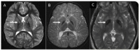
Neuropathology of MPAN is characterized by increased iron deposition in the globus pallidus and substantia nigra.

Genotype-Phenotype Correlations

The phenotypes of AR and AD MPAN are indistinguishable, both arising from loss of function of the C19orf12 protein.

Prevalence

The prevalence of MPAN is roughly estimated at less than one in 1,000,000.

The prevalence may be higher in the Turkish population due to the c.32C>T (p.Thr11Met) founder variant [Olgiati et al 2017] (see Table 6).

Differential Diagnosis

Other genetic types of neurodegeneration with brain iron accumulation (NBIA). Ten genes in total are known to be associated with NBIA: ATP13A2, C19orf12, COASY, CP, DCAF17, FA2H, FTL, PANK2, PLA2G6, and WDR45. Of note, C19orf12-related NBIA (MPAN) – accounting for 5%-10% of all NBIA – is the fourth most common genetic type of NBIA, after beta-propeller protein-associated neurodegeneration (BPAN), pantothenate kinase-associated neurodegeneration (PKAN), and PLA2G6-associated neurodegeneration (PLAN).

  • Neuronal loss, gliosis, widespread iron deposits, and eosinophilic spheroidal structures in the globus pallidus in MPAN are similar to the neuropathologic changes seen in PKAN. In MPAN, however, widespread Lewy bodies throughout the neocortex, deep gray matter, and midbrain are more prominent than the findings observed in PLAN and other forms of NBIA [Hartig et al 2011, Hogarth et al 2013].
  • Peripheral axonal spheroids, previously thought to be limited to PLAN, may be detected in skin or nerve biopsies of individuals with MPAN [Kurian et al 2008, Hogarth et al 2013].
  • The high frequency of optic atrophy in MPAN helps distinguish it from other forms of NBIA, in which optic atrophy is more rare.

See Neurodegeneration with Brain Iron Accumulation (NBIA) Overview for the typical presentation and other key clinical manifestations of each NBIA genetic type and for a review of other disorders to consider in the differential diagnosis of NBIA.

Management

Evaluations Following Initial Diagnosis

To establish the extent of disease and needs in an individual diagnosed with mitochondrial membrane protein-associated neurodegeneration (MPAN), the evaluations summarized in Table 3 (if not performed as part of the evaluation that led to the diagnosis) are recommended.

Table 3.

Recommended Evaluations Following Initial Diagnosis in Individuals with Mitochondrial Membrane Protein-Associated Neurodegeneration

System/ConcernEvaluationComment
Neurologic For children: pediatric neurology assessmentIt is important to establish care & work for continuity as disease is slowly progressive.
For adults: referral to movement disorders neurologistProgressive parkinsonism is more common in adult-onset MPAN.
Referral to neuromuscular clinic (OT/PT / rehab specialist)To assess gross motor & fine motor skills, gait, ambulation & need for adaptive devices, PT/OT
Cognitive decline & neuropsychiatric abnormalities Referral to psychiatrist, psychologist, or neuropsychologistTo assess cognitive dysfunction & neuropsychiatric involvement
For school-age children: developmental assessment
  • To incl motor, adaptive, cognitive, & speech/language eval
  • Eval for early intervention / special education
Optic atrophy Complete eye examTo incl:
  • BCVA
  • Refractive error
  • Color vision testing
  • Slit lamp exam
  • Dilated funduscopic exam
Dysarthria Speech/language evalFor those w/severe dysarthria: assess need for means of alternative communication.
Dysphagia For those w/frequent choking, severe dysphagia, or significant weight loss: assess nutritional status & aspiration risk.Consider involving a gastroenterology/nutrition/feeding team.
Genetic
counseling
By genetics professionals 1To inform patients & families re nature, MOI, & implications of MPAN to facilitate medical & personal decision making
Family support/
resources
Assess need for:
  • Community resources & support/advocacy organizations (e.g., Parent to Parent);
  • Social work involvement for parental support;
  • Home nursing referral.

BCVA = best-corrected Snellen visual acuity; MOI = mode of inheritance; OT = occupational therapy; PT = physical therapy

1.

Medical geneticist, certified genetic counselor, or certified advanced genetic nurse

Treatment of Manifestations

Table 4.

Treatment of Manifestations in Individuals with Mitochondrial Membrane Protein-Associated Neurodegeneration

Manifestation/
Concern
TreatmentConsiderations/Other
Cognitive decline
in adults
Progressive supports will be needed to assist w/ADL.Most persons in end stages of disease require full-time care.
DD/ID in children See Developmental Delay / Intellectual Disability Management Issues.
Neuropsychiatric
complications
By psychiatristTreatment is generally symptomatic & follows standard practice.
Optic atrophy If symptomatic, magnifying visual aids
  • Use of low vision aids 1
  • Work w/agencies for visually impaired 2
Dystonia/
Spasticity
Pharmacologic treatmentConsider oral baclofen, trihexyphenidyl, intramuscular botulinum toxin, & a trial of intrathecal baclofen if indicated.
Parkinsonism Pharmacologic treatmentResponse to levodopa & other medications is variable, but sometimes quite good for several years.
Dysarthria Speech/language resources for means of alternative communicationCognitive decline may complicate treatment.
Dysphagia
& related
complications
  • Consider nutritional & vitamin supplementation to meet dietary needs.
  • Gastric feeding tube as needed to minimize weight loss & ↓ risk of aspiration
  • Glycopyrrolate or transdermal scopolamine patch to ↓ volume of secretions
  • Consider tracheostomy.
The authors encourage providers to open an ongoing dialog about feeding tubes. While some may at first resist using a feeding tube, many families later indicate they wish they had done so sooner.
Constipation Over-the-counter fiber supplements &/or stool softenersCommon; likely caused by a combination of immobility, medications, diet, & the disease itself
Family/
Community
Ensure appropriate social work involvement to connect families w/local resources, respite, & support.Coordinate care to manage multiple subspecialty appointments, equipment, medications, & supplies.
Ethics
consultation
Clinical ethics servicesAssess health care decisions in context of the best interest of the child & values & preferences of the family.

ADL = activities of daily living; DD = developmental delay; ID = intellectual disability

1.

Low vision aids such as magnifiers and closed circuit television may provide useful reading vision for individuals with reduced central acuity and constricted visual fields.

2.

In the US, publicly funded agencies at the state level provide services for the blind or those with progressive eye disorders; services include vocational training, mobility training, and skills for independent living.

Developmental Delay / Intellectual Disability Management Issues

The following information represents typical management recommendations for individuals with developmental delay / intellectual disability in the United States; standard recommendations may vary from country to country.

Ages 0-3 years. Referral to an early intervention program is recommended for access to occupational, physical, speech, and feeding therapy as well as infant mental health services, special educators, and sensory impairment specialists. In the US, early intervention is a federally funded program available in all states that provides in-home services to target individual therapy needs.

Ages 3-5 years. In the US, developmental preschool through the local public school district is recommended. Before placement, an evaluation is made to determine needed services and therapies and an individualized education plan (IEP) is developed for those who qualify based on established motor, language, social, or cognitive delay. The early intervention program typically assists with this transition. Developmental preschool is center based; for children too medically unstable to attend, home-based services are provided.

All ages. Consultation with a developmental pediatrician is recommended to ensure the involvement of appropriate community, state, and educational agencies (US) and to support parents in maximizing quality of life. Some issues to consider:

  • IEP services:
    • An IEP provides specially designed instruction and related services to children who qualify.
    • IEP services will be reviewed annually to determine whether any changes are needed.
    • Special education law requires that children participating in an IEP be in the least restrictive environment feasible at school and included in general education as much as possible, when and where appropriate.
    • Vision consultants should be a part of the child's IEP team to support access to academic material.
    • PT, OT, and speech services will be provided in the IEP to the extent that the need affects the child's access to academic material. Beyond that, private supportive therapies based on the affected individual's needs may be considered. Specific recommendations regarding type of therapy can be made by a developmental pediatrician.
    • As a child enters the teen years, a transition plan should be discussed and incorporated in the IEP. For those receiving IEP services, the public school district is required to provide services until age 21.
  • A 504 plan (Section 504: a US federal statute that prohibits discrimination based on disability) can be considered for those who require accommodations or modifications such as front-of-class seating, assistive technology devices, classroom scribes, extra time between classes, modified assignments, and enlarged text.
  • Developmental Disabilities Administration (DDA) enrollment is recommended. DDA is a US public agency that provides services and support to qualified individuals. Eligibility differs by state but is typically determined by diagnosis and/or associated cognitive/adaptive disabilities.
  • Families with limited income and resources may also qualify for supplemental security income (SSI) for their child with a disability.

Surveillance

Table 5.

Recommended Surveillance for Individuals with Mitochondrial Membrane Protein-Associated Neurodegeneration

System/ConcernEvaluationFrequency
Neurologic
manifestations
  • Neurologic assessment for progression
  • Assess response to medications, side effects, &/or need for new medications or dosage adjustments.
  • When stable: annually
  • Others: may need at 3-6-mo intervals
Musculoskeletal/
ADL
OT/PT assessment of ambulation, gross motor skills, fine motor skills, ADLAnnually until later disease stage, when these become unnecessary
Cognitive/
Psychiatric
Evaluate mood, signs of psychosis, cognitive complaints to identify need for pharmacologic & psychotherapeutic interventions.When indicated by onset of new manifestations
DD/ID in children To assess educational needsAnnually
Optic atrophy Eye exam, visual acuity, color vision, visual field testing, dilated examEvery 1-2 yrs
Monitor effectiveness of visual aids.Annually
Dysarthria Assessment of communication needs
  • Children: annually
  • Adults: every 1-3 yrs
Dysphagia Assessment of swallowing, feeding, nutrition (often by feeding team)As indicated by onset of new manifestations
Social support Assessment of family's access to resources & support (e.g., in-home nursing, respite care)Annually

ADL = activities of daily living; DD = developmental delay; ID = intellectual disability

Evaluation of Relatives at Risk

See Genetic Counseling for issues related to testing of at-risk relatives for genetic counseling purposes.

Therapies Under Investigation

Iron chelation using deferiprone has been investigated in a randomized, double-blind, placebo-controlled trial in a distinct NBIA disorder, PKAN [Klopstock et al 2019]. Results indicate that deferiprone treatment in PKAN did not lead to statistically significant clinical change, although there was a trend towards slower progression on the dystonia scale used. Since other MPAN treatments are symptomatic, some individuals may elect to try deferiprone off label based on these results in a related disorder. Deferiprone treatment in MPAN has been reported in two patients: (1) Two-year treatment in a 13-year-old led to reduction of iron content in the substantia nigra, while pallidal iron depositions and clinical status remained unchanged [Löbel et al 2014]; (2) In another patient with MPAN who received deferiprone, treatment had to be discontinued because of gastrointestinal side effects [Gore et al 2016].

Search ClinicalTrials.gov in the US and EU Clinical Trials Register in Europe for access to information on clinical studies for a wide range of diseases and conditions.

Genetic Counseling

Genetic counseling is the process of providing individuals and families with information on the nature, mode(s) of inheritance, and implications of genetic disorders to help them make informed medical and personal decisions. The following section deals with genetic risk assessment and the use of family history and genetic testing to clarify genetic status for family members; it is not meant to address all personal, cultural, or ethical issues that may arise or to substitute for consultation with a genetics professional. —ED.

Mode of Inheritance

Mitochondrial membrane protein-associated neurodegeneration (MPAN) is inherited in an autosomal recessive or, less commonly, an autosomal dominant manner.

Autosomal Recessive Inheritance – Risk to Family Members

Parents of a proband

  • The parents of an affected individual are obligate heterozygotes (i.e., presumed to be carriers of one C19orf12 pathogenic variant based on family history).
  • Molecular genetic testing is recommended for the parents of a proband to confirm that both parents are heterozygous for a C19orf12 pathogenic variant and to allow reliable recurrence risk assessment. If a pathogenic variant is detected in only one parent, the following possibilities should be considered:
  • Heterozygotes (carriers) are asymptomatic and are not at risk of developing the disorder.

Sibs of a proband

  • If both parents are known to be heterozygous for a C19orf12 pathogenic variant, each sib of an affected individual has at conception a 25% chance of being affected, a 50% chance of being an asymptomatic carrier, and a 25% chance of being unaffected and not a carrier.
  • Heterozygotes (carriers) are asymptomatic and are not at risk of developing the disorder.

Offspring of a proband. Unless an individual with autosomal recessive MPAN has children with an affected individual or a carrier, his/her offspring will be obligate heterozygotes (carriers) for a pathogenic variant in C19orf12.

Other family members. Each sib of the proband's parents is at a 50% risk of being a carrier of a C19orf12 pathogenic variant.

Carrier Detection

Carrier testing for at-risk relatives requires prior identification of the autosomal recessive C19orf12 pathogenic variants in the family.

Autosomal Dominant Inheritance – Risk to Family Members

Parents of a proband

Sibs of a proband. The risk to the sibs of the proband depends on the genetic status of the proband's parents:

Offspring of a proband

  • Each child of an individual with autosomal dominant MPAN has a 50% risk of inheriting the pathogenic variant.
  • While physical and cognitive impairment in MPAN reduces the possibility of having children, some individuals with late-onset MPAN will reproduce before the onset of symptoms [Gregory et al 2019].

Other family members. The risk to other family members depends on the status of the proband's parents: if a parent is affected or has the C19orf12 pathogenic variant, his or her family members are at risk.

Predictive testing of at-risk asymptomatic adult family members requires prior identification of the autosomal dominant C19orf12 pathogenic variant in the family.

Potential consequences of such testing (including but not limited to socioeconomic changes and the need for long-term follow up and evaluation arrangements for individuals with a positive test result) as well as the capabilities and limitations of predictive testing should be discussed in the context of formal genetic counseling prior to testing.

Related Genetic Counseling Issues

Family planning

  • The optimal time for determination of genetic risk and discussion of the availability of prenatal/preimplantation genetic testing is before pregnancy.
  • It is appropriate to offer genetic counseling (including discussion of potential risks to offspring and reproductive options) to young adults who are affected, are carriers, or are at risk of being carriers or affected.

DNA banking is the storage of DNA (typically extracted from white blood cells) for possible future use. Because it is likely that testing methodology and our understanding of genes, allelic variants, and diseases will improve in the future, consideration should be given to banking DNA of affected individuals.

Prenatal Testing and Preimplantation Genetic Testing

Once the pathogenic C19orf12 variant(s) have been identified in an affected family member, prenatal testing for a pregnancy at increased risk and preimplantation genetic testing for MPAN are possible.

Differences in perspective may exist among medical professionals and within families regarding the use of prenatal testing. While most centers would consider use of prenatal testing to be a personal decision, discussion of these issues may be helpful.

Resources

GeneReviews staff has selected the following disease-specific and/or umbrella support organizations and/or registries for the benefit of individuals with this disorder and their families. GeneReviews is not responsible for the information provided by other organizations. For information on selection criteria, click here.

  • NBIA Alliance
    Email: Info@NBIAalliance.org
  • NBIA Disorders Association
  • NBIAcure
    Center of Excellence for NBIA Clinical Care and Research
    International Registry for NBIA and Related Disorders
    Oregon Health & Science University
    Email: info@nbiacure.org
  • Treat Iron-Related Childhood Onset Neurodegeneration (TIRCON)
    Germany
    Email: TIRCON@med.uni-muenchen.de

Molecular Genetics

Information in the Molecular Genetics and OMIM tables may differ from that elsewhere in the GeneReview: tables may contain more recent information. —ED.

Table A.

Mitochondrial Membrane Protein-Associated Neurodegeneration: Genes and Databases

GeneChromosome LocusProteinHGMDClinVar
C19orf12 19q12 Protein C19orf12 C19orf12 C19orf12

Data are compiled from the following standard references: gene from HGNC; chromosome locus from OMIM; protein from UniProt. For a description of databases (Locus Specific, HGMD, ClinVar) to which links are provided, click here.

Table B.

OMIM Entries for Mitochondrial Membrane Protein-Associated Neurodegeneration (View All in OMIM)

614297CHROMOSOME 19 OPEN READING FRAME 12; C19ORF12
614298NEURODEGENERATION WITH BRAIN IRON ACCUMULATION 4; NBIA4

Molecular Pathogenesis

C19orf12 encodes C19orf12, a protein that localizes predominantly to the mitochondria [Hartig et al 2011]. The two protein isoforms in humans have 141 and 152 amino acids, NP_001242976.1 and NP_001026896.2, respectively.

Association with the mitochondrial membrane and co-regulation with proteins of fatty acid biogenesis and branched chain amino acid degradation expression profiles suggest similarities to other proteins known to be defective in NBIA. C19orf12 is suspected of playing a role in lipid homeostasis [Hartig et al 2011].

Mechanism of disease causation

C19orf12-specific laboratory technical considerations. Heterozygous pathogenic variants that cause AD MPAN are located in the last exon of C19orf12 and are predicted to escape nonsense-mediated decay, resulting in a truncated protein after amino acid 79 (with wild type residues 69-76 intact) [Gregory et al 2019]. This finding should help inform interpretation of results by clinical laboratories.

Table 6.

Notable C19orf12 Pathogenic Variants

Reference SequencesDNA Nucleotide ChangePredicted Protein ChangeComment [Reference]
NM_001031726​.3
NP_001026896​.2
c.32C>Tp.Thr11Met
c.187G>Cp.Ala63ProMRI did not show brain iron accumulation in some persons [Landouré et al 2013].
c.204_214del11p.Gly69ArgfsTer10
  • Founder variant in Eastern European (Polish) population
  • Assoc w/juvenile onset [Hartig et al 2011]

Variants listed in the table have been provided by the authors. GeneReviews staff have not independently verified the classification of variants.

GeneReviews follows the standard naming conventions of the Human Genome Variation Society (varnomen​.hgvs.org). See Quick Reference for an explanation of nomenclature.

Chapter Notes

Author Notes

Investigators and clinicians at Oregon Health & Science University have studied the NBIA disorders, including MPAN, for more than 25 years. Our program includes an NBIA Center of Excellence committed to providing comprehensive care for patients with NBIA around the world (nbiacure.org/nbia-clinic). Our multidisciplinary team investigates NBIA disorders broadly, from gene discovery to clinical trials for rational therapeutics, to improve the lives of those with NBIA.

For more information, please contact us:
Phone: 503-494-4344
Fax: 503-494-6886
Email: ude.usho@ayrogerg
Website: nbiacure.org

Acknowledgments

The authors acknowledge funding from the European Commission Seventh Framework Programme (FP7/2007-2013, HEALTH-F2-2011, grant agreement No. 277984, TIRCON).

Author History

Allison Gregory, MS, CGC (2014-present)
Monika Hartig, MD; Technische Universität München (2014-2021)
Susan J Hayflick, MD (2014-present)
Penelope Hogarth, MD (2014-present)
Thomas Klopstock, MD (2021-present)
Tomasz Kmiec, MD (2014-present)
Holger Prokisch, PhD; Technische Universität München (2014-2021)

Revision History

  • 4 March 2021 (bp) Comprehensive update posted live
  • 27 February 2014 (me) Review posted live
  • 3 September 2013 (ag) Original submission

References

Literature Cited

  • Al Macki N, Rashdi I. A novel deletion mutation of exon 2 of the C19orf12 gene in an Omani family with mitochondrial membrane protein-associated neurodegeneration (MPAN). Oman Med J. 2017;32:66–8. [PMC free article: PMC5187394] [PubMed: 28042406]
  • Bayram E, Peker E, Metzger S, Akbostanct M. Myoclonus, hydrocephalus in mitochondrial protein-associated neurodegeneration. Can J Neurol Sci. 2019;46:628–30. [PubMed: 31327327]
  • Deschauer M, Gaul C, Behrmann C, Prokisch H, Zierz S, Haack TB. C19orf12 mutations in neurodegeneration with brain iron accumulation mimicking juvenile amyotrophic lateral sclerosis. J Neurol. 2012;259:2434–9. [PubMed: 22584950]
  • Dogu O, Krebs CE, Kaleagasi H, Demirtas Z, Oksuz N, Walker RH, Paisán-Ruiz C. Rapid disease progression in adult-onset mitochondrial membrane protein-associated neurodegeneration. Clin Genet. 2013;84:350–5. [PubMed: 23278385]
  • Gagliardi M, Annesi G, Lesca G, Broussolle E, Iannello G, Vaiti V, Gambardella A, Quattrone A. C19orf12 gene mutations in patients with neurodegeneration with brain iron accumulation. Parkinsonism Relat Disord. 2015;21:813–6. [PubMed: 25962551]
  • Gore E, Appleby BS, Cohen ML, DeBrosse SD, Leverenz JB, Miller BL, Siedlak SL, Zhu X, Lerner AJ. Clinical and imaging characteristics of late onset mitochondrial membrane protein-associated neurodegeneration (MPAN). Neurocase. 2016;22:476–83. [PMC free article: PMC5568540] [PubMed: 27801611]
  • Gregory A, Lotia M, Jeong SY, Fox R, Zhen D, Sanford L, Hamada J, Jahic A, Beetz C, Freed A, Kurian MA, Cullup T, van der Weijden MCM, Nguyen V, Setthavongsack N, Garcia D, Krajbich V, Pham T, Woltjer R, George BP, Minks KQ, Paciorkowski AR, Hogarth P, Jankovic J, Hayflick SJ. Autosomal dominant mitochondrial membrane protein-associated neurodegeneration (MPAN). Mol Genet Genomic Med. 2019;7:e00736. [PMC free article: PMC6625130] [PubMed: 31087512]
  • Hartig M, Prokisch H, Meitinger T, Klopstock T. Mitochondrial membrane protein-associated neurodegeneration (MPAN). Int Rev Neurobiol. 2013;110:73–84. [PubMed: 24209434]
  • Hartig MB, Iuso A, Haack T, Kmiec T, Jurkiewicz E, Heim K, Roeber S, Tarabin V, Dusi S, Krajewska-Walasek M, Jozwiak S, Hempel M, Winkelmann J, Elstner M, Oexle K, Klopstock T, Mueller-Felber W, Gasser T, Trenkwalder C, Tiranti V, Kretzschmar H, Schmitz G, Strom TM, Meitinger T, Prokisch H. Absence of an orphan mitochondrial protein, c19orf12, causes a distinct clinical subtype of neurodegeneration with brain iron accumulation. Am J Hum Genet. 2011;89:543–50. [PMC free article: PMC3188837] [PubMed: 21981780]
  • Hogarth P, Gregory A, Kruer MC, Sanford L, Wagoner W, Natowicz MR, Egel RT, Subramony SH, Goldman JG, Berry-Kravis E, Foulds NC, Hammans SR, Desguerre I, Rodriguez D, Wilson C, Diedrich A, Green S, Tran H, Reese L, Woltjer RL, Hayflick SJ. New NBIA subtype: genetic, clinical, pathologic, and radiographic features of MPAN. Neurology. 2013;80:268–75. [PMC free article: PMC3589182] [PubMed: 23269600]
  • Jónsson H, Sulem P, Kehr B, Kristmundsdottir S, Zink F, Hjartarson E, Hardarson MT, Hjorleifsson KE, Eggertsson HP, Gudjonsson SA, Ward LD, Arnadottir GA, Helgason EA, Helgason H, Gylfason A, Jonasdottir A, Jonasdottir A, Rafnar T, Frigge M, Stacey SN, Th Magnusson O, Thorsteinsdottir U, Masson G, Kong A, Halldorsson BV, Helgason A, Gudbjartsson DF, Stefansson K. Parental influence on human germline de novo mutations in 1,548 trios from Iceland. Nature. 2017;549:519–22. [PubMed: 28959963]
  • Kim J, Liao Y, Ionita C, Bale AE, Daras B, Ascadi G. Mitochondrial membrate protein-associated neurodegeneration mimicking juvenile amyotrophic lateral sclerosis. Pediatr Neurol. 2016;64:83–6. [PubMed: 27671242]
  • Klopstock T, Tricta F, Neumayr L, Karin I, Zorzi G, Fradette C, Kmiec T, Buchner B, Steele HE, Horvath R, Chinnery PF, Basu A, Kupper C, Neuhofer C, Kalman B, Dusek P, Yapici Z, Wilson I, Zhao F, Zibordi F, Nardocci N, Aguilar C, Hayflick SJ, Spino M, Blamire AM, Hogarth P, Vichinsky E. Safety and efficacy of deperiprone for pantothenate kinase-associated neurodegeneration: a randomised, double-blind, controlled trial and an open-label extension study. Lancet Neurol. 2019;18:631–42. [PubMed: 31202468]
  • Kurian MA, Morgan NV, MacPherson L, Foster K, Peake D, Gupta R, Philip SG, Hendriksz C, Morton JE, Kingston HM, Rosser EM, Wassmer E, Gissen P, Maher ER. Phenotypic spectrum of neurodegeneration associated with mutations in the PLA2G6 gene (PLAN). Neurology. 2008;70:1623–9. [PubMed: 18443314]
  • Landouré G, Zhu PP, Lourenço CM, Johnson JO, Toro C, Bricceno KV, Rinaldi C, Meilleur KG, Sangaré M, Diallo O, Pierson TM, Ishiura H, Tsuji S, Hein N, Fink JK, Stoll M, Nicholson G, Gonzalez MA, Speziani F, Dürr A, Stevanin G, Biesecker LG, Accardi J, Landis DM, Gahl WA, Traynor BJ, Marques W Jr, Züchner S, Blackstone C, Fischbeck KH, Burnett BG. Hereditary spastic paraplegia type 43 (SPG43) is caused by mutation in C19orf12. Hum Mutat. 2013;34:1357–60. [PMC free article: PMC3819934] [PubMed: 23857908]
  • Langwinska-Wosko E, Skowronska M, Kmiec T, Czlonkowska A. Retinal and optic nerve abnormalities in neurodegeneration associated with mutations in C19orf12 (MPAN). J Neurol Sci. 2016;370:237–240. [PubMed: 27772766]
  • Löbel U, Schweser F, Nickel M, Deistung A, Grosse R, Hagel C, et al. Brain iron quantification by MRI in mitochondrial membrane protein-associated neurodegeneration under iron-chelating therapy. Ann Clin Transl Neurol. 2014;1:1041–6. [PMC free article: PMC4284129] [PubMed: 25574478]
  • Olgiati S, Doğu O, Tufekcioglu Z, Diler Y, Saka E, Gultekin M, Kaleagasi H, Kuipers D, Graafland J, Breedveld GJ, Quadri M, Sürmeli R, Sünter G, Doğan T, Yalçın AD, Bilgiç B, Elibol B, Emre M, Hanagasi HA, Bonifati V. The p.Thr11Met mutation in c19orf12 is frequent among adult Turkish patients with MPAN. Parkinsonism Relat Disord. 2017;39:64–70. [PubMed: 28347615]
  • Rahbari R, Wuster A, Lindsay SJ, Hardwick RJ, Alexandrov LB, Turki SA, Dominiczak A, Morris A, Porteous D, Smith B, Stratton MR, Hurles ME, et al. uycdxTiming, rates and spectra of human germline mutation. Nat Genet. 2016;48:126–33. [PMC free article: PMC4731925] [PubMed: 26656846]
  • Schottmann G, Stenzel W, Lützkendorf S, Schuelke M, Knierim E. A novel frameshift mutation of C19ORF12 causes NBIA4 with cerebellar atrophy and manifests with severe peripheral motor axonal neuropathy. Clin Genet. 2014;85:290–2. [PubMed: 23521069]
  • Schulte EC, Claussen MC, Jochim A, Haack T, Hartig M, Hempel M, Prokisch H, Haun-Jünger U, Winkelmann J, Hemmer B, Förschler A, Ilg R. Mitochondrial membrane protein associated neurodegeneration: a novel variant of neurodegeneration with brain iron accumulation. Mov Disord. 2013;28:224–7. [PubMed: 23436634]
  • Selikhova M, Fedotova E, Wiethoff S, Schottlaender LV, Klyushnikov S, Illarioshkin SN, Houlden H. A 30-year history of MPAN case from Russia. Clin Neurol Neurosurg. 2017;159:111–3. [PubMed: 28641177]
  • Skowronska M, Kmiec T, Jurkiewicz E, Malczyk K, Kurkowska-Jastrzebska I, Czlonkowska A. Evolution and novel radiological changes of neurodegeneration associated with mutations in C19orf12. Parkinsonism Relat Disord. 2017;39:71–76. [PubMed: 28347614]
  • Stenson PD, Mort M, Ball EV, Evans K, Hayden M, Heywood S, Hussain M, Phillips AD, Cooper DN. The Human Gene Mutation Database: towards a comprehensive repository of inherited mutation data for medical research, genetic diagnosis and next-generation sequencing studies. Hum Genet. 2017;136:665–77. [PMC free article: PMC5429360] [PubMed: 28349240]
Copyright © 1993-2024, University of Washington, Seattle. GeneReviews is a registered trademark of the University of Washington, Seattle. All rights reserved.

GeneReviews® chapters are owned by the University of Washington. Permission is hereby granted to reproduce, distribute, and translate copies of content materials for noncommercial research purposes only, provided that (i) credit for source (http://www.genereviews.org/) and copyright (© 1993-2024 University of Washington) are included with each copy; (ii) a link to the original material is provided whenever the material is published elsewhere on the Web; and (iii) reproducers, distributors, and/or translators comply with the GeneReviews® Copyright Notice and Usage Disclaimer. No further modifications are allowed. For clarity, excerpts of GeneReviews chapters for use in lab reports and clinic notes are a permitted use.

For more information, see the GeneReviews® Copyright Notice and Usage Disclaimer.

For questions regarding permissions or whether a specified use is allowed, contact: ude.wu@tssamda.

Bookshelf ID: NBK185329PMID: 24575447

Views

Tests in GTR by Gene

Related information

  • OMIM
    Related OMIM records
  • PMC
    PubMed Central citations
  • PubMed
    Links to PubMed
  • Gene
    Locus Links

Similar articles in PubMed

See reviews...See all...

Recent Activity

Your browsing activity is empty.

Activity recording is turned off.

Turn recording back on

See more...