U.S. flag

An official website of the United States government

NCBI Bookshelf. A service of the National Library of Medicine, National Institutes of Health.

Adam MP, Feldman J, Mirzaa GM, et al., editors. GeneReviews® [Internet]. Seattle (WA): University of Washington, Seattle; 1993-2024.

Cover of GeneReviews®

GeneReviews® [Internet].

Show details

ESCO2 Spectrum Disorder

, MD, PhD, , PhD, and , MD.

Author Information and Affiliations

Initial Posting: ; Last Update: March 26, 2020.

Estimated reading time: 27 minutes

Summary

Clinical characteristics.

ESCO2 spectrum disorder is characterized by mild-to-severe prenatal growth restriction, limb malformations (which can include bilateral symmetric tetraphocomelia or hypomelia caused by mesomelic shortening), hand anomalies (including oligodactyly, thumb aplasia or hypoplasia, and syndactyly), elbow and knee flexion contractures (involving elbows, wrists, knees, ankles, and feet [talipes equinovarus]), and craniofacial abnormalities (which can include bilateral cleft lip and/or cleft palate, micrognathia, widely spaced eyes, exophthalmos, downslanted palpebral fissures, malar flattening, and underdeveloped ala nasi), ear malformation, and corneal opacities. Intellectual disability (ranging from mild to severe) is common. Early mortality is common among severely affected pregnancies and newborns; mildly affected individuals may survive to adulthood.

Diagnosis/testing.

The diagnosis of ESCO2 spectrum disorder is established in a proband with suggestive clinical findings by identification of either biallelic pathogenic variants in ESCO2 by molecular genetic testing or premature centromere separation (PCS) by cytogenetic testing.

Management.

Treatment of manifestations: Individualized treatment aimed to improve quality of life; surgery for cleft lip and/or palate, for correction of limb abnormalities, and to improve proper development of the prehensile hand grasp. Prostheses, speech assessment and therapy, special education for developmental delays, and standard treatment for ophthalmologic, cardiac, and renal abnormalities as indicated.

Surveillance: Periodic assessment of: growth and weight gain; motor and language development; speech development and hearing (in those with cleft lip and palate); and educational needs. Follow up for ophthalmologic, cardiac, and/or renal anomalies per treating physicians.

Genetic counseling.

ESCO2 spectrum disorder is inherited in an autosomal recessive manner. If both parents are known to be heterozygous for an ESCO2 pathogenic variant, each sib of an affected individual has at conception a 25% chance of inheriting both pathogenic variants and being affected, a 50% chance of inheriting one pathogenic variant and being an unaffected carrier, and a 25% chance of inheriting both normal alleles. When the ESCO2 pathogenic variants have been identified in an affected family member, carrier testing of at-risk relatives, prenatal testing for pregnancies at increased risk, and preimplantation genetic testing are possible.

GeneReview Scope

ESCO2 Spectrum Disorder: Included Phenotypes 1
  • Roberts syndrome
  • SC phocomelia syndrome

For synonyms and outdated names see Nomenclature.

1.

For other genetic causes of these phenotypes, see Differential Diagnosis.

Diagnosis

ESCO2 spectrum disorder comprises a phenotypic continuum that ranges from Roberts syndrome at the severe end to SC phocomelia at the milder end.

Suggestive Findings

ESCO2 spectrum disorder should be suspected in an individual with the following clinical findings and family history.

Clinical findings [Hennekam et al 2010, Vega et al 2010, Vega et al 2016]:

  • Prenatal growth restriction ranging from mild to severe. Mean birth length and weight is below the third centile in most term and prematurely born affected infants.
  • Limb malformations including bilateral symmetric tetraphocomelia or hypomelia caused by mesomelic shortening. Upper limbs are more severely affected than lower limbs.
  • Hand abnormalities. Most commonly oligodactyly with thumb aplasia or hypoplasia, followed by fifth finger clinodactyly or hypoplasia
  • Flexion contractures of the knees, ankles, wrists, and elbows; talipes equinovarus
  • Craniofacial abnormalities including bilateral cleft lip and/or palate, micrognathia, widely spaced eyes, exophthalmos, downslanted palpebral fissures, malar flattening, underdeveloped ala nasi, and ear malformation
  • Developmental delay / intellectual disability is not always present, segregates by families, and probably correlates with the presence of corneal opacities. There are families with severe phocomelia and bilateral cleft lip and palate with no corneal opacities and no intellectual disability. When present, intellectual disability ranges from mild to severe.
  • Other
    • Urogenital abnormalities: cryptorchidism, enlarged phallus
    • Renal anomalies: horseshoe kidney, polycystic kidney
    • Heart defects: ventricular septal defect
    • Eye abnormalities: corneal opacities
    • Sparse hair

Family history consistent with autosomal recessive inheritance, including consanguinity

Establishing the Diagnosis

The diagnosis ESCO2 spectrum disorder is established in a proband with suggestive clinical findings by identification of either biallelic pathogenic (or likely pathogenic) variants in ESCO2 by molecular genetic testing (see Table 1) or premature centromere separation (PCS) by cytogenetic testing.

Note: (1) Per ACMG/AMP variant interpretation guidelines, the terms "pathogenic variants" and "likely pathogenic variants" are synonymous in a clinical setting, meaning that both are considered diagnostic and both can be used for clinical decision making [Richards et al 2015]. Reference to "pathogenic variants" in this section is understood to include any likely pathogenic variants. (2) Identification of biallelic ESCO2 variants of uncertain significance (or of one known ESCO2 pathogenic variant and one ESCO2 variant of uncertain significance) does not establish or rule out a diagnosis.

Molecular genetic testing approaches can include a combination of gene-targeted testing (single-gene testing, multigene panel) and comprehensive genomic testing (exome sequencing, genome sequencing) depending on the phenotype.

Gene-targeted testing requires that the clinician determine which gene(s) are likely involved, whereas genomic testing does not. Because the phenotype of ESCO2 spectrum disorder is broad, individuals with the distinctive findings described in Suggestive Findings are likely to be diagnosed using gene-targeted testing (see Option 1), whereas those in whom the diagnosis of ESCO2 spectrum disorder has not been considered are more likely to be diagnosed using genomic testing (see Option 2).

Option 1

Single-gene testing. Sequence analysis of ESCO2 is performed first to detect small intragenic deletions/insertions and missense, nonsense, and splice site variants. Note: Depending on the sequencing method used, single-exon, multiexon, or whole-gene deletions/duplications may not be detected. If only one or no variant is detected by the sequencing method used, the next step is typically to perform gene-targeted deletion/duplication analysis to detect exon and whole-gene deletions or duplications; to date, however, such variants have not been identified as a cause of ESCO2 spectrum disorder.

A limb malformation or cleft palate multigene panel that includes ESCO2 and other genes of interest (see Differential Diagnosis) is most likely to identify the genetic cause of the condition while limiting identification of variants of uncertain significance and pathogenic variants in genes that do not explain the underlying phenotype. Note: (1) The genes included in the panel and the diagnostic sensitivity of the testing used for each gene vary by laboratory and are likely to change over time. (2) Some multigene panels may include genes not associated with the condition discussed in this GeneReview. Given the rarity of ESCO2 spectrum disorder some panels may not include this gene. (3) In some laboratories, panel options may include a custom laboratory-designed panel and/or custom phenotype-focused exome analysis that includes genes specified by the clinician. (4) Methods used in a panel may include sequence analysis, deletion/duplication analysis, and/or other non-sequencing-based tests.

For an introduction to multigene panels panels click here. More detailed information for clinicians ordering genetic tests can be found here.

Option 2

Comprehensive genomic testing. Exome sequencing is the most commonly used genomic testing method; genome sequencing is also possible.

If exome sequencing is not diagnostic, exome array (when clinically available) may be considered to detect (multi)exon deletions or duplications that cannot be detected by sequence analysis; however, to date such variants have not been identified as a cause of ESCO2 spectrum disorder.

For an introduction to comprehensive genomic testing click here. More detailed information for clinicians ordering genomic testing can be found here.

Table 1.

Molecular Genetic Testing Used in ESCO2 Spectrum Disorder

Gene 1MethodProportion of Pathogenic Variants 2 Detectable by Method
ESCO2 Sequence analysis 3100% 4
Gene-targeted deletion/duplication analysis 5None reported 6
1.
2.

See Molecular Genetics for information on variants detected in this gene.

3.

Sequence analysis detects variants that are benign, likely benign, of uncertain significance, likely pathogenic, or pathogenic. Variants may include small intragenic deletions/insertions and missense, nonsense, and splice site variants; typically, exon or whole-gene deletions/duplications are not detected. For issues to consider in interpretation of sequence analysis results, click here.

4.

Data derived from subscription-based professional version of Human Gene Mutation Database [Stenson et al 2020]

5.

Gene-targeted deletion/duplication analysis detects intragenic deletions or duplications. Methods used may include a range of techniques such as quantitative PCR, long-range PCR, multiplex ligation-dependent probe amplification (MLPA), and a gene-targeted microarray designed to detect single-exon deletions or duplications.

6.

No data on detection rate of gene-targeted deletion/duplication analysis are available.

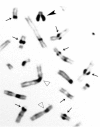
Cytogenetic testing. Standard cytogenetic preparations stained with Giemsa or C-banding techniques show the characteristic chromosome abnormality of premature centromere separation (PCS) and separation of the heterochromatic regions (also termed heterochromatin repulsion [HR]) in most chromosomes in all metaphases (Figure 1).

Figure 1. . C-banding of metaphase chromosomes.

Figure 1.

C-banding of metaphase chromosomes. Arrows show selected chromosomes with premature centromere separation. Solid black arrowhead points to "splitting" of the Y chromosome heterochromatic region. Open arrowheads show selected chromosomes with normal C-banded (more...)

Note on terminology used in RBS: The centromere and the heterochromatin are affected in RBS. (1) The term "premature centromere separation" (PCS) describes the cytogenetic abnormalities observed in standard cytogenetic preparations and the prematurely separating centromeres during metaphase rather than in anaphase. PCS is related to the most probable pathologic mechanism and associated spindle checkpoint activation and impaired cell proliferation. (2) The term "heterochromatin repulsion" (HR) only describes the cytogenetic abnormality of the heterochromatin and does not describe the abnormal process of sister chromatid cohesion, which is fundamental to the pathophysiology of RBS. (3) Until a better term is available to define the structural and functional characteristics of RBS, the authors prefer to use the combined term "PCS/HR."

  • Many chromosomes display a "railroad track" appearance as a result of the absence of the primary constriction and presence of "puffing" or "repulsion" at the heterochromatic regions around the centromeres and nucleolar organizers.
  • The heterochromatic region of the long arm of the Y chromosome is often widely separated in metaphase spreads.

Note: PCS/HR is different from premature sister chromatid separation (PSCS) described in Cornelia de Lange syndrome and premature centromere division (PCD) associated with mosaic variegated aneuploidy syndrome, in which separation and splaying involves not only the centromeric regions but also the entire sister chromatids [Plaja et al 2001, Kaur et al 2005].

Aneuploidy, micronucleation, and multilobulated nuclei are also common findings in RBS cell cultures.

Clinical Characteristics

Clinical Description

ESCO2 spectrum disorder is characterized by mild-to-severe prenatal growth restriction, limb malformations (which can include bilateral symmetric tetraphocomelia or hypomelia caused by mesomelic shortening), hand anomalies, multiple joint contractures, craniofacial abnormalities, and often corneal opacities. Intellectual disability is common.

To date 150 individuals have been identified with ESCO2 spectrum disorder [Ismail et al 2016]. The following description of the phenotypic features associated with this condition is based on that report and on Vega et al [2010] and Vega et al [2016].

Table 2.

Select Clinical Features of ESCO2 Spectrum Disorder

FeatureProportion of Individuals w/Feature 1Comment
Growth deficiency49/49 (100%)
Microcephaly38/40 (95%)
Phocomelia53/53 (100%)
  • Upper limbs only (21%)
  • Upper & lower limbs (79%)
Bone fusions15/23 (65%)
  • Knees, ankles, wrists, elbows, talipes equinovarus, syndactyly (18%)
  • Flexion contractures (16%)
Cleft lip & palate29/48 (56%)Cleft palate only (5%)
DD/ID23/38 (61%)
Ocular
abnormalities
(37%)
  • Corneal opacities (33%)
  • Microphthalmia (8%)
  • Nystagmus (8%)
  • Glaucoma (8%)
Urogenital
anomalies
14/30 (48%)
  • Cryptorchidism (22%)
  • Enlarged phallus (33%)
  • Enlarged clitoris (45%)
Renal anomalies1/8 (12%)Polycystic kidney; horseshoe kidney
Cardiac anomalies10/34 (29%)ASD, VSD

ASD = atrial septal defect; DD/ID = developmental delay / intellectual disability; VSD = ventricular septal defect

1.

Numerator = number of individuals with the feature; denominator = number of individuals with information on the feature.

Growth restriction of prenatal onset is the most consistent finding in all affected individuals. Postnatal growth restriction can be moderate to severe and correlates with the severity of the limb and craniofacial malformations.

Limb malformations include symmetric mesomelic shortening and anterior-posterior axis involvement in which the frequency and degree of involvement of long bones is, in decreasing order:

  • Upper limbs. Radii, ulnae, and humeri in the upper limbs;
  • Lower limbs: Fibulae, tibiae, and femur.

The degree of limb abnormality follows a cephalo-caudal pattern: the upper limbs are more severely affected than the lower, with several cases of upper limb-only malformations.

Hand malformations include brachydactyly and oligodactyly. The thumb is most often affected by proximal positioning or digitalization, hypoplasia, or agenesis. The fifth finger is the next most affected digit with clinodactyly, hypoplasia, or agenesis. In those with severe involvement, only three fingers are present (and rarely, only one finger).

Limb bone fusions, evident in individuals with mild phocomelia, are more common in the knees and ankles, although also present in hands.

Craniofacial abnormalities include: cleft lip and/or cleft palate, premaxillary prominence, micrognathia (77%), microcephaly, brachycephaly (63%), midfacial capillary hemangioma (71%), malar flattening (88%), downslanted palpebral fissures (57%), widely spaced eyes (85%), exophthalmos resulting from shallow orbits (59%), corneal opacities (33%), underdeveloped ala nasi (77%), convex nasal ridge, and ear malformations (69%).

Mildly affected individuals have no palatal abnormalities or only a high-arched palate. The most severely affected individuals have fronto-ethmoid-nasal-maxillary encephalocele.

Correlation between the degree of limb and facial involvement is observed: individuals with mild limb abnormalities also have mild craniofacial malformations, whereas those with severely affected limbs have extensive craniofacial abnormalities.

Intellectual disability is present in the majority of affected individuals; normal intellectual and social development have also been reported [Petrinelli et al 1984, Stanley et al 1988, Maserati et al 1991, Holden et al 1992].

Other findings that may be observed:

  • Abnormal genitalia
    • Males. Enlarged penis (30%), relatively large appearance in relation to the reduced limbs; cryptorchidism, and hypospadias [Satar et al 1994]
    • Females. Enlarged clitoris (46%)
  • Renal anomalies. Polycystic kidney (2%), horseshoe kidney, hydronephrosis (2%)
  • Heart defects. Atrial septal defect, ventricular septal defect, patent ductus arteriosus
  • Eye abnormalities. Commonly, corneal opacities with occasional involvement of the crystalline lens; other ocular findings include microphthalmia and glaucoma [Ismail et al 2016].
  • Hair. Sparse hair, silvery blonde scalp hair
  • Cranial nerve paralysis. Occasional; two patients have had third cranial nerve paralysis related to cavernous angioma [Ogilvy et al 1993]; additional extracranial neoplasms have been reported (1 case with malignant melanoma, 1 with rhabdomyosarcoma) [Parry et al 1986, Wenger et al 1988, Feingold 1992].
  • Moyamoya disease (2%) and stroke secondary to intracranial aneurysms (4%) occurred during late adolescence and young adulthood [Herrmann et al 1969, Vega et al 2010].

Prognosis. Little is known about the natural history of ESCO2 spectrum disorder. Wide clinical variability is observed among affected individuals, including sibs. The prognosis depends on the malformations present: the severity of manifestations correlates with survival. Mortality is high among most of the severely affected pregnancies and newborns. Mildly affected children are more likely to survive to adulthood.

The cause of death has not been reported for most affected individuals. Reported causes are infection (5 individuals), aneurysm/hemorrhage (3 individuals), and malignancy (3 individuals) [Herrmann & Opitz 1977, Vega et al 2010]. Perinatal mortality is correlated with severity of the malformations; early death is usually secondary to respiratory complications and infection [Van Den Berg & Francke 1993].

Genotype-Phenotype Correlations

To date, correlation of ESCO2 variants with specific phenotypic features has not been established.

Nomenclature

In 1919, John B Roberts reported phocomelia, bilateral cleft lip and cleft palate, and protrusion of the intermaxillary region in three children of an Italian couple who were first cousins [Roberts 1919].

In 1969, J Herrmann and colleagues described a syndrome of intrauterine and postnatal growth retardation, mild symmetric reduction of the limbs, flexion contractures of various joints, multiple minor anomalies (including capillary hemangiomas of the face, cloudy corneas, hypoplastic cartilages of the ears and nose, micrognathia, and scanty, silvery-blond hair), and autosomal recessive inheritance. They named this condition the pseudothalidomide or SC syndrome (for the initials of the surnames of the 2 families described) [Herrmann et al 1969].

Clinical evidence that these two phenotypes are allelic was supported by the observations that both were caused by biallelic pathogenic variants in ESCO2 [Schüle et al 2005, Vega et al 2005].

Other synonyms used for Roberts syndrome in the past are Appelt-Gerken-Lenz syndrome, hypomelia-hypotrichosis-facial hemangioma syndrome, tetraphocomelia-cleft palate syndrome, and pseudothalidomide syndrome.

Prevalence

ESCO2 spectrum disorder is rare; no accurate estimates of prevalence have been published. Approximately 150 individuals of diverse racial and ethnic backgrounds have been reported.

Parental consanguinity is common.

Differential Diagnosis

While some syndromes share some of the clinical features of ESCO2 spectrum disorder, a physical examination and skeletal survey should allow for differentiation between individuals with ESCO2 spectrum disorder and those with conditions that are clinically similar.

Table 3.

Genes of Interest in the Differential Diagnosis of ESCO2 Spectrum Disorder

Gene(s)DisorderMOILimb MalformationsOther Clinical FeaturesComment
BRCA2
BRIP1
FANCA
FANCB
FANCC
FANCD2
FANCE
FANCF
FANCG
FANCI 1
Fanconi anemia AR
XL 2
Unilateral or bilateral malformations of upper limbs (e.g., hypoplastic thumb & hypoplastic radius) & lower limbsBone marrow failure, ↑ risk for malignancy; physical abnormalities (e.g., short stature, abnormal skin pigmentation, microcephaly; & ophthalmic & genitourinary tract anomalies)Disorder w/preaxial reduction defects to consider in individuals w/mild manifestations
BUB1B
CEP57
TRIP13
Mosaic variegated aneuploidy syndrome (OMIM PS257300)ARSevere microcephaly, growth restriction, ID, childhood cancer predisposition
  • Constitutional mosaicism for chromosome gains & losses
  • Cytogenetic findings are similar (not =) to RBS & incl premature centromere division (mitotic cells show split centromeres & splayed chromatids in all or most chromosomes). 3
HDAC8
NIPBL
RAD21
SMC1A
SMC3
Cornelia de Lange syndrome AD
XL
Upper-limb reduction defects ranging from subtle phalangeal abnormalities to oligodactylyDistinctive facial features, growth restriction (prenatal onset; <5th %ile throughout life), hirsutism, craniofacial features 4, cardiac septal defects, gastrointestinal dysfunction, hearing loss, myopia, & cryptorchidism or hypoplastic genitaliaCytogenetic findings are similar (not =) to RBS & incl premature sister chromatid separation (separation & splaying involves not only the centromeric regions but also the entire sister chromatids). 5
RBM8A Thrombocytopenia-absent radius (TAR) syndrome ARBilateral absence of radii w/presence of both thumbs; other upper- & lower-limb skeletal anomaliesGenerally transient thrombocytopenia (<50 platelets/nL); & anomalies of the vertebrae, heart, & genitourinary system
  • Disorder to consider in individuals w/severe manifestations
  • Cleft lip & palate assoc w/skeletal changes such as absent radius suggests RBS rather than TAR syndrome.
RECQL4 Baller-Gerold syndrome 6ARRadial ray defect manifest as: oligodactyly (↓ number of digits); aplasia or hypoplasia of the thumb &/or of the radiusCoronal craniosynostosis (brachycephaly) w/ocular proptosis & prominent forehead, growth restriction, & poikilodermaDisorder w/preaxial reduction defects to consider in individuals w/mild manifestations
TBX5 Holt-Oram syndrome (HOS)ADUpper-extremity malformations involving radial, thenar, or carpal bones; occasionally, phocomeliaCongenital heart malformation &/or cardiac conduction disease
  • Disorder to consider in individuals w/severe manifestations
  • HOS can be excluded in individuals w/congenital malformations of: ulnar ray only, kidney, vertebra, craniofacies, auditory system (hearing loss or ear malformations), lower limb, anus, or eye
WNT3 Tetra amelia syndrome 1 (OMIM 273395)ARComplete absence of all 4 limbsAnomalies of the cranium, face, eyes, urogenital system, anus, heart, lungs, skeleton, & CNS 7Disorder to consider in individuals w/severe manifestations

AD = autosomal dominant; AR = autosomal recessive; CNS = central nervous system; DD = developmental delay; ID = intellectual disability; MOI = mode of inheritance; RBS = Roberts syndrome; XL = X-linked

1.

Listed genes represent the most common genetic causes; other genes known to be associated with Fanconi anemia are: ERCC4, FANCL, FANCM, MAD2L2, PALB2, RAD51, RAD51C, RFWD3, SLX4, UBE2T, and XRCC2.

2.

Fanconi anemia (FA) is inherited in an autosomal recessive manner with the exception of FANCB-FA (inherited in X-linked manner) and RAD51-FA (inherited in an autosomal dominant manner).

3.
4.

Craniofacial features include: synophrys, arched eyebrows, long eyelashes, small nose with anteverted nares, small widely spaced teeth, and microcephaly.

5.
6.

Phenotypic overlap of Baller-Gerold and Roberts syndrome was noted in an individual with bicoronal synostosis and bilateral radial hypoplasia, initially diagnosed with Baller-Gerold syndrome and later found to have premature centromere separation [Huson et al 1990].

7.

Other anomalies in tetra amelia syndrome:
 • Cranium and face (cleft lip/cleft palate, micrognathia, microtia, single naris, choanal atresia, absence of nose)
 • Eyes (microphthalmia, microcornea, cataract, coloboma, palpebral fusion)
 • Urogenital system (renal agenesis, persistence of cloaca, absence of external genitalia, atresia of vagina); anus (atresia); heart; lungs (hypoplasia/aplasia)
 • Skeleton (hypoplasia/absence of pelvic bones, absence of ribs, absence of vertebrae)
 • Central nervous system (agenesis of olfactory nerves, optic nerves, and corpus callosum; hydrocephalus)

Other disorders to consider in the differential diagnosis of individuals with severe manifestations

  • Zimmer phocomelia, characterized by tetra amelia, facial clefts, absence of ears and nose, and anal atresia. Other findings include: absence of frontal bones; pulmonary hypoplasia with adenomatoid malformation; absence of thyroid; dysplastic kidneys, gallbladder, spleen, uterus, and ovaries; and imperforate vagina. The molecular basis of this phenotype has not been confirmed (see OMIM 273395).
  • Splenogonadal fusion with limb defects and micrognathia, characterized by abnormal fusion between the spleen and the gonad or the remnants of the mesonephros. Tetra amelia and mild mandibular and oral abnormalities (micrognathia; multiple unerupted teeth; crowding of the upper incisors; and deep, narrow, V-shaped palate without cleft) have also been observed. The molecular basis of this phenotype has not been confirmed (see OMIM 183300).
  • DK phocomelia syndrome, characterized by phocomelia, thrombocytopenia, encephalocele, and urogenital abnormalities. Additional malformations include: cleft palate, absence of radius and digits, anal atresia, abnormal lobation of the lungs, and diaphragmatic agenesis. The molecular basis of this phenotype has not been confirmed (see OMIM 223340).
  • Thalidomide embryopathy, characterized by abnormalities of the long bones of the extremities. Upper limb bones are affected in an order of frequency starting with the thumb, followed by the radius, the humerus, the ulna, and finally the fingers on the ulnar side of the hand. In extreme cases, the radius, ulna, and humerus are lacking; and the hand bud arises from the shoulders. Legs may be affected but less severely. The second major group of defects involves the ears (anotia, microtia, accessory auricles) and the eyes (coloboma of the iris, anophthalmia, microphthalmia). Internal defects commonly involve the heart, kidneys, and urinary, alimentary, and genital tracts.
    First introduced as a sedative agent, thalidomide was also used to treat morning sickness. It was withdrawn from the market in the 1960s because of reports of teratogenicity. Currently, thalidomide is used to treat various cancers and dermatologic, neurologic, and inflammatory diseases [Franks et al 2004].
    To reduce the risk of fetal exposure, the marketing and use of thalidomide in the United States is restricted through the mandatory System for Thalidomide Education and Prescribing Safety program [Zeldis et al 1999]. As of January 2005, more than 100,000 individuals have been prescribed thalidomide without any instances of drug-related birth defects [Uhl et al 2006].

Management

Evaluations Following Initial Diagnosis

To establish the extent of disease and needs in an individual diagnosed with ESCO2 spectrum disorder the evaluations summarized in Table 4 (if not performed as part of the evaluation that led to the diagnosis) are recommended.

Note: No published guidelines to evaluate the clinical manifestations contributing to morbidity and mortality exist. The recommendations given are based on the literature and the experience of clinical geneticists.

Table 4.

Recommended Evaluations Following Initial Diagnosis in Individuals with ESCO2 Spectrum Disorder

System/ConcernEvaluationComment
Growth
restriction
Measure height, weight, head circumference.Assess for evidence of failure to thrive.
Limb
malformations
Multidisciplinary clinic assessment by orthopedist, physical medicine, OT, PTAssess:
  • Gross motor & fine motor skills, contractures;
  • Need for adaptive devices to improve fine motor & gross motor skills;
  • Possible need for: surgery to improve prehensile hand grasp; prostheses.
Cleft lip/palate Multidisciplinary craniofacial team assessmentAssess:
  • Effect of lip & palatal anomalies on feeding, speech development;
  • Need for surgical interventions.
DD/ID Developmental assessmentTo incl:
  • Motor, speech-language evaluation, & general cognitive skills
  • Evaluation for early intervention/special education
Genital
abnormalities
Assessment by pediatricianReferral to pediatric urologist for further evaluation of cryptorchidism & hypospadias
Renal anomalies Ultrasound examinationTo assess for polycystic or horseshoe kidney
Heart defects Assessment by pediatric cardiologist for structural heart defectsAtrial & ventricular septal defects, PDA
Eye
abnormalities
Assessment by pediatric ophthalmologist
  • Newborn: corneal opacities
  • Adolescence, early adulthood: cranial nerve III cavernous angioma
Infection
susceptibility
Assessment by pediatric immunologistNeonatal: ↑ risk of infections, Ig levels in serum
Intracranial
aneurysms
Assessment by neurologistBrain scan for vascular malformation & moyamoya diseases
Malignancy Assessment by dermatologistMelanoma, rhabdomyosarcoma
Genetic
counseling
By genetics professionals 1To inform affected persons & families re nature, MOI, & implications of ESCO2 spectrum disorder to facilitate medical & personal decision making
Family support
& resources
Assess:
  • Use of community or online resources such as Parent to Parent;
  • Need for social work involvement for parental support;
  • Need for home nursing referral.

DD = developmental delay; ID = intellectual disability; MOI = mode of inheritance; OT = occupational therapist; PT = physical therapist

1.

Medical geneticist, certified genetic counselor, certified genetic nurse

Treatment of Manifestations

Treatment is based on the affected individual's specific needs and may include the issues in Table 5.

Table 5.

Treatment of Manifestations in Individuals with ESCO2 Spectrum Disorder

Manifestation/
Concern
TreatmentConsiderations/Other
Poor weight gain /
Weight-faltering
Assessment of caloric intake; high-calorie foods/formulas &/or supplementation via nasogastric or enteral feeding may be necessary.
Limb
malformations
Multidisciplinary neuromuscular clinic physical medicine, OT/PTMaximize gross motor & fine motor skills through PT/OT & use of adaptive devices; prostheses as indicated.
OrthopedicsConsider:
  • PT to improve range of motion;
  • Stretching, night splints, serial casts, surgery.
Avoid periods of prolonged immobilization (e.g., following surgery).
Hand
malformations
Hand surgeonHand surgery as needed for early & proper development of prehensile grasp
Cleft palate Specialized cleft bottles; timing & type of surgical repair determined by craniofacial team 1If micrognathia also present, may be evaluated for Pierre Robin sequence
DD/ID See DD/ID Management Issues.
Speech issues Assessment by speech language pathologist as part of multidisciplinary craniofacial teamInterventions depend on etiology of speech issues & may incl speech therapy, surgery, &/or use of assistive communication devices.
Urogenital
abnormalities
Per treating nephrologist/urologist
Renal anomalies
Congenital
heart defects
Per treating cardiologist
Eye abnormalities Per treating ophthalmologist
Stroke Per treating neurologist
Family/
Community
  • Ensure appropriate social work involvement to connect families w/local resources, respite, & support.
  • Coordinate care to manage multiple subspecialty appointments, equipment, medications, & supplies.
  • Ongoing assessment of need for palliative care involvement &/or home nursing
  • Consider involvement in adaptive sports or Special Olympics.

DD/ID = developmental delay / intellectual disability; OT = occupational therapy; PT = physical therapy

Developmental Delay / Intellectual Disability Management Issues

The following information represents typical management recommendations for individuals with developmental delay / intellectual disability in the United States; standard recommendations may vary from country to country.

Ages 0-3 years. Referral to an early intervention program is recommended for access to occupational, physical, speech, and feeding therapy as well as infant mental health services, special educators, and sensory impairment specialists. In the US, early intervention is a federally funded program available in all states that provides in-home services to target individual therapy needs.

Ages 3-5 years. In the US, developmental preschool through the local public school district is recommended. Before placement, an evaluation is made to determine needed services and therapies and an individualized education plan (IEP) is developed for those who qualify based on established motor, language, social, or cognitive delay. The early intervention program typically assists with this transition. Developmental preschool is center based; for children too medically unstable to attend, home-based services are provided.

All ages. Consultation with a developmental pediatrician is recommended to ensure the involvement of appropriate community, state, and educational agencies (US) and to support parents in maximizing quality of life. Some issues to consider:

  • IEP services:
    • An IEP provides specially designed instruction and related services to children who qualify.
    • IEP services will be reviewed annually to determine whether any changes are needed.
    • As required by special education law, children should be in the least restrictive environment feasible at school and included in general education as much as possible and when appropriate.
    • Vision and hearing consultants should be a part of the child's IEP team to support access to academic material.
    • PT, OT, and speech services will be provided in the IEP to the extent that the need affects the child's access to academic material. Beyond that, private supportive therapies based on the affected individual's needs may be considered. Specific recommendations regarding type of therapy can be made by a developmental pediatrician.
    • As a child enters the teen years, a transition plan should be discussed and incorporated in the IEP. For those receiving IEP services, the public school district is required to provide services until age 21.
  • A 504 plan (Section 504: a US federal statute that prohibits discrimination based on disability) can be considered for those who require accommodations or modifications such as front-of-class seating, assistive technology devices, classroom scribes, extra time between classes, modified assignments, and enlarged text.
  • Developmental Disabilities Administration (DDA) enrollment is recommended. DDA is a US public agency that provides services and support to qualified individuals. Eligibility differs by state but is typically determined by diagnosis and/or associated cognitive/adaptive disabilities.
  • Families with limited income and resources may also qualify for supplemental security income (SSI) for their child with a disability.

Motor Dysfunction

Gross motor dysfunction

  • Physical therapy is recommended to maximize mobility and to reduce the risk for later-onset orthopedic complications (e.g., contractures, scoliosis, hip dislocation).
  • Consider use of durable medical equipment and positioning devices as needed (e.g., wheelchairs, walkers, bath chairs, orthotics, adaptive strollers).

Fine motor dysfunction. Occupational therapy is recommended for difficulty with fine motor skills that affect adaptive function such as feeding, grooming, dressing, and writing.

Communication issues. Consider evaluation for alternative means of communication (e.g., augmentative and alternative communication [AAC]) for individuals who have expressive language difficulties. An AAC evaluation can be completed by a speech-language pathologist who has expertise in the area. The evaluation will consider cognitive abilities and sensory impairments to determine the most appropriate form of communication. AAC devices can range from low-tech, such as picture exchange communication, to high-tech, such as voice-generating devices. Contrary to popular belief, AAC devices do not hinder verbal development of speech, but rather support optimal speech and language development.

Social/Behavioral Concerns

Consultation with a developmental pediatrician may be helpful in guiding parents through appropriate behavior management strategies or providing prescription medications, when necessary.

Concerns about serious aggressive or destructive behavior can be addressed by a pediatric psychiatrist.

Surveillance

Table 6.

Recommended Surveillance for Individuals with an ESCO2 Spectrum Disorder

System/ConcernEvaluationFrequency
Growth Primary care provider: monitor growth incl weight, height, & head circumference, blood pressure, risk of infection.At every visit
Development Monitor developmental progress & educational needs, mobility & self-care needs. Assess for household barriers to mobility/self-help.At every visit
Limb
malformations
Physical medicine, OT/PT assessment of mobility, self-help skills, and need for architectural modifications at homeAt every visit
Cleft lip/palate Multidisciplinary craniofacial team:
  • Assess equipment & techniques for feeding infants w/cleft palate.
  • Determine surgical repair timing & type of procedure.
  • Audiologic evaluation as needed
  • In infants: visit frequency determined by feeding & respiratory issues
  • In children: varies depending on comorbidities; at least annually
Speech
  • Speech assessment by speech language pathologist familiar w/cleft palate & neuromuscular contributors to speech issues
  • Consider speech therapy, surgical interventions, & augmentative communication devices.
At least annually
Renal anomalies Renal function test, blood pressure measurementAnnually
Congenital
heart defects
Per treating cardiologist / cardiac surgeonPer treating cardiologist / cardiac surgeon
Eye abnormalities Visual acuityPer treating ophthalmologist
Malignancies Skin, musculoskeletal, & intracranialAnnually
Stroke Aneurysms, vascular malformation, & intracranial neoplasmsStarting in adolescence
Infection risk Ig serum levelDuring infancy
Family/
Community
Architectural adaptations at home based on level of physical & intellectual disabilityAccording to developmental & physical needs

OT/PT = occupational therapy / physical therapy

Evaluation of Relatives at Risk

See Genetic Counseling for issues related to testing of at-risk relatives for genetic counseling purposes.

Therapies Under Investigation

Search ClinicalTrials.gov in the US and EU Clinical Trials Register in Europe for information on clinical studies for a wide range of diseases and conditions. Note: There may not be clinical trials for this disorder.

Genetic Counseling

Genetic counseling is the process of providing individuals and families with information on the nature, mode(s) of inheritance, and implications of genetic disorders to help them make informed medical and personal decisions. The following section deals with genetic risk assessment and the use of family history and genetic testing to clarify genetic status for family members; it is not meant to address all personal, cultural, or ethical issues that may arise or to substitute for consultation with a genetics professional. —ED.

Mode of Inheritance

An ESCO2 spectrum disorder is inherited in an autosomal recessive manner.

Risk to Family Members

Parents of a proband

  • The parents of an affected child are obligate heterozygotes (i.e., presumed to be carriers of one ESCO2 pathogenic variant based on family history).
  • Molecular genetic testing is recommended for the parents of a proband to confirm that both parents are heterozygous for an ESCO2 pathogenic variant and allow reliable recurrence risk assessment. (Although a de novo pathogenic variant has not been reported in ESCO2 spectrum disorder to date, de novo variants are known to occur at a low but appreciable rate in autosomal recessive disorders [Jónsson et al 2017]).
  • Heterozygotes (carriers) are asymptomatic and are not at risk of developing the disorder.

Sibs of a proband

  • If both parents are known to be heterozygous for an ESCO2 pathogenic variant, each sib of an affected individual has at conception a 25% chance of being affected, a 50% chance of being an asymptomatic carrier, and a 25% chance of being unaffected and not a carrier.
  • Heterozygotes (carriers) are asymptomatic and are not at risk of developing the disorder.

Offspring of a proband

  • Pregnancies in individuals with an ESCO2 spectrum disorder are rare; two have been reported, one resulting in an unaffected girl and the other resulting in a second-trimester miscarriage [Parry et al 1986].
  • The offspring of an individual with an ESCO2 spectrum disorder are obligate heterozygotes (carriers) for a pathogenic variant in ESCO2.

Other family members. Each sib of the proband's parents is at a 50% risk of being a carrier of an ESCO2 pathogenic variant.

Carrier Detection

Carrier testing for at-risk relatives requires prior identification of the ESCO2 pathogenic variants in the family.

Related Genetic Counseling Issues

Family planning

  • The optimal time for determination of genetic risk, clarification of carrier status, and discussion of the availability of prenatal/preimplantation genetic testing is before pregnancy.
  • It is appropriate to offer genetic counseling (including discussion of potential risks to offspring and reproductive options) to the parents of affected children and young adults who are affected, are carriers, or are at risk of being carriers.

DNA banking. Because it is likely that testing methodology and our understanding of genes, pathogenic mechanisms, and diseases will improve in the future, consideration should be given to banking DNA from probands in whom a molecular diagnosis has not been confirmed (i.e., the causative pathogenic mechanism is unknown). For more information, see Huang et al [2022].

Prenatal Testing and Preimplantation Genetic Testing

Molecular genetic testing. Once the ESCO2 pathogenic variants have been identified in an affected family member, prenatal testing for a pregnancy at increased risk for an ESCO2 spectrum disorder and preimplantation genetic testing are possible.

Ultrasound examination combined with cytogenetic testing. Prenatal testing for pregnancies at increased risk is possible by cytogenetic testing of fetal cells obtained by amniocentesis or chorionic villus sampling. These findings in conjunction with ultrasound examination to follow growth and to evaluate the limbs, heart, palate, and other organs or structures affected in ESCO2 spectrum disorder can be used for prenatal diagnosis of the syndrome [Kennelly & Moran 2007].

Differences in perspective may exist among medical professionals and within families regarding the use of prenatal testing. While most centers would consider use of prenatal testing to be a personal decision, discussion of these issues may be helpful.

Resources

GeneReviews staff has selected the following disease-specific and/or umbrella support organizations and/or registries for the benefit of individuals with this disorder and their families. GeneReviews is not responsible for the information provided by other organizations. For information on selection criteria, click here.

  • Children's Craniofacial Association
    Phone: 800-535-3643
    Email: contactCCA@ccakids.com
  • Face Equality International
    United Kingdom
  • REACH
    Helping children with upper limb differences live life without limits.
    United Kingdom
    Phone: 0845 1306 225; 020 3478 0100

Molecular Genetics

Information in the Molecular Genetics and OMIM tables may differ from that elsewhere in the GeneReview: tables may contain more recent information. —ED.

Table A.

ESCO2 Spectrum Disorder: Genes and Databases

Data are compiled from the following standard references: gene from HGNC; chromosome locus from OMIM; protein from UniProt. For a description of databases (Locus Specific, HGMD, ClinVar) to which links are provided, click here.

Table B.

OMIM Entries for ESCO2 Spectrum Disorder (View All in OMIM)

268300ROBERTS-SC PHOCOMELIA SYNDROME; RBS
269000none found
609353ESTABLISHMENT OF SISTER CHROMATID COHESION N-ACETYLTRANSFERASE 2; ESCO2

Molecular Pathogenesis

ESCO2 encodes ESCO2, an acetyltransferase [Gordillo et al 2008] associated with two different complexes:

ESCO2 travels with the MCM and PCNA complexes during DNA replication and acetylates the cohesion complex, a ring-shaped complex involved in sister chromatid cohesion and control of gene transcription. ESCO2 acetylates the cohesin complex subunit SMC3 at lysine residues, contributing to the stability of the binding of the cohesion complex with the DNA, holding together just-replicated sister chromatids [Yatskevich et al 2019].

Without ESCO2, metaphase chromosomes lack primary constrictions and show impaired centromeric cohesion; however, the sister chromatids are held together by arm cohesion allowing their metaphase alignment. Cells proceed to anaphase despite the lack of centromeric cohesion [Vega et al 2005]. Cell division is highly inefficient with low cell viability, high frequency of aneuploidy with micronucleation, and increased apoptosis of progenitor cells [Gordillo et al 2008, Monnich et al 2011].

There is evidence that cohesin binding contributes to the formation of DNA loops, allowing the physical association of distant promoters or enhancers that affect gene transcription. Although cells in ESCO2 spectrum disorder have an abnormal gene expression profile [Liu et al 2009, Xu et al 2013, Banerji et al 2016], the clinical significance of disruption of gene expression is unknown.

Mechanism of disease causation. ESCO2 loss of function

Table 7.

Notable ESCO2 Pathogenic Variants

Reference SequencesDNA Nucleotide ChangePredicted Protein ChangeComment [Reference]
NM_001017420​.2
NP_001017420​.1
c.505C>Tp.Arg169TerFounder variants in Colombia [Vega et al 2005]
c.750_751insGp.Glu251GlyfsTer30
c.879_880delAGp.Arg293SerfsTer7Recurrent variant in Turkey [Vega et al 2005]
c.1131+1G>Ap.Arg338fsTer17Recurrent variant found in multiple unrelated individuals [Schüle et al 2005, Dogan et al 2014, Colombo et al 2019]

Variants listed in the table have been provided by the authors. GeneReviews staff have not independently verified the classification of variants.

GeneReviews follows the standard naming conventions of the Human Genome Variation Society (varnomen​.hgvs.org). See Quick Reference for an explanation of nomenclature.

Chapter Notes

Revision History

  • 26 March 2020 (bp) Comprehensive update posted live
  • 14 November 2013 (me) Comprehensive update posted live
  • 14 April 2009 (cd) Revision: sequence analysis and prenatal testing available for ESCO2 mutations
  • 2 October 2008 (me) Comprehensive update posted live
  • 18 April 2006 (me) Review posted live
  • 2 December 2005 (ewj) Original submission

References

Literature Cited

  • Banerji R, Eble DM, Iovine MK, Skibbens RV. Esco2 regulates cx43 expression during skeletal regeneration in the zebrafish fin. Dev Dyn. 2016;245:7–21. [PubMed: 26434741]
  • Bender D, Da Silva EML, Chen J, Poss A, Gawey L, Rulon Z, Rankin S. Multivalent interaction of ESCO2 with the replication machinery is required for sister chromatid cohesion in vertebrates. Proc Natl Acad Sci U S A. 2020;117:1081–9. [PMC free article: PMC6969535] [PubMed: 31879348]
  • Colombo EA, Mutlu-Albayrak H, Shafeghati Y, Balasar M, Piard J, Gentilini D, Di Blasio AM, Gervasini C, Van Maldergem L, Larizza L. Phenotypic overlap of Roberts and Baller-Gerold syndromes in two patients with craniosynostosis, limb reductions, and ESCO2 mutations. Front Pediatr. 2019;7:210. [PMC free article: PMC6546804] [PubMed: 31192177]
  • Dogan M, Firinci F, Balci YI, Zeybek S, Ozgürler F, Erdogan I, Varan B, Semerci CN. The Roberts syndrome: a case report of an infant with valvular aortic stenosis and mutation in ESCO2. JPMA. J Pakistan Med Assoc. 2014;64:457–60. [PubMed: 24864645]
  • Feingold M. History of C-patient with SC-Roberts/pseudothalidamide syndrome. Am J Med Genet. 1992;43:898–9. [PubMed: 1642282]
  • Franks ME, Macpherson GR, Figg WD. Thalidomide. Lancet. 2004;363:1802–11. [PubMed: 15172781]
  • Gordillo M, Vega H, Trainer AH, Hou F, Sakai N, Luque R, Kayserili H, Basaran S, Skovby F, Hennekam RC, Uzielli ML, Schnur RE, Manouvrier S, Chang S, Blair E, Hurst JA, Forzano F, Meins M, Simola KO, Raas-Rothschild A, Schultz RA, McDaniel LD, Ozono K, Inui K, Zou H, Jabs EW. The molecular mechanism underlying Roberts syndrome involves loss of ESCO2 acetyltransferase activity. Hum Mol Genet. 2008;17:2172–80. [PubMed: 18411254]
  • Hennekam RCM, Krantz ID, Allanson JE. Gorlin's Syndromes of the Head and Neck. 5 ed. New York, NY: Oxford University Press; 2010:999-1003.
  • Herrmann J, Feingold M, Tuffli GA, Opitz JM. A familial dysmorphogenetic syndrome of limb deformities, characteristic facial appearance and associated anomalies: the 'pseudothalidomide' or 'SC-syndrome'. Birth Defects Orig Art Ser. 1969;5:81–9.
  • Herrmann J, Opitz JM. The SC phocomelia and the Roberts syndrome: nosologic aspects. Eur J Pediatr. 1977;125:117–34. [PubMed: 872834]
  • Holden KR, Jabs EW, Sponseller PD. Roberts/pseudothalidomide syndrome and normal intelligence: approaches to diagnosis and management. Dev Med Child Neurol. 1992;34:534–9. [PubMed: 1612213]
  • Huang SJ, Amendola LM, Sternen DL. Variation among DNA banking consent forms: points for clinicians to bank on. J Community Genet. 2022;13:389–97. [PMC free article: PMC9314484] [PubMed: 35834113]
  • Huson SM, Rodgers CS, Hall CM, Winter RM. The Baller-Gerold syndrome: phenotypic and cytogenetic overlap with Roberts syndrome. J Med Genet. 1990;27:371–5. [PMC free article: PMC1017134] [PubMed: 2359099]
  • Ismail S, Essawi M, Sedky N, Hassan H, Fayez A, Helmy N, Temtamy SA. Roberts syndrome: clinical and cytogenetic studies in 8 egyptian patients and molecular studies in 4 patients with genotype/phenotype correlation. Genet Couns. 2016;27:305–23. [PubMed: 30204960]
  • Ivanov MP, Ladurner R, Poser I, Beveridge R, Rampler E, Hudecz O, Peters JM. The replicative helicase MCM recruits cohesin acetyltransferase ESCO2 to mediate centromeric sister chromatid cohesion. EMBO J. 2018;37:e97150. [PMC free article: PMC6068434] [PubMed: 29930102]
  • Jónsson H, Sulem P, Kehr B, Kristmundsdottir S, Zink F, Hjartarson E, Hardarson MT, Hjorleifsson KE, Eggertsson HP, Gudjonsson SA, Ward LD, Arnadottir GA, Helgason EA, Helgason H, Gylfason A, Jonasdottir A, Jonasdottir A, Rafnar T, Frigge M, Stacey SN, Th Magnusson O, Thorsteinsdottir U, Masson G, Kong A, Halldorsson BV, Helgason A, Gudbjartsson DF, Stefansson K. Parental influence on human germline de novo mutations in 1,548 trios from Iceland. Nature. 2017;549:519–22. [PubMed: 28959963]
  • Kaur M, DeScipio C, McCallum J, Yaeger D, Devoto M, Jackson LG, Spinner NB, Krantz ID. Precocious sister chromatid separation (PSCS) in Cornelia de Lange syndrome. Am J Med Genet A. 2005;138:27–31. [PMC free article: PMC2766539] [PubMed: 16100726]
  • Kennelly MM, Moran P. A clinical algorithm of prenatal diagnosis of Radial Ray Defects with two and three dimensional ultrasound. Prenat Diagn. 2007;27:730–7. [PubMed: 17533626]
  • Liu J, Zhang Z, Bando M, Itoh T, Deardorff MA, Clark D, Krantz ID. Transcriptional dysregulation in NIPBL and cohesin mutant human cells. PLoS Biol. 2009;7:e1000119. [PMC free article: PMC2680332] [PubMed: 19468298]
  • Maserati E, Pasquali F, Zuffardi O, Buttitta P, Cuoco C, Defant G, Gimelli G, Fraccaro M. Roberts syndrome: phenotypic variation, cytogenetic definition and heterozygote detection. Ann Genet. 1991;34:239–46. [PubMed: 1809233]
  • Monnich M, Kuriger Z, Print CG, Horsfield JA. A zebrafish model of roberts syndrome reveals that esco2 depletion interferes with development by disrupting the cell cycle. PLoS One. 2011;6:e20051. [PMC free article: PMC3102698] [PubMed: 21637801]
  • Ogilvy CS, Pakzaban P, Lee JM. Oculomotor nerve cavernous angioma in a patient with Roberts syndrome. Surg Neurol. 1993;40:39–42. [PubMed: 8322177]
  • Parry DM, Mulvihill JJ, Tsai SE, Kaiser-Kupfer MI, Cowan JM. SC phocomelia syndrome, premature centromere separation, and congenital cranial nerve paralysis in two sisters, one with malignant melanoma. Am J Med Genet. 1986;24:653–72. [PubMed: 3740099]
  • Petrinelli P, Antonelli A, Marcucci L, Dallapiccola B. Premature centromere splitting in a presumptive mild form of Roberts syndrome. Hum Genet. 1984;66:96–9. [PubMed: 6698562]
  • Plaja A, Vendrell T, Smeets D, Sarret E, Gili T, Catala V, Mediano C, Scheres JM. Variegated aneuploidy related to premature centromere division (PCD) is expressed in vivo and is a cancer-prone disease. Am J Med Genet. 2001;98:216–23. [PubMed: 11169558]
  • Richards S, Aziz N, Bale S, Bick D, Das S, Gastier-Foster J, Grody WW, Hegde M, Lyon E, Spector E, Voelkerding K, Rehm HL, et al. Standards and guidelines for the interpretation of sequence variants: a joint consensus recommendation of the American College of Medical Genetics and Genomics and the Association for Molecular Pathology. Genet Med. 2015;17:405–24. [PMC free article: PMC4544753] [PubMed: 25741868]
  • Roberts JB. A child with double cleft of lip and palate, protrusion of the intermaxillary portion of the upper jab and imperfect development of the bones of the four extremeties. Ann Surg. 1919;70:252–3.
  • Satar M, Atici A, Bişak U, Tunali N. Roberts-SC phocomelia syndrome: a case with additional anomalies. Clin Genet. 1994;45:107–8. [PubMed: 8004795]
  • Schüle B, Oviedo A, Johnston K, Pai S, Francke U. Inactivating mutations in ESCO2 cause SC phocomelia and Roberts syndrome: no phenotype-genotype correlation. Am J Hum Genet. 2005;77:1117–28. [PMC free article: PMC1285169] [PubMed: 16380922]
  • Stanley WS, Pai GS, Horger EO 3rd, Yan YS, McNeal KS. Incidental detection of premature centromere separation in amniocytes associated with a mild form of Roberts syndrome. Prenat Diagn. 1988;8:565–9. [PubMed: 3205861]
  • Stenson PD, Mort M, Ball EV, Chapman M, Evans K, Azevedo L, Hayden M, Heywood S, Millar DS, Phillips AD, Cooper DN. The Human Gene Mutation Database (HGMD®): optimizing its use in a clinical diagnostic or research setting. Hum Genet. 2020;139:1197–207. [PMC free article: PMC7497289] [PubMed: 32596782]
  • Uhl K, Cox E, Rogan R, Zeldis JB, Hixon D, Furlong LA, Singer S, Holliman T, Beyer J, Woolever W. Thalidomide use in the US: experience with pregnancy testing in the S.T.E.P.S. programme. Drug Saf. 2006;29:321–9. [PubMed: 16569081]
  • Van Den Berg DJ, Francke U. Roberts syndrome: a review of 100 cases and a new rating system for severity. Am J Med Genet. 1993;47:1104–23. [PubMed: 8291532]
  • Vega H, Gordillo M, Jabs EW. Inborn Errors of Development. 3 ed. New York, NY: Oxford University Press; 2016:1001-4.
  • Vega H, Trainer AH, Gordillo M, Crosier M, Kayserili H, Skovby F, Uzielli ML, Schnur RE, Manouvrier S, Blair E, Hurst JA, Forzano F, Meins M, Simola KO, Raas-Rothschild A, Hennekam RC, Jabs EW. Phenotypic variability in 49 cases of ESCO2 mutations, including novel missense and codon deletion in the acetyltransferase domain, correlates with ESCO2 expression and establishes the clinical criteria for Roberts syndrome. J Med Genet. 2010;47:30–7. [PubMed: 19574259]
  • Vega H, Waisfisz Q, Gordillo M, Sakai N, Yanagihara I, Yamada M, van Gosliga D, Kayserili H, Xu C, Ozono K, Jabs EW, Inui K, Joenje H. Roberts syndrome is caused by mutations in ESCO2, a human homolog of yeast ECO1 that is essential for the establishment of sister chromatid cohesion. Nat Genet. 2005;37:468–70. [PubMed: 15821733]
  • Wenger SL, Blatt J, Steele MW, Lloyd DA, Bellinger M, Phebus CK, Horn M, Jaffe R. Rhabdomyosarcoma in Roberts syndrome. Cancer Genet Cytogenet. 1988;31:285–9. [PubMed: 3349442]
  • Xu B, Lee KK, Zhang L, Gerton JL. Stimulation of mTORC1 with L-leucine rescues defects associated with Roberts syndrome. PLoS Genet. 2013;9:e1003857. [PMC free article: PMC3789817] [PubMed: 24098154]
  • Yatskevich S, Rhodes J, Nasmyth K. Organization of chromosomal DNA by SMC complexes. Annu Rev Genet. 2019;53:445–82. [PubMed: 31577909]
  • Zeldis JB, Williams BA, Thomas SD, Elsayed ME. S.T.E.P.S.: a comprehensive program for controlling and monitoring access to thalidomide. Clin Ther. 1999;21:319–30. [PubMed: 10211535]
Copyright © 1993-2024, University of Washington, Seattle. GeneReviews is a registered trademark of the University of Washington, Seattle. All rights reserved.

GeneReviews® chapters are owned by the University of Washington. Permission is hereby granted to reproduce, distribute, and translate copies of content materials for noncommercial research purposes only, provided that (i) credit for source (http://www.genereviews.org/) and copyright (© 1993-2024 University of Washington) are included with each copy; (ii) a link to the original material is provided whenever the material is published elsewhere on the Web; and (iii) reproducers, distributors, and/or translators comply with the GeneReviews® Copyright Notice and Usage Disclaimer. No further modifications are allowed. For clarity, excerpts of GeneReviews chapters for use in lab reports and clinic notes are a permitted use.

For more information, see the GeneReviews® Copyright Notice and Usage Disclaimer.

For questions regarding permissions or whether a specified use is allowed, contact: ude.wu@tssamda.

Bookshelf ID: NBK1153PMID: 20301332

Views

Tests in GTR by Gene

Related information

  • OMIM
    Related OMIM records
  • PMC
    PubMed Central citations
  • PubMed
    Links to PubMed
  • Gene
    Locus Links

Similar articles in PubMed

See reviews...See all...

Recent Activity

Your browsing activity is empty.

Activity recording is turned off.

Turn recording back on

See more...