U.S. flag

An official website of the United States government

NCBI Bookshelf. A service of the National Library of Medicine, National Institutes of Health.

Adam MP, Feldman J, Mirzaa GM, et al., editors. GeneReviews® [Internet]. Seattle (WA): University of Washington, Seattle; 1993-2024.

Cover of GeneReviews®

GeneReviews® [Internet].

Show details

SCN3A-Related Neurodevelopmental Disorder

, MS, CGC and , MD, PhD.

Author Information and Affiliations

Initial Posting: ; Last Revision: November 4, 2021.

Estimated reading time: 32 minutes

Summary

Clinical characteristics.

SCN3A-related neurodevelopmental disorder (SCN3A-ND) encompasses a spectrum of clinical severity associated with epilepsy and/or brain malformation. Affected individuals may have (a) developmental and epileptic encephalopathy (DEE) (i.e., intractable seizures with developmental delays associated with ongoing epileptiform EEG activity) with or without malformations of cortical development; or (b) malformations of cortical development with or without mild focal epilepsy. Some degree of early childhood developmental delay is seen in all affected individuals; the severity varies widely, ranging from isolated speech delay to severe developmental delay. Infantile hypotonia is common but may be mild or absent in those without DEE. In those with DEE, seizure onset is typically in the first six to 12 months of life. A variety of seizure types have been described. Seizures remain intractable to multiple anti-seizure medications in approximately 50% of individuals with DEE without malformations of cortical development (MCD) and in 90% of individuals with DEE and MCD. Seizures may be absent or infrequent in those without DEE. Brain MRI findings range from normal to showing thinning or hypoplasia of the corpus callosum, to various malformations of cortical development. Autonomic dysregulation, oromotor dysfunction leading to the need for gastrostomy tube placement, progressive microcephaly, hyperkinetic movement disorder, and cortical visual impairment can also be seen in those with DEE.

Diagnosis/testing.

The diagnosis of SCN3A-related neurodevelopmental disorder is established in a proband with suggestive findings and a heterozygous pathogenic variant in SCN3A identified by molecular genetic testing.

Management.

Treatment of manifestations: Empiric treatment of seizures with standard anti-seizure medications (no specific anti-seizure medication has been shown to be more efficacious than another); feeding therapy and consideration of gastrostomy tube placement in those with dysphagia; standard treatment for developmental delay / intellectual disability, spasticity, hyperkinetic movements, autonomic dysfunction, and central visual impairment.

Surveillance: At each visit: measurement of growth parameters; evaluation for signs/symptoms of a movement disorder or autonomic dysfunction; monitor for new seizures or changes in seizures; assessment of developmental progress, educational needs, behavioral issues, mobility and self-help skills; annual ophthalmology evaluation to assess for visual impairment.

Agents/circumstances to avoid: There is no evidence that specific anti-seizure medications can worsen seizures associated with SCN3A-ND. There is no evidence that sleep deprivation or fever exacerbate seizures associated with SCN3A-ND.

Evaluation of relatives at risk: It is appropriate to clarify the genetic status of apparently asymptomatic younger at-risk relatives of an affected individual in order to identify as early as possible those who would benefit from early treatment.

Pregnancy management: Use of anti-seizure medication during pregnancy reduces the risk for mortality during pregnancy, but exposure to anti-seizure medication may increase the risk for adverse fetal outcome (depending on the drug used, the dose, and the stage of pregnancy at which medication is taken). The risk of an adverse outcome to the fetus from anti-seizure medication exposure is often less than that associated with exposure to an untreated maternal seizure disorder. Discussion of the risks and benefits of using a given anti-seizure medication during pregnancy should ideally take place prior to conception. Transitioning to a lower-risk medication prior to pregnancy may be possible.

Genetic counseling.

SCN3A-ND is inherited in an autosomal dominant manner. The majority of probands diagnosed with SCN3A-ND have the disorder as the result of a de novo pathogenic variant. If a parent of the proband is affected and/or is known to have the SCN3A pathogenic variant identified in the proband, the risk to the sibs of inheriting the pathogenic variant is 50%. Once the SCN3A pathogenic variant has been identified in an affected family member, prenatal testing for a pregnancy at increased risk and preimplantation genetic testing are possible.

Diagnosis

Clinical diagnostic criteria for SCN3A-related neurodevelopmental disorder (SCN3A-ND) have not been published.

Suggestive Findings

SCN3A-ND should be considered in individuals with the following clinical features, seizure types, brain MRI findings, and/or family history.

Clinical features

  • Intractable seizures beginning in the first year of life, particularly in the first month of life (median age 2 weeks)
  • Developmental delay or intellectual disability, often in the severe-to-profound range in those with early-onset developmental and epileptic encephalopathy (DEE)
  • Progressive microcephaly
  • Ictal or non-ictal autonomic disturbances, including facial flushing, sweating, apnea and desaturation, anisocoria, and bradycardia
  • Significant axial hypotonia, often progressing to spastic quadriplegia
  • Oromotor dyspraxia and dysarthria in individuals with malformations of cortical development with or without mild focal epilepsy.

Seizure types

  • Generalized and focal tonic seizures (the most common presenting seizure type)
  • Focal autonomic seizures
  • Focal motor seizures
  • Epileptic spasms

Brain MRI findings

  • Bilateral dysgyria, pachygyria, or polymicrogyria that can be focal, multifocal, or diffuse (affecting all lobes of the neocortex)
  • Less commonly, other malformations of cortical development with features of lissencephaly
  • Hypoplasia of the corpus callosum with or without malformation of cortical development
  • Cerebral atrophy

Family history is consistent with autosomal dominant inheritance (e.g., affected males and females in multiple generations). Note: (1) A positive family history is often seen in individuals without DEE. (2) Absence of a known family history does not preclude the diagnosis.

Establishing the Diagnosis

The diagnosis of SCN3A-ND is established in a proband with suggestive findings and a heterozygous pathogenic (or likely pathogenic) variant in SCN3A identified by molecular genetic testing (see Table 1).

Note: (1) Per ACMG/AMP variant interpretation guidelines, the terms "pathogenic variants" and "likely pathogenic variants" are synonymous in a clinical setting, meaning that both are considered diagnostic and both can be used for clinical decision making [Richards et al 2015]. Reference to "pathogenic variants" in this section is understood to include any likely pathogenic variants. (2) Identification of a heterozygous SCN3A variant of uncertain significance does not establish or rule out the diagnosis of this disorder.

Because the phenotype of SCN3A-ND is broad and sometimes indistinguishable from other inherited disorders with seizures and/or malformations of cortical development, molecular genetic testing typically includes either a multigene panel or exome sequencing.

Note: Single-gene testing (sequence analysis of SCN3A, followed by gene-targeted deletion/duplication analysis) is typically NOT recommended.

An epilepsy or brain malformation multigene panel that includes SCN3A and other genes of interest (see Differential Diagnosis) is most likely to identify the genetic cause of the condition while limiting identification of variants of uncertain significance and pathogenic variants in genes that do not explain the underlying phenotype. Note: (1) The genes included in the panel and the diagnostic sensitivity of the testing used for each gene vary by laboratory and are likely to change over time. (2) Some multigene panels may include genes not associated with the condition discussed in this GeneReview. (3) In some laboratories, panel options may include a custom laboratory-designed panel and/or custom phenotype-focused exome analysis that includes genes specified by the clinician. (4) Methods used in a panel may include sequence analysis, deletion/duplication analysis, and/or other non-sequencing-based tests.

For an introduction to multigene panels click here. More detailed information for clinicians ordering genetic tests can be found here.

Comprehensive genomic testing does not require the clinician to determine which gene(s) are likely involved. Exome sequencing is most commonly used; genome sequencing is also possible.

For an introduction to comprehensive genomic testing click here. More detailed information for clinicians ordering genomic testing can be found here.

Table 1.

Molecular Genetic Testing Used in SCN3A-Related Neurodevelopmental Disorder

Gene 1MethodProportion of Probands with a Pathogenic Variant 2 Detectable by Method
SCN3A Sequence analysis 331/31 4
Gene-targeted deletion/duplication analysis 5None reported 6
1.
2.

See Molecular Genetics for information on variants detected in this gene.

3.

Sequence analysis detects variants that are benign, likely benign, of uncertain significance, likely pathogenic, or pathogenic. Variants may include small intragenic deletions/insertions and missense, nonsense, and splice site variants; typically, exon or whole-gene deletions/duplications are not detected. For issues to consider in interpretation of sequence analysis results, click here.

4.
5.

Gene-targeted deletion/duplication analysis detects intragenic deletions or duplications. Methods used may include a range of techniques such as quantitative PCR, long-range PCR, multiplex ligation-dependent probe amplification (MLPA), and a gene-targeted microarray designed to detect single-exon deletions or duplications.

6.

All pathogenic variants identified in individuals with SCN3A-ND are missense variants, with the exception of one in-frame deletion of a single amino acid [Inuzuka et al 2020].

Clinical Characteristics

Clinical Description

SCN3A-related neurodevelopmental disorder (SCN3A-ND) encompasses a spectrum of clinical severity associated with epilepsy and/or malformation of cortical development (MCD). Affected individuals may have developmental and epileptic encephalopathy (DEE) (i.e., intractable seizures with developmental delays associated with ongoing epileptiform EEG activity) with or without MCD, or MCD with or without mild focal epilepsy.

To date, 38 individuals from 31 families have been identified with a pathogenic variant in SCN3A [Iossifov et al 2014, Lamar et al 2017, Trujillano et al 2017, Miyatake et al 2018, Smith et al 2018, Tumienė et al 2018, Zaman et al 2018, Li et al 2019, Inuzuka et al 2020, Zaman et al 2020, Ziats et al 2020]. The following summarizes the clinical findings in these individuals.

Table 2.

Select Features of SCN3A-Related Neurodevelopmental Disorder

FeatureProportion of
Persons w/Feature
Comment
Developmental delay 38/38 (100%)Severity ranges widely from mild isolated speech delay to profound developmental delays.
Malformation of cortical development 31/38 (82%)
Epilepsy 26/38 (68%)A common but not obligatory feature
Autonomic dysregulation 40%-60%Features typically assoc w/DEE
Infantile hypotonia 16/30 (53%)
Oromotor dysfunction 13/32 (41%)Most prominent in those w/o DEE
Progressive microcephaly 12/30 (40%)Features typically assoc w/DEE
Hyperkinetic movement disorder 6/33 (18%)

DEE = developmental and epileptic encephalopathy

SCN3A-ND represents a clinical spectrum in which three main phenotypes are described:

Note: One known affected individual was a fetus whose clinical features could not be further categorized. Two additional individuals with pathogenic SCN3A variants could not be categorized because of incomplete clinical and neuroimaging data [Iossifov et al 2014, Zaman et al 2020].

Because the small number of recognized affected individuals with each phenotype and the fact that the clinical overlap between these phenotypes is broad, the following discussion applies to all three phenotypes.

Developmental delay (DD) / intellectual disability (ID). Some degree of early childhood DD is seen in all affected individuals – although the severity varies widely, ranging from isolated speech delay to severe developmental delay.

In those with intractable seizures with ongoing epileptiform EEG activity:

  • All affected individuals have severe-to-profound developmental delays and are nonverbal and nonambulatory.
  • Ongoing ID is present in older individuals, ranging from severe to profound.

In those who have mild focal epilepsy or no epilepsy, long-term cognitive outcomes include:

  • Normal cognition in 2/11 (18%)
  • Borderline/mild ID in 7/11 (64%)
  • Moderate/severe ID in 2/11 (18%)

Neurologic. Most affected individuals have infantile hypotonia, although this may be absent or mild in those who have mild focal epilepsy or no epilepsy.

  • In individuals with DEE, neurologic examination typically reveals significant truncal or generalized hypotonia with prominent head lag in younger children.
  • In those with malformation of cortical development:
    • Hypotonia often progresses to spastic quadriplegia in later childhood in those who also have DEE;
    • In those without DEE, older individuals may have pyramidal signs, mild spasticity, or mild hemiparesis.

Epilepsy. The type and course of seizures are variable, and depend on whether an affected individual has DEE and on the presence or absence of a malformation of cortical development.

In those with DEE:

  • Seizure onset is typically in the first six to 12 months of life, most often in the first week of life (median age 1-2 weeks), although seizure onset can range from day of birth to age five years.
  • The most common presenting seizure types in those without MCD are generalized tonic seizures or epileptic spasms. In those with MCD, the most common presenting seizure types are generalized tonic and focal autonomic seizures.
    Tonic seizures may have a prominent autonomic or apneic component, consistent with apparent life-threatening events (ALTEs) in infants.
  • Affected individuals typically develop additional seizure types, which can include the following:
    • Epileptic spasms
    • Generalized tonic-clonic seizures
    • Myoclonic seizures
    • Focal tonic seizures
    • Focal impaired awareness seizures
    • Focal autonomic seizures
    • Focal motor seizures
  • Seizures remain intractable to multiple anti-seizure medications in approximately 50% of individuals without MCD and in 90% of individuals with MCD.

In those with MCD without DEE:

  • A majority (11/13; 85%) do not have a history of seizures or epilepsy;
  • A minority (2/13; 15%) have a single unprovoked focal or generalized tonic-clonic seizure in late childhood or adolescence not requiring anti-seizure medication.

EEG findings. The DEEs include many defined syndromes characterized by a range of seizure types and EEG findings. Interictal EEG patterns include diffuse, multifocal, or focal slowing; generalized or multifocal epileptiform discharges; burst-suppression seen in Ohtahara syndrome; attenuation/low voltage; generalized polyspike-wave or slow spike-wave discharges; hypsarrhythmia as seen in association with infantile spasms syndrome; and others. EEG findings seen in SCN3A-ND may include:

  • Multifocal epileptiform discharges or hypsarrhythmia in those with DEE but without MCD;
  • Burst-suppression early in the disease course, then multifocal epileptiform discharges, and hypsarrhythmia in those with MCD;
  • Focal epileptiform abnormalities on EEG even in the absence of clinical seizures in 70% (10/13) of reported individuals with SCN3A-ND with MCD without DEE.

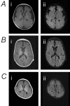
Brain MRI findings can vary from normal to showing thinning or hypoplasia of the corpus callosum to various malformations of cortical development (see Suggestive Findings, Brain MRI findings and Figure 1) [Smith et al 2018, Zaman et al 2020]. As some individuals with severe DEE are without MCD, the presence and extent of MCD does not appear to correlate with clinical phenotype. However, diffuse dysgyria is typically associated with severe/profound DEE (14/15; 93%), while focal unilateral or bilateral perisylvian polymicrogyria (PMG) is most often seen in those with a more mild phenotype ranging from normal neurocognitive function to mild/moderate ID, and no history or seizures or infrequent seizures only (11/13; 85%).

Figure 1.

Figure 1.

Magnetic resonance imaging scans of individuals with SCN3A-related neurodevelopmental disorder A. MRI of the brain of a person with profound global developmental delay and treatment-resistant epilepsy at age two years who had a de novo heterozygous p.Ile875Thr (more...)

Autonomic dysregulation. Ictal and/or non-ictal autonomic disturbances are observed in 45% (9/20) of individuals with DEE [Lamar et al 2017, Zaman et al 2020]. Features include episodes of the following:

  • Asymmetric or unilateral flushing / color change of the face, which may be confused with Harlequin syndrome
  • Brady- or tachycardia
  • Apnea and cyanosis with oxygen desaturation
  • Excessive sweating
  • Anisocoria with sluggish pupillary response to light

These episodes may occur several times per day and may or may not be associated with ictal abnormalities on EEG.

Oropharyngeal/Feeding

  • Oromotor dysfunction is seen in a majority of individuals [Smith et al 2018, Zaman et al 2020], and represents a prominent component of the clinical presentation in those with SCN3A-ND who have bilateral perisylvian PMG. Clinical features may include the following:
    • Abnormal tongue movements
    • Speech/language difficulties, including dysarthria
    • Difficulties whistling, blowing
    • Abnormal brisk jaw jerk
  • Some affected individuals with the severe SCN3A-ND phenotype exhibit chronic failure to thrive with G-tube dependence for feeding.

Other associated features in some individuals with DEE:

  • Progressive microcephaly
  • Dyskinesia, including choreoathetosis in those without MCD and dystonia and/or hyperkinetic movements in those with MCD [Zaman et al 2020]
  • Cortical visual impairment
  • Features of autism spectrum disorder
  • Early-childhood death in two individuals, one due to sudden unexpected death in epilepsy and the other of an undetermined cause [Zaman et al 2020]

Genotype-Phenotype Correlations

c.2624T>C (p.Ile875Thr) is the most common recurrent pathogenic variant, identified in 26% of individuals with SCN3A-ND [Miyatake et al 2018, Smith et al 2018, Zaman et al 2018, Li et al 2019, Zaman et al 2020]. The phenotype associated with this variant is relatively homogeneous, severe, and with poor outcomes from a developmental and epilepsy management perspective. Clinical features include:

  • Profound developmental delay / intellectual disability (in 10/10 individuals; 100%)
  • Neonatal onset intractable seizures (median onset 2 weeks) (8/9; 89%)
  • Multiple seizure types, often generalized tonic seizures
  • Diffuse, bilateral polymicrogyria (10/10; 100%)
  • Microcephaly (7/9;78%)
  • Paroxysmal ictal and non-ictal autonomic dysregulation (4/9; 44%)

A clear genotype-phenotype correlation between other variants or classes of variants has not yet been established.

Nomenclature

Outdated terms previously used to describe SCN3A-ND:

  • Cryptogenic pediatric partial epilepsy
  • Epilepsy, familial focal, with variable foci 4
  • Epileptic encephalopathy, early infantile, 62

Prevalence

The prevalence of SCN3A-ND is not known. Approximately 38 individuals have been reported in the literature to date. Due to the de novo nature of SCN3A pathogenic variants associated with DEE phenotypes, it is expected that the prevalence is consistent across populations.

Differential Diagnosis

The phenotype(s) observed in SCN3A-ND are not sufficient to diagnose an SCN3A-ND without genetic testing, as other conditions may be associated with clinical features and neuroimaging findings within the same range of phenotypes.

Epilepsy. The epilepsy phenotype of SCN3A-ND cannot be definitively distinguished from other causes of early-infantile epileptic encephalopathies (>80 genetic causes have been identified; see OMIM Phenotypic Series).

Malformation of cortical development (MCD). A key element of the differential diagnosis is to distinguish SCN3A-ND from other causes of MCD as these may be associated with a different clinical course and/or mode of inheritance. Other causes of MCD include:

Treatment-resistant epilepsy (with onset in the first month of life) plus MCD (pachy/polymicrogyria or dysgyria). Approximately 90% of individuals with SCN3A-ND have epilepsy with onset in the first year of life (mean age of onset is 2 weeks for individuals with early-onset epilepsy) [Zaman et al 2020]. Approximately 80% of individuals with SCN3A-ND have MCD [Miyatake et al 2018, Smith et al 2018, Zaman et al 2018, Li et al 2019, Zaman et al 2020]. Hence, the combination of treatment-resistant epilepsy with onset in the first month of life with MCD (pachy/polymicrogyria or dysgyria) is suggestive of SCN3A-ND.

SCN3A-ND can be distinguished from the most common epileptic encephalopathy, SCN1A-related Dravet syndrome (see SCN1A Seizure Disorders), by a typically earlier age of onset, a lack of clear temperature sensitivity, and the frequent co-occurrence of diffuse or multifocal MCD (see Table 3).

Table 3.

SCN3A-Related Neurodevelopmental Disorder and SCN1A-Related Dravet Syndrome

Clinical
Characteristic
SCN3A-NDSCN1A-Related Dravet Syndrome
Age of onset In ~75%, onset in 1st yr of life (mean age of onset in those w/early-onset epilepsy: 2 wks)Age 1-18 mos 1 w/most persons presenting at age 4-12 mos (median age ~5 mos) 2, although a rare, very severe early-onset form has been seen. 3
Temperature
sensitivity
Febrile seizures have been described in <10% of all persons w/SCN3A-ND. 4Dravet syndrome is defined by temperature-sensitive & febrile seizures.
Infantile spasms
syndrome
~15% of persons present w/infantile spasms. 5Rare 6
EEG findings Diffuse slowing; slow spike & wave discharges; multifocal or diffuse epileptiform discharges; hypsarrhythmia; burst suppressionAlthough findings vary, initial EEGs are often normal or show mild/nonspecific abnormalities & often evolve to incl generalized spike & wave or polyspike & wave accompanied by multifocal spikes.

SCN3A-ND can resemble the phenotypes seen in SCN2A-related encephalopathy (OMIM 613721) or SCN8A-related epilepsy with encephalopathy in the absence of brain malformation, although there are rare reports of individuals with SCN2A-related encephalopathy associated with MCD [Vlachou et al 2019, Allen et al 2021]. To date three individuals have been reported with SCN2A-related DEE with MCD. Given limited clinical information, detailed comparisons between SCN2A-related DEE with MCD and SCN3A-ND are not possible.

Treatable causes of early infantile epileptic encephalopathy. It is important to distinguish SCN3A-ND from treatable causes of early infantile epileptic encephalopathy including neurometabolic disorders and the following:

Management

Treatment of SCN3A-related neurodevelopmental disorder (SCN3A-ND) is best led by a physician familiar with pharmacotherapy of treatment-resistant epilepsy, such as a pediatric neurologist or pediatric epileptologist.

No clinical practice guidelines for SCN3A-ND have been published.

Evaluations Following Initial Diagnosis

To establish the extent of disease and needs in an individual diagnosed with SCN3A-ND, the evaluations summarized in Table 4 (if not performed as part of the evaluation that led to the diagnosis) are recommended.

Table 4.

Recommended Evaluations Following Initial Diagnosis in Individuals with SCN3A-Related Neurodevelopmental Disorder

System/ConcernEvaluationComment
Neurologic Neurologic eval
  • To incl brain MRI & EEG
  • Consider video EEG to assess seizure frequency, define seizure type(s), & assess for autonomic dysfunction. 1
  • To incl assessment for hyperkinetic movements
Development Developmental assessment
  • To incl motor, adaptive, cognitive, & speech/language eval
  • Eval for early intervention / special education
Psychiatric/
Behavioral
Neuropsychiatric evalIn persons age >12 mos: screen for behavior concerns incl sleep disturbances, ADHD, anxiety, &/or traits suggestive of ASD.
Musculoskeletal Orthopedics / physical medicine & rehab / PT & OT evalTo incl assessment of:
  • Gross motor & fine motor skills
  • Contractures & kyphoscoliosis
  • Mobility, activities of daily living, & need for adaptive devices
  • Need for PT (to improve gross motor skills) &/or OT (to improve fine motor skills)
Gastrointestinal/
Feeding
Gastroenterology / nutrition / feeding team eval
  • To incl eval of oromotor dysfunction & nutritional status
  • Consider eval for gastric tube placement in persons w/dysphagia &/or aspiration risk.
Eyes Ophthalmologic evalTo assess for ↓ vision
Genetic
counseling
By genetics professionals 2To inform affected persons & their families re nature, MOI, & implications of SCN3A-ND to facilitate medical & personal decision making
Family support
& resources
Assess need for:

MOI = mode of inheritance; OT = occupational therapy; PT = physical therapy

1.

Continuous video EEG may be used to help clarify events of unclear nature, such as autonomic manifestations [Zaman et al 2020] or myoclonus.

2.

Medical geneticist, certified genetic counselor, certified advanced genetic nurse

Treatment of Manifestations

Table 5.

Treatment of Manifestations in Individuals with SCN3A-Related Neurodevelopmental Disorder

Manifestation/
Concern
TreatmentConsiderations/Other
Epilepsy Empiric treatment of seizures w/standard ASM 1
  • Incl lacosamide, phenytoin, & carbamazepine 2, 3
  • No one ASM has been shown to be more efficacious than another.
  • Education of parents/caregivers 4
Hyperkinetic
movements
Standard treatment per neurologist
Autonomic
dysfunction
DD/ID See Developmental Delay / Intellectual Disability Management Issues.
Spasticity Orthopedics / physical medicine & rehab / PT & OT incl stretching to help avoid contractures & fallsConsider need for positioning & mobility devices, disability parking placard.
Poor weight
gain / FTT
Feeding therapy; gastrostomy tube placement may be required for persistent feeding issues.Low threshold for clinical feeding eval &/or radiographic swallowing study if clinical signs or symptoms of dysphagia
Central visual
impairment
No specific treatment; early intervention to stimulate visual development
Family/
Community
  • Ensure appropriate social work involvement to connect families w/local resources, respite, & support.
  • Coordinate care to manage multiple subspecialty appointments, equipment, medications, & supplies.
  • Ongoing assessment of need for home nursing
  • Consider involvement in adaptive sports or Special Olympics.

ASM = anti-seizure medication; DD/ID = developmental delay / intellectual disability; FTT = failure to thrive; OT = occupational therapy; PT = physical therapy

1.

Based on the finding that many/most variants (>90%) associated with epileptic encephalopathy in cases of SCN3A-related neurodevelopmental disorders were shown to exhibit gain of channel function [Zaman et al 2018, Zaman et al 2020], therapeutic trials of anti-seizure medications that target sodium channels may be indicated [Brunklaus 2020, Musto et al 2020]. However, clinical data on treatment efficacy are limited at this time.

2.
3.

Of the 15 affected individuals in the Zaman et al [2020] study for whom detailed treatment data were available, six were treated at some point in the disease course with anti-seizure drugs with a prominent mechanism of action of sodium channel blockade (oxcarbazepine, phenytoin, lacosamide, lamotrigine), and these drugs alone or in various combinations were ineffective at achieving seizure freedom or marked reduction in seizure frequency.

4.

Education of parents/caregivers regarding common seizure presentations is appropriate. For information on non-medical interventions and coping strategies for children diagnosed with epilepsy, see Epilepsy Foundation Toolbox.

Developmental Delay / Intellectual Disability Management Issues

The following information represents typical management recommendations for individuals with developmental delay / intellectual disability in the United States; standard recommendations may vary from country to country.

Ages 0-3 years. Referral to an early intervention program is recommended for access to occupational, physical, speech, and feeding therapy as well as infant mental health services, special educators, and sensory impairment specialists. In the US, early intervention is a federally funded program available in all states that provides in-home services to target individual therapy needs.

Ages 3-5 years. In the US, developmental preschool through the local public school district is recommended. Before placement, an evaluation is made to determine needed services and therapies and an individualized education plan (IEP) is developed for those who qualify based on established motor, language, social, or cognitive delay. The early intervention program typically assists with this transition. Developmental preschool is center based; for children too medically unstable to attend, home-based services are provided.

All ages. Consultation with a developmental pediatrician is recommended to ensure the involvement of appropriate community, state, and educational agencies (US) and to support parents in maximizing quality of life. Some issues to consider:

  • IEP services:
    • An IEP provides specially designed instruction and related services to children who qualify.
    • IEP services will be reviewed annually to determine whether any changes are needed.
    • Special education law requires that children participating in an IEP be in the least restrictive environment feasible at school and included in general education as much as possible, when and where appropriate.
    • Vision and hearing consultants should be a part of the child's IEP team to support access to academic material.
    • PT, OT, and speech services will be provided in the IEP to the extent that the need affects the child's access to academic material. Beyond that, private supportive therapies based on the affected individual's needs may be considered. Specific recommendations regarding type of therapy can be made by a developmental pediatrician.
    • As a child enters the teen years, a transition plan should be discussed and incorporated in the IEP. For those receiving IEP services, the public school district is required to provide services until age 21.
  • A 504 plan (Section 504: a US federal statute that prohibits discrimination based on disability) can be considered for those who require accommodations or modifications such as front-of-class seating, assistive technology devices, classroom scribes, extra time between classes, modified assignments, and enlarged text.
  • Developmental Disabilities Administration (DDA) enrollment is recommended. DDA is a US public agency that provides services and support to qualified individuals. Eligibility differs by state but is typically determined by diagnosis and/or associated cognitive/adaptive disabilities.
  • Families with limited income and resources may also qualify for supplemental security income (SSI) for their child with a disability.

Motor Dysfunction

Gross motor dysfunction

  • Physical therapy is recommended to maximize mobility and to reduce the risk for later-onset orthopedic complications (e.g., contractures, scoliosis, hip dislocation).
  • Consider use of durable medical equipment and positioning devices as needed (e.g., wheelchairs, walkers, bath chairs, orthotics, adaptive strollers).
  • For muscle tone abnormalities including hypertonia or dystonia, consider involving appropriate specialists to aid in management of baclofen, tizanidine, Botox®, anti-parkinsonian medications, or orthopedic procedures.

Fine motor dysfunction. Occupational therapy is recommended for difficulty with fine motor skills that affect adaptive function such as feeding, grooming, dressing, and writing.

Oral motor dysfunction should be assessed at each visit and clinical feeding evaluations and/or radiographic swallowing studies should be obtained for choking/gagging during feeds, poor weight gain, frequent respiratory illnesses, or feeding refusal that is not otherwise explained. Assuming that the child is safe to eat by mouth, feeding therapy (typically from an occupational or speech therapist) is recommended to help improve coordination or sensory-related feeding issues. Feeds can be thickened or chilled for safety. When feeding dysfunction is severe, an NG-tube or G-tube may be necessary.

Communication issues. Consider evaluation for alternative means of communication (e.g., augmentative and alternative communication [AAC]) for individuals who have expressive language difficulties. An AAC evaluation can be completed by a speech-language pathologist who has expertise in the area. The evaluation will consider cognitive abilities and sensory impairments to determine the most appropriate form of communication. AAC devices can range from low-tech, such as picture exchange communication, to high-tech, such as voice-generating devices. Contrary to popular belief, AAC devices do not hinder verbal development of speech, but rather support optimal speech and language development.

Social/Behavioral Concerns

Children may qualify for and benefit from interventions used in treatment of autism spectrum disorder, including applied behavior analysis (ABA). ABA therapy is targeted to the individual child's behavioral, social, and adaptive strengths and weaknesses and typically performed one on one with a board-certified behavior analyst.

Consultation with a developmental pediatrician may be helpful in guiding parents through appropriate behavior management strategies or providing prescription medications, such as medication used to treat attention-deficit/hyperactivity disorder (ADHD), when necessary.

Concerns about serious aggressive or destructive behavior can be addressed by a pediatric psychiatrist.

Surveillance

Table 6.

Recommended Surveillance for Individuals with SCN3A-Related Neurodevelopmental Disorder

System/ConcernEvaluationFrequency
Constitutional/
Growth
Measurement of weight, length/height, & head circumferenceAt each visit
Neurologic 1
  • Monitor those w/seizures as clinically indicated. 2
  • Monitor for signs & symptoms of a movement disorder & of autonomic dysfunction.
Development 1 Monitor developmental progress & educational needs.
Psychiatric/
Behavioral
Behavioral assessment for anxiety, attention, & aggressive or self-injurious behavior
Musculoskeletal Physical medicine, OT/PT assessment of mobility, self-help skills
Eyes Ophthalmology eval to assess for visual impairmentAnnually
Miscellaneous/
Other
Assess family need for social work support (e.g., palliative/respite care, home nursing, other local resources) & care coordination.At each visit

OT = occupational therapy; PT = physical therapy

1.

Serial neurologic examination and neurodevelopmental assessment is appropriate.

2.

EEG monitoring is appropriate when new or different seizure type(s) are suspected.

Agents/Circumstances to Avoid

There is NO evidence that:

  • Specific anti-seizure medications can worsen seizures associated with SCN3A-N;
  • Sleep deprivation or fever exacerbates seizures associated with SCN3A-ND.

Evaluation of Relatives at Risk

It is appropriate to clarify the genetic status of apparently asymptomatic younger at-risk relatives of an affected individual in order to identify as early as possible those who would benefit from early treatment.

See Genetic Counseling for issues related to testing of at-risk relatives for genetic counseling purposes.

Pregnancy Management

In general, women with epilepsy or a seizure disorder of any cause are at greater risk for mortality during pregnancy than pregnant women without a seizure disorder; use of anti-seizure medication during pregnancy reduces this risk. However, exposure to anti-seizure medication may increase the risk for adverse fetal outcome (depending on the drug used, the dose, and the stage of pregnancy at which medication is taken). Nevertheless, the risk of an adverse outcome to the fetus from anti-seizure medication exposure is often less than that associated with exposure to an untreated maternal seizure disorder. Therefore, use of anti-seizure medication to treat a maternal seizure disorder during pregnancy is typically recommended. Discussion of the risks and benefits of using a given anti-seizure drug during pregnancy should ideally take place prior to conception. Transitioning to a lower-risk medication prior to pregnancy may be possible [Sarma et al 2016].

See MotherToBaby for further information on medication use during pregnancy.

Therapies Under Investigation

Therapeutic trials of anti-seizure medications that target sodium channels may be indicated in cases of severe, treatment-resistant epilepsy or epileptic encephalopathy associated with SCN3A-ND based on theoretic considerations [Zaman et al 2018, Brunklaus 2020, Musto et al 2020, Zaman et al 2020].

Search ClinicalTrials.gov in the US and EU Clinical Trials Register in Europe for access to information on clinical studies for a wide range of diseases and conditions. Note: There may not be clinical trials for this disorder.

Genetic Counseling

Genetic counseling is the process of providing individuals and families with information on the nature, mode(s) of inheritance, and implications of genetic disorders to help them make informed medical and personal decisions. The following section deals with genetic risk assessment and the use of family history and genetic testing to clarify genetic status for family members; it is not meant to address all personal, cultural, or ethical issues that may arise or to substitute for consultation with a genetics professional. —ED.

Mode of Inheritance

SCN3A-ND is inherited in an autosomal dominant manner.

Risk to Family Members

Parents of a proband

  • All probands with SCN3A-related developmental and epileptic encephalopathy (DEE) reported to date have represented simplex cases (i.e., the only affected family member) and are presumed to have the disorder as the result of a de novo pathogenic variant.
  • Vertical transmission of an SCN3A pathogenic variant from an affected parent to an affected child has been reported in one family. In this family, the proband inherited a pathogenic SCN3A variant from a similarly affected parent [Zaman et al 2020]. The affected parent had bilateral perisylvian polymicrogyria, a single generalized tonic-clonic seizure at age five years, oromotor dysfunction, and mild intellectual disability.
  • Transmission of an SCN3A pathogenic variant from an apparently unaffected parent to a proband has been reported in three families [Smith et al 2018, Zaman et al 2020]. (Note: True penetrance estimates cannot be calculated based on these families because the apparently asymptomatic heterozygous family members did not undergo complete neurologic, neuroimaging, and EEG examinations to evaluate for more subtle presentations of disease.)
  • Molecular genetic testing is recommended for the parents of the proband to confirm their genetic status and to allow reliable recurrence risk counseling.
  • If the pathogenic variant identified in the proband is not identified in either parent, the following possibilities should be considered:
    • The proband has a de novo pathogenic variant. Note: A pathogenic variant is reported as "de novo" if: (1) the pathogenic variant found in the proband is not detected in parental DNA; and (2) parental identity testing has confirmed biological maternity and paternity. If parental identity testing is not performed, the variant is reported as "assumed de novo" [Richards et al 2015].
    • The proband inherited a pathogenic variant from a parent with germline (or somatic and germline) mosaicism. Note: Testing of parental leukocyte DNA may not detect all instances of somatic mosaicism and will not detect a pathogenic variant that is present in the germ cells only.
      Parental mosaicism not been reported in SCN3A-ND to date; however, it is estimated that approximately 10% of individuals (who represent simplex cases) with any form of genetic developmental and epileptic encephalopathy have the disorder as the result of a pathogenic variant inherited from an asymptomatic, mosaic parent [Myers et al 2018].
  • The family history of some individuals diagnosed with SCN3A-ND may appear to be negative because of failure to recognize the disorder in family members (possibly because family members have not undergone neuroimaging or a thorough neurologic examination to evaluate for oromotor dysfunction). Therefore, an apparently negative family history cannot be confirmed unless molecular genetic testing has demonstrated that neither parent is heterozygous for the pathogenic variant identified in the proband.

Sibs of a proband. The risk to the sibs of the proband depends on the genetic status of the proband's parents:

  • If a parent of the proband is affected and/or is known to have the SCN3A pathogenic variant identified in the proband, the risk to the sibs of inheriting the pathogenic variant is 50%.
  • The likelihood that a sib who inherits an SCN3A pathogenic variant will have manifestations similar to those of the proband is unknown. Although three multiplex families have been reported with heterozygous family members with no apparent manifestations of SCN3A-ND [Smith et al 2018, Zaman et al 2020], penetrance cannot be estimated based on these families because the purportedly asymptomatic heterozygous family members did not undergo detailed evaluation (including neurologic examination, neuroimaging, and EEG), which would detect milder manifestations of the disorder such as early speech/language delay and focal brain malformation.
  • If the proband has a known SCN3A pathogenic variant that cannot be detected in the leukocyte DNA of either parent, the recurrence risk to sibs is estimated to be 1%-2% because of the theoretic possibility of parental germline mosaicism [Rahbari et al 2016]. Although parental mosaicism has not been reported in SCN3A-ND, it is estimated that approximately 10% of individuals (who represent simplex cases) with any form of genetic developmental and epileptic encephalopathy have the disorder as the result of a pathogenic variant inherited from an asymptomatic, mosaic parent [Myers et al 2018].
  • If the parents have not been tested for the SCN3A pathogenic variant but have undergone detailed evaluation (including neurologic examination, neuroimaging, and EEG) and are clinically unaffected, the risk to the sibs of a proband appears to be low; however, sibs are still presumed to be at increased risk for SCN3A-ND because of the possibility of reduced penetrance in a heterozygous parent or the theoretic possibility of parental germline mosaicism.

Offspring of a proband. Each child of an individual with SCN3A-ND has a 50% chance of inheriting the pathogenic variant.

Other family members. The risk to other family members depends on the status of the proband's parents: if a parent has the SCN3A pathogenic variant, the parent's family members may be at risk.

Related Genetic Counseling Issues

See Management, Evaluation of Relatives at Risk for information on evaluating at-risk relatives for the purpose of early diagnosis and treatment.

Family planning

  • The optimal time for determination of genetic risk and discussion of the availability of prenatal/preimplantation genetic testing is before pregnancy.
  • It is appropriate to offer genetic counseling (including discussion of potential risks to offspring and reproductive options) to parents of affected individuals and young adults who are affected.

DNA banking. Because it is likely that testing methodology and our understanding of genes, pathogenic mechanisms, and diseases will improve in the future, consideration should be given to banking DNA from probands in whom a molecular diagnosis has not been confirmed (i.e., the causative pathogenic mechanism is unknown). For more information, see Huang et al [2022].

Prenatal Testing and Preimplantation Genetic Testing

Once the SCN3A pathogenic variant has been identified in an affected family member, prenatal and preimplantation genetic testing are possible.

Differences in perspective may exist among medical professionals and within families regarding the use of prenatal testing. While most centers would consider use of prenatal testing to be a personal decision, discussion of these issues may be helpful.

Resources

GeneReviews staff has selected the following disease-specific and/or umbrella support organizations and/or registries for the benefit of individuals with this disorder and their families. GeneReviews is not responsible for the information provided by other organizations. For information on selection criteria, click here.

Molecular Genetics

Information in the Molecular Genetics and OMIM tables may differ from that elsewhere in the GeneReview: tables may contain more recent information. —ED.

Table A.

SCN3A-Related Neurodevelopmental Disorder: Genes and Databases

Data are compiled from the following standard references: gene from HGNC; chromosome locus from OMIM; protein from UniProt. For a description of databases (Locus Specific, HGMD, ClinVar) to which links are provided, click here.

Table B.

OMIM Entries for SCN3A-Related Neurodevelopmental Disorder (View All in OMIM)

182391SODIUM VOLTAGE-GATED CHANNEL, ALPHA SUBUNIT 3; SCN3A
617935EPILEPSY, FAMILIAL FOCAL, WITH VARIABLE FOCI 4; FFEVF4
617938DEVELOPMENTAL AND EPILEPTIC ENCEPHALOPATHY 62; DEE62

Molecular Pathogenesis

SCN3A encodes the type III voltage-gated sodium (Na+) α subunit Nav1.3. Voltage-gated Na+ channels are molecular complexes formed by one pore-forming α-subunit, two non-pore-forming auxiliary β-subunits, and accessory proteins that regulate localization, cell surface expression, and channel physiology. The α subunits – including Nav1.3 – are 24-pass transmembrane proteins with four domains (DI-IV), each of which contains six subunits (S1-6); subunit 4 of each domain is known to be critical for sensing transmembrane voltage (the "voltage sensor") and transducing this into mechanical changes that alter channel gating. Subunits S5-6 of each domain form the channel pore through which Na+ flux occurs [Catterall 2000, Catterall 2012].

Pathogenic missense variants that cause SCN3A-related neurodevelopmental disorder are located throughout the Nav1.3 channel protein, although the pathogenic variants identified in cases of severe or profound developmental disability appear to be clustered in S4-6 of DII-IV (Figure 2) [Miyatake et al 2018, Zaman et al 2018, Inuzuka et al 2020, Zaman et al 2020].

Figure 2. . Schematic of the Nav1.

Figure 2.

Schematic of the Nav1.3 protein showing pathogenic variants associated with SCN3A-ND Shown are locations of all missense variants corresponding to disease-associated pathogenic or likely pathogenic variants in SCN3A, with the number of cases indicated (more...)

SCN3A is known to be expressed in the developing brain of rodents [Beckh et al 1989, Felts et al 1997] and humans [Whitaker et al 2000] including in radial glia, in intermediate progenitor cells, and in developing neurons [Pollen et al 2015, Smith et al 2018]. In utero electroporation of disease-associated variant SCN3A DNA into fetal ferret brain produced polymicrogyria. How pathogenic variants in a Na+ channel might lead to malformation of cortical development (MCD) such as polymicrogyria, pachygyria, or other dysgyria, is not known, but may relate to abnormal electrogenesis in developing/migrating neurons or neuronal progenitors.

Pathogenic variants in SCN3A lead to alterations in the amino acid sequence of Nav1.3 and have been shown to alter the biophysical function of the channel in various ways. Electrophysiologic recordings in heterologous systems demonstrate increased slowly-inactivating ("persistent") Na+ current and impaired fast inactivation, consistent with gain of function at the ion channel level. How dysfunction at the channel level impacts the function of neurons and neuronal circuits to produce seizures and epilepsy is unclear.

Mechanism of disease causation. Gain of function

Table 7.

Notable SCN3A Pathogenic Variants

Reference SequencesDNA Nucleotide
Change
Predicted
Protein Change
Comment
NM_006922​.3
NP_008853​.3
c.2624T>Cp.Ile875ThrMost common recurrent pathogenic variant; assoc w/treatment-resistant seizures, severe/profound DD/ID, & malformation of cortical development (pachy- or polymicrogyria)

DD/ID = developmental delay / intellectual disability

Variants listed in the table have been provided by the authors. GeneReviews staff have not independently verified the classification of variants.

GeneReviews follows the standard naming conventions of the Human Genome Variation Society (varnomen​.hgvs.org). See Quick Reference for an explanation of nomenclature.

Chapter Notes

Author Notes

Ethan M Goldberg is Assistant Professor in the Division of Neurology at the Children's Hospital of Philadelphia and Department of Neurology & Neuroscience at the University of Pennsylvania Perelman School of Medicine and Attending Physician in the Neurogenetics Clinic at the Children's Hospital of Philadelphia. His laboratory studies mechanisms of epilepsy in experimental models of genetic epilepsies including of SCN3A-related neurodevelopmental disorder.

The Epilepsy Neurogenetics Initiative at Children's Hospital of Philadelphia integrates genetic testing into the comprehensive care plan of children with difficult-to-treat or unexplained epilepsies and provides access to expert care for children with genetic epilepsy syndromes. Our team of child neurologists and genetic counselors has particular expertise in the clinical care of children with SCN3A-related neurodevelopmental disorder. More information can be found at www.chop.edu.

Revision History

  • 4 November 2021 (ma) Revision: frequency of de novo SCN3A-ND clarified (Risk to Family Members)
  • 3 June 2021 (ma) Review posted live
  • 9 November 2020 (klh) Original submission

References

Literature Cited

  • Allen AS, Aggarwal V, Berkovic SF, Cossette P, Delanty N, Dlugos D, Eichler EE, Epstein MP, Freyer C, Goldstein DB, et al. Epilepsy Phenome/Genome Project, Epi4K Consortium. Diverse genetic causes of polymicrogyria with epilepsy. Epilepsia. 2021;62:973–83. [PMC free article: PMC10838185] [PubMed: 33818783]
  • Beckh S, Noda M, Lübbert H, Numa S. Differential regulation of three sodium channel messenger RNAs in the rat central nervous system during development. EMBO J. 1989;8:3611–6. [PMC free article: PMC402042] [PubMed: 2555170]
  • Boles RG, Pober BR, Gibson LH, Willis CR, McGrath J, Roberts DJ, Yang-Feng TL. Deletion of chromosome 2q24-q31 causes characteristic digital anomalies: case report and review. Am J Med Genet. 1995;55:155–60. [PubMed: 7717414]
  • Brunklaus A. Precision medicine in sodium channelopathies – moving beyond seizure control towards disease modification. Eur J Paediatr Neurol. 2020;24:7. [PubMed: 31980364]
  • Catterall WA. From ionic currents to molecular mechanisms: the structure and function of voltage-gated sodium channels. Neuron. 2000;26:13–25. [PubMed: 10798388]
  • Catterall WA. Voltage-gated sodium channels at 60: structure, function and pathophysiology. J Physiol. 2012;590:2577–89. [PMC free article: PMC3424717] [PubMed: 22473783]
  • Felts PA, Yokoyama S, Dib-Hajj S, Black JA, Waxman SG. Sodium channel alpha-subunit MRNAs I, II, III, NaG, Na6 and HNE (PN1): different expression patterns in developing rat nervous system. Brain Res Mol Brain Res. 1997;45:71–82. [PubMed: 9105672]
  • Goeggel Simonetti B, Rieubland C, Courage C, Strozzi S, Tschumi S, Gallati S, Lemke JR. Duplication of the sodium channel gene cluster on 2q24 in children with early onset epilepsy. Epilepsia. 2012;53:2128–34. [PubMed: 23016767]
  • Grosso S, Orrico A, Galli L, Di Bartolo R, Sorrentino V, Balestri P. SCN1A mutation associated with atypical Panayiotopoulos syndrome. Neurology. 2007;69:609–11. [PubMed: 17679682]
  • Huang SJ, Amendola LM, Sternen DL. Variation among DNA banking consent forms: points for clinicians to bank on. J Community Genet. 2022;13:389–97. [PMC free article: PMC9314484] [PubMed: 35834113]
  • Inuzuka LM, Macedo-Souza LI, Della-Ripa B, Cabral KS, Monteiro F, Kitajima JP, de Souza Godoy LF, de Souza Delgado D, Kok F, Garzon E. Neurodevelopmental disorder associated with de novo SCN3A pathogenic variants: two new cases and review of the literature. Brain Dev. 2020;42:211–16. [PubMed: 31677917]
  • Iossifov I, O’Roak BJ, Sanders SJ, Ronemus M, Krumm N, Levy D, Stessman HA, Witherspoon KT, Vives L, Patterson KE, Smith JD, Paeper B, Nickerson DA, Dea J, Dong S, Gonzalez LE, Mandell JD, Mane SM, Murtha MT, Sullivan CA, Walker MF, Waqar Z, Wei L, Willsey AJ, Yamrom B, Lee YH, Grabowska E, Dalkic E, Wang Z, Marks S, Andrews P, Leotta A, Kendall J, Hakker I, Rosenbaum J, Ma B, Rodgers L, Troge J, Narzisi G, Yoon S, Schatz MC, Ye K, McCombie WR, Shendure J, Eichler EE, State MW, Wigler M. The contribution of de novo coding mutations to autism spectrum disorder. Nature. 2014;515:216–21. [PMC free article: PMC4313871] [PubMed: 25363768]
  • Lamar T, Vanoye CG, Calhoun J, Wong JC, Dutton SB, Jorge BS, Velinov M, Escayg A, Kearney JA. SCN3A deficiency associated with increased seizure susceptibility. Neurobiol Dis. 2017;102:38–48. [PMC free article: PMC5446790] [PubMed: 28235671]
  • Li J, Gao K, Yan H, Xiangwei W, Liu N, Wang T, Xu H, Lin Z, Xie H, Wang J, Wu Y, Jiang Y. Reanalysis of whole exome sequencing data in patients with epilepsy and intellectual disability/mental retardation. Gene. 2019;700:168–75. [PubMed: 30904718]
  • Miyatake S, Kato M, Sawaishi Y, Saito T, Nakashima M, Mizuguchi T, Mitsuhashi S, Takata A, Miyake N, Saitsu H, Matsumoto N. Recurrent SCN3A p.Ile875Thr variant in patients with polymicrogyria. Ann Neurol. 2018;84:159–61. [PubMed: 29740860]
  • Musto E, Gardella E, Møller RS. Recent advances in treatment of epilepsy-related sodium channelopathies. Eur J Paediatr Neurol. 2020;24:123–128. [PubMed: 31889633]
  • Myers CT, Hollingsworth G, Muir AM, Schneider AL, Thuesmunn Z, Knupp A, King C, Lacroix A, Mehaffey MG, Berkovic SF, Carvill GL, Sadleir LG, Scheffer IE, Mefford HC. Parental mosaicism in "de novo" epileptic encephalopathies. New Engl J Med. 2018;378:1646–8. [PMC free article: PMC5966016] [PubMed: 29694806]
  • Okumura A, Yamamoto T, Shimojima K, Honda Y, Abe S, Ikeno M, Shimizu T. Refractory neonatal epilepsy with a de novo duplication of chromosome 2q24.2q24.3. Epilepsia. 2011;52:e66–9. [PubMed: 21692795]
  • Pescucci C, Caselli R, Grosso S, Mencarelli MA, Mari F, Farnetani MA, Piccini B, Artuso R, Bruttini M, Priolo M, Zuffardi O, Gimelli S, Balestri P, Renieri A. 2q24-q31 deletion: report of a case and review of the literature. Eur J Med Genet. 2007;50:21–32. [PubMed: 17088112]
  • Pollen AA, Nowakowski TJ, Chen J, Retallack H, Sandoval-Espinosa C, Nicholas CR, Shuga J, Liu SJ, Oldham MC, Diaz A, Lim DA, Leyrat AA, West JA, Kriegstein AR. Molecular identity of human outer radial glia during cortical development. Cell. 2015;163:55–67. [PMC free article: PMC4583716] [PubMed: 26406371]
  • Rahbari R, Wuster A, Lindsay SJ, Hardwick RJ, Alexandrov LB, Al Turki S, Dominiczak A, et al. Timing, rates and spectra of human germline mutation. Nat Genet. 2016;48:126–33. [PMC free article: PMC4731925] [PubMed: 26656846]
  • Richards S, Aziz N, Bale S, Bick D, Das S, Gastier-Foster J, Grody WW, Hegde M, Lyon E, Spector E, Voelkerding K, Rehm HL, et al. Standards and guidelines for the interpretation of sequence variants: a joint consensus recommendation of the American College of Medical Genetics and Genomics and the Association for Molecular Pathology. Genet Med. 2015;17:405–24. [PMC free article: PMC4544753] [PubMed: 25741868]
  • Sadleir LG, Mountier EI, Gill D, Davis S, Joshi C, Devile C, Kurian MA, Mandelstam S, Wirrell E, Nickels KC, Murali HR, Carvill G, Myers CT, Mefford HC, Scheffer IE, et al. Not all SCN1A epileptic encephalopathies are Dravet syndrome. Neurology. 2017;89:1035–42. [PMC free article: PMC5589790] [PubMed: 28794249]
  • Sarma AK, Khandker N, Kurczewski L, Brophy GM. Medical management of epileptic seizures: challenges and solutions. Neuropsychiatr Dis Treat. 2016;12:467–85. [PMC free article: PMC4771397] [PubMed: 26966367]
  • Smith RS, Kenny CJ, Ganesh V, Jang A, Borges-Monroy R, Partlow JN, Hill RS, Shin T, Chen AY, Doan RN, Anttonen AK, Ignatius J, Medne L, Bönnemann CG, Hecht JL, Salonen O, Barkovich AJ, Poduri A, Wilke M, de Wit MCY, Mancini GMS, Sztriha L, Im K, Amrom D, Andermann E, Paetau R, Lehesjoki AE, Walsh CA, Lehtinen MK. Sodium channel SCN3A (NaV1.3) regulation of human cerebral cortical folding and oral motor development. Neuron. 2018;99:905–913.e7. [PMC free article: PMC6226006] [PubMed: 30146301]
  • Takayama R, Fujiwara T, Shigematsu H, Imai K, Takahashi Y, Yamakawa K, Inoue Y. Long-term course of Dravet syndrome: a study from an epilepsy center in Japan. Epilepsia. 2014;55:528–38. [PubMed: 24502503]
  • Trujillano D, Bertoli-Avella AM, Kandaswamy KK, Weiss MER, Köster J, Marais A, Paknia O, Schröder R, Garcia-Aznar JM, Werber M, Brandau O, Calvo Del Castillo M, Baldi C, Wessel K, Kishore S, Nahavandi N, Eyaid W, Al Rifai MT, Al-Rumayyan A, Al-Twaijri W, Alothaim A, Alhashem A, Al-Sannaa N, Al-Balwi M, Alfadhel M, Rolfs A, Abou Jamra R. Clinical exome sequencing: results from 2819 samples reflecting 1000 families. Eur J Hum Genet. 2017;25:176–82. [PMC free article: PMC5255946] [PubMed: 27848944]
  • Tumienė B, Maver A, Writzl K, Hodžić A, Čuturilo G, Kuzmanić-Šamija R, Čulić V, Peterlin B. Diagnostic exome sequencing of syndromic epilepsy patients in clinical practice. Clin Genet. 2018;93:1057–62. [PubMed: 29286531]
  • Vlachou V, Larsen L, Pavlidou E, Ismayilova N, Mazarakis ND, Scala M, Pantazi M, Mankad K, Kinali M. SCN2A mutation in an infant with Ohtahara syndrome and neuroimaging findings: expanding the phenotype of neuronal migration disorders. J Genet. 2019;98:54. [PubMed: 31204721]
  • Wallace RH, Hodgson BL, Grinton BE, Gardiner RM, Robinson R, Rodriguez-Casero V, Sadleir L, Morgan J, Harkin LA, Dibbens LM, Yamamoto T, Andermann E, Mulley JC, Berkovic SF, Scheffer IE. Sodium channel A1-subunit mutations in severe myoclonic epilepsy of infancy and infantile spasms. Neurology. 2003;61:765–69. [PubMed: 14504318]
  • Whitaker WR, Clare JJ, Powell AJ, Chen YH, Faull RL, Emson PC. Distribution of voltage-gated sodium channel alpha-subunit and beta-subunit MRNAs in human hippocampal formation, cortex, and cerebellum. J Comp Neurol. 2000;422:123–39. [PubMed: 10842222]
  • Wirrell EC, Laux L, Donner E, Jette N, Knupp K, Meskis MA, Miller I, Sullivan J, Welborn M, Berg AT. Optimizing the diagnosis and management of Dravet syndrome: recommendations from a North American consensus panel. Pediatr Neurol. 2017;68:18–34.e3. [PubMed: 28284397]
  • Yoshitomi S, Takahashi Y, Ishizuka M, Yamaguchi T, Watanabe A, Nasu H, Ueda Y, Ohtani H, Ikeda H, Imai K, Shigematsu H, Inoue Y, Tanahashi Y, Aiba K, Ohta H, Shimada S, Yamamoto T. Three patients manifesting early infantile epileptic spasms associated with 2q24.3 microduplications. Brain Dev. 2015;37:874–9. [PubMed: 25843248]
  • Zaman T, Helbig I, Babić Božovi I, DeBrosse SD, Bergqvist AC, Wallis K, Medne L, Maver A, Peterlin B, Helbig KL, Zhang X, Goldberg EM. Mutations in SCN3A cause early infantile epileptic encephalopathy. Ann Neurol. 2018;83:703–17. [PMC free article: PMC5912987] [PubMed: 29466837]
  • Zaman T, Helbig KL, Clatot J, Thompson CH, Kang SK, Stouffs K, Jansen AE, Verstraete L, Jacquinet A, Parrini E, Guerrini R, Fujiwara Y, Miyatake S, Ben-Zeev B, Bassan H, Reish O, Marom D, Hauser N, Vu TA, Ackermann S, Spencer CE, Lippa N, Srinivasan S, Charzewska A, Hoffman-Zacharska D, Fitzpatrick D, Harrison V, Vasudevan P, Joss S, Pilz DT, Fawcett KA, Helbig I, Matsumoto N, Kearney JA, Fry AE, Goldberg EM. SCN3A-related neurodevelopmental disorder: a spectrum of epilepsy and brain malformation. Ann Neurol. 2020;88:348–62. [PMC free article: PMC8552104] [PubMed: 32515017]
  • Ziats MN, Ahmad A, Bernat JA, Fisher R, Glassford M, Hannibal MC, Jacher JE, Weiser N, Keegan CE, Lee KN, Marzulla TB, O'Connor BC, Quinonez SC, Seemann L, Turner L, Bielas S, Harris NL, Ogle JD, Innis JW, Martin DM. Genotype–phenotype analysis of 523 patients by genetics evaluation and clinical exome sequencing. Pediatr Res. 2020;87:735–39. [PMC free article: PMC7082194] [PubMed: 31618753]
Copyright © 1993-2024, University of Washington, Seattle. GeneReviews is a registered trademark of the University of Washington, Seattle. All rights reserved.

GeneReviews® chapters are owned by the University of Washington. Permission is hereby granted to reproduce, distribute, and translate copies of content materials for noncommercial research purposes only, provided that (i) credit for source (http://www.genereviews.org/) and copyright (© 1993-2024 University of Washington) are included with each copy; (ii) a link to the original material is provided whenever the material is published elsewhere on the Web; and (iii) reproducers, distributors, and/or translators comply with the GeneReviews® Copyright Notice and Usage Disclaimer. No further modifications are allowed. For clarity, excerpts of GeneReviews chapters for use in lab reports and clinic notes are a permitted use.

For more information, see the GeneReviews® Copyright Notice and Usage Disclaimer.

For questions regarding permissions or whether a specified use is allowed, contact: ude.wu@tssamda.

Bookshelf ID: NBK570882PMID: 34081427

Views

Tests in GTR by Gene

Related information

  • OMIM
    Related OMIM records
  • PMC
    PubMed Central citations
  • PubMed
    Links to PubMed
  • Gene
    Locus Links

Similar articles in PubMed

See reviews...See all...

Recent Activity

Your browsing activity is empty.

Activity recording is turned off.

Turn recording back on

See more...