U.S. flag

An official website of the United States government

NCBI Bookshelf. A service of the National Library of Medicine, National Institutes of Health.

Adam MP, Feldman J, Mirzaa GM, et al., editors. GeneReviews® [Internet]. Seattle (WA): University of Washington, Seattle; 1993-2024.

Cover of GeneReviews®

GeneReviews® [Internet].

Show details

FLNA Deficiency

, MD, MMSc and , MD, PhD.

Author Information and Affiliations

Initial Posting: ; Last Update: September 30, 2021.

Estimated reading time: 29 minutes

Summary

Clinical characteristics.

FLNA deficiency is associated with a phenotypic spectrum that includes FLNA-related periventricular nodular heterotopia (Huttenlocher syndrome), congenital heart disease (patent ductus arteriosus, atrial and ventricular septal defects), valvular dystrophy, dilatation and rupture of the thoracic aortic, pulmonary disease (pulmonary hypertension, alveolar hypoplasia, emphysema, asthma, chronic bronchitis), gastrointestinal dysmotility and obstruction, joint hypermobility, and macrothrombocytopenia.

Diagnosis/testing.

The diagnosis of FLNA deficiency is established in a proband by identification of a heterozygous FLNA pathogenic variant in a female or a hemizygous FLNA pathogenic variant in a male.

Management.

Treatment of manifestations: Treatment of epilepsy generally follows principles for a seizure disorder caused by a known structural brain abnormality. Anti-seizure medication is typically selected based on specific attributes (e.g., teratogenic risk during pregnancy, tolerability, and efficacy). Standard treatment for aortic or carotid dissection, congenital heart disease, and valvular disease. Because of the risk for aortic or carotid dissection, ensure good blood pressure control. Good pulmonary toilet is recommended to preserve lung function. Sildenafil and medical management for pulmonary hypertension. Standard treatments for impaired bowel motility. Treatment of joint hypermobility as recommended by orthopedist, physical therapist, and/or occupational therapist. Treatment of bleeding diathesis per hematologist. If needed, early intervention services and special education support may be considered.

Surveillance: Monitor for seizures, constipation, joint issues, bleeding diathesis, and developmental issues at each visit. Cardiology evaluations, echocardiogram, stress testing, and cardiac MRI as recommended by cardiologist. Follow up with pulmonologist for any lung issues.

Evaluation of relatives at risk: Given the risk for vascular disease in neurologically asymptomatic individuals, it is appropriate to evaluate the older and younger at-risk relatives of an affected individual in order to identify as early as possible those who would benefit from echocardiogram and cardiac MRI to screen for FLNA-associated structural heart disease and thoracic aortic aneurysms.

Pregnancy management: The teratogenic risk to the fetus associated with the use of anti-seizure medication during pregnancy depends on the type of anti-seizure medication used, the dose, and the gestational age of the fetus. There are currently no guidelines regarding the most appropriate surveillance for and management of cardiac, vascular, and connective tissue problems during pregnancy.

Genetic counseling.

FLNA deficiency is inherited in an X-linked manner. The condition is prenatally or neonatally lethal in most males; therefore, the majority of affected individuals are female. About 50% of affected females inherit the pathogenic variant from their mother and at least 50% have a de novo pathogenic variant. For women with FLNA deficiency, the risk of passing the pathogenic variant to each child is 50%. Because of the high rate of prenatal lethality in males, most sons born to women with FLNA deficiency are unaffected. Prenatal diagnosis by molecular genetic testing is possible once the pathogenic variant has been identified in an affected family member. Periventricular nodules may be visualized by imaging as early as 24 weeks' gestation; however, the sensitivity of imaging for the prenatal detection of PVNH is not known.

GeneReview Scope

FLNA Deficiency: Phenotypic spectrum 1
  • FLNA-related periventricular nodular heterotopia (FLNA-related PVNH; Huttenlocher syndrome)
  • Isolated X-linked cardiac valvular dysplasia
  • Isolated gastrointestinal manifestations
  • Isolated macrothrombocytopenia

For synonyms and outdated names see Nomenclature.

1.

For other genetic causes of these phenotypes, see Differential Diagnosis.

Diagnosis

Suggestive Findings

FLNA deficiency should be suspected in an individual with the following clinical features, neuroimaging findings, and family history. (Note: Affected males frequently show male lethality.)

Clinical features

  • Seizure disorder
  • Cardiovascular findings: dilated aortic root or thoracic ascending aorta, valvular heart disease, structural heart disease
  • Pulmonary findings: pulmonary hypertension, alveolar hypoplasia, emphysema, asthma, chronic bronchitis
  • Gastrointestinal manifestations: feeding difficulties, constipation, progressive weight loss, congenital short bowel, chronic intestinal pseudo-obstruction
  • Joint hypermobility

Neuroimaging features

  • On brain MRI, bilateral, nearly contiguous periventricular nodular heterotopia (ectopic collections of neurons) lining the lateral ventricles beneath an otherwise normal-appearing cortex; occasionally, mild abnormalities of cerebral cortical gyri are present.
    Note: CT does not allow visualization of brain structures as clearly as MRI; therefore, heterotopia may be missed by CT imaging.
  • Thinning of the corpus callosum and malformations of the posterior fossa (e.g., mild cerebellar hypoplasia, enlarged cisterna magna) in some individuals (See Figure 1.)
Figure 1.

Figure 1.

Anatomic phenotype of PVNH in an individual with a heterozygous pathogenic variant in FLNA A. MRI of the head demonstrating characteristic periventricular nodular heterotopia

Family history consistent with X-linked inheritance with male lethality is strongly suggestive of FLNA deficiency.

Establishing the Diagnosis

Female proband. The diagnosis of FLNA deficiency is established in a female proband by identification of a heterozygous loss-of-function variant in FLNA on molecular genetic testing (see Table 1).

Male proband. Male lethality is common. However, the diagnosis of FLNA deficiency is established in a male proband by identification of a hemizygous loss-of-function variant in FLNA on molecular genetic testing (see Table 1).

Molecular testing approaches can include single-gene testing or use of a multigene panel:

  • Single-gene testing. Sequence analysis of FLNA is performed first followed by gene-targeted deletion/duplication analysis if no pathogenic variant is found.
  • A multigene panel that includes FLNA and other genes of interest (see Differential Diagnosis) may also be considered. Note: (1) The genes included in the panel and the diagnostic sensitivity of the testing used for each gene vary by laboratory and are likely to change over time. (2) Some multigene panels may include genes not associated with the condition discussed in this GeneReview; thus, clinicians need to determine which multigene panel is most likely to identify the genetic cause of the condition while limiting identification of variants of uncertain significance and pathogenic variants in genes that do not explain the underlying phenotype. (3) In some laboratories, panel options may include a custom laboratory-designed panel and/or custom phenotype-focused exome analysis that includes genes specified by the clinician. (4) Methods used in a panel may include sequence analysis, deletion/duplication analysis, and/or other non-sequencing-based tests.
    For an introduction to multigene panels click here. More detailed information for clinicians ordering genetic tests can be found here.

Table 1.

Molecular Genetic Testing Used in FLNA Deficiency

Gene 1MethodProportion of Probands with a Pathogenic Variant 2 Detectable by Method
FLNA Sequence analysis 3~90% 4, 5
Gene-targeted deletion/duplication analysis 6~10% 4
1.
2.

See Molecular Genetics for information on variants detected in this gene.

3.

Sequence analysis detects variants that are benign, likely benign, of uncertain significance, likely pathogenic, or pathogenic. Variants may include small intragenic deletions/insertions and missense, nonsense, and splice site variants; typically, exon or whole-gene deletions/duplications are not detected. For issues to consider in interpretation of sequence analysis results, click here.

4.

Data derived from the subscription-based professional view of Human Gene Mutation Database [Stenson et al 2020]

5.

Sequence analysis of genomic DNA cannot detect deletion of one or more exons or the entire X-linked gene in a heterozygous female.

6.

Gene-targeted deletion/duplication analysis detects intragenic deletions or duplications. Methods used may include a range of techniques such as quantitative PCR, long-range PCR, multiplex ligation-dependent probe amplification (MLPA), and a gene-targeted microarray designed to detect single-exon deletions or duplications.

Clinical Characteristics

Clinical Description

FLNA deficiency is prenatally or neonatally lethal in most males; therefore, the majority of affected individuals are female. To date, more than 100 individuals (both males and females) have been identified with loss-of-function variants in FLNA. The following description of the phenotypic features associated with this condition is based on these reports.

Table 2.

FLNA Deficiency: Frequency of Select Features (Males and Females)

Feature% of Persons w/FeatureComment
Seizure disorder75%-90%
Cardiovascular65%Patent ductus arteriosus; dilatation & rupture of thoracic aorta; atrial & ventricular septal defects; valvular dystrophy; vasculopathy &/or coagulopathy → stroke
Pulmonary disease25%Pulmonary hypertension, alveolar hypoplasia, emphysema, asthma, chronic bronchitis
↓ gastric motility6%Chronic intestinal pseudo-obstruction, feeding difficulties
Joint hypermobility<15%
Distally shortened digits<5%

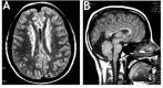
Seizure disorder. Approximately 88% of individuals (including males and females) with FLNA-related periventricular nodular heterotopia (PVNH) have presented with a seizure disorder [Guerrini & Carrozzo 2001]; in other series that included investigation of asymptomatic heterozygous mothers, the percentage was 63.3% [Lange et al 2015]. Many individuals are ascertained by MRI scan following a first seizure. As individuals are increasingly identified based on non-neurologic findings, a lower proportion of individuals with seizures will likely be confirmed. Age of seizure onset may be as early as the first years of life, but typically individuals present during childhood or adolescence, or even as adults, with an overall average age of seizure onset in the mid-teens [Fox et al 1998, Sheen et al 2001, Lange et al 2015]. The severity of the seizure disorder may range from mild (i.e., rare events, not requiring anti-seizure medication) to intractable seizures.

No correlation exists between the extent and severity of the nodular heterotopia seen radiographically and the clinical manifestations, though early seizure onset correlates with poorer developmental outcome [Lange et al 2015]. The ectopic heterotopias in some individuals act as foci for abnormal neuronal activity. Anatomic studies have shown aberrant projections extending from the periventricular heterotopias. Depth electrode recordings have demonstrated epileptogenic discharges from these nodules [Kothare et al 1998]. Thus, the seizure disorder appears to arise from the heterotopias in most individuals.

Cardiovascular findings. In a large cohort of 114 individuals from 80 families with loss-of-function FLNA variants and PVNH, 64.9% had a cardiovascular anomaly. Two thirds were structural cardiac defects, with patent ductus arteriosus, ventricular septal defects, and valve abnormalities being the most common [Chen et al 2018].

Almost 20% of individuals reported by Chen et al [2018] had thoracic aortic dilatation or aneurysm (TAA). Individuals with TAA typically had involvement of the aortic root and ascending aorta, and half of individuals with TAA had a sinus of Valsalva aneurysm [Feng et al 2006].

Isolated X-linked cardiac valvular dysplasia (XCVD) has been identified in individuals with FLNA missense variants associated with some retained FLNA function, typically resulting in myxomatous valvular dystrophy most commonly involving the mitral valve [Kyndt et al 2007, Le Tourneau et al 2018] although other valvular involvement was also noted. XCVD may necessitate cardiac surgery in some individuals. This phenotype was identified in individuals with one of three FLNA missense variants: p.Pro637Gln, p.His743Pro, or p.Gly288Arg. Affected males in the cohort had more severe valvular disease than females, who were sometimes unaffected [Le Tourneau et al 2018].

Pulmonary findings. Individuals with FLNA deficiency may develop varying degrees of pulmonary hypertension, which when severe has necessitated treatment with sildenafil. Several reported individuals (both male and female) had cardiorespiratory failure before age one year. The severity of the poorly defined respiratory disease, resembling bronchopulmonary dysplasia, has led to lung transplantation in several of these young children [Masurel-Paulet et al 2011, Clapham et al 2012, Lord et al 2014, Burrage et al 2017, Kremer et al 2019, Sasaki et al 2019].

Gastrointestinal manifestations. The spectrum of disease severity has ranged from constipation to chronic intestinal pseudo-obstruction (CIPO). Several individuals (primarily males), have been reported with delayed gastric motility [Negri et al 2020]. Clinical presentation typically includes feeding difficulties at birth, constipation, progressive weight loss, and congenital short bowel seen in isolation or in combination with intestinal malrotation [Oegema et al 2013]. Feeding issues may be present and improve with time, resolving after the first year of life. Conservative treatments including diet management were successful in some symptomatic older individuals [Oegema et al 2013]. Prolonged nutrition supplementation, gastrotomy, and/or jejunostomy tube feeding may be necessary in some individuals.

Males with FLNA loss-of-function variants have presented with severe abdominal wall defects, resulting in prune belly syndrome; characterized by: wrinkled, lax abdominal walls with partial or complete skeletal muscle deficiency; urinary tract dilatation with poorly contractile smooth muscle; and undescended testes [Iqbal et al 2020].

Isolated gastrointestinal manifestations. Loss-of-function FLNA variants have also been described as a cause of congenital short bowel syndrome in males and females. Males with X-linked congenital intestinal pseudo-obstruction (CIPO) have been found to have FLNA loss-of-function variants [van der Werf et al 2015, Negri et al 2020, Wade et al 2020]. CIPO includes reduced intestinal motility and a short, malrotated, and dilated small intestine. Severe intestinal dysmotility resulted in the need for long-term parenteral nutrition in several individuals [Jenkins et al 2018].

Joint hypermobility. In a case series of 47 individuals (2 males, 45 females) with FLNA-related PVNH, 14.7% presented with joint laxity. Joint dislocation can also occur, involving the hips and other joints [Lange et al 2015].

Skin abnormalities. Cutis laxa, hyperextensibility, and thin, soft, or translucent skin [Reinstein et al 2013, Ritelli et al 2017] may also be present, occurring independently or in combination.

Shortened digits. Some individuals are reported to have hypoplasia of the distal phalanges in general or of the great toe, or short proximal thumbs.

Macrothrombocytopenia. Individuals with FLNA deficiency can present with large platelets that may also be reduced in number, resulting in excessive bruising, bleeding, and slow wound healing [Nurden et al 2011]. Macrothombocytopenia can occur with missense, deletion, exon-skipping [Oda et al 2016], or truncation variants of FLNA. Isolated macrothrombocytopenia was reported in an individual with thrombocytopenia, bleeding, and an FLNA missense variant (p.Glu1795Lys) without other features of FLNA deficiency [Nurden et al 2011].

Cognitive function / dyslexia. Intelligence is normal to borderline. Formal cognitive testing of ten affected individuals (2 males, 8 females) with FLNA loss-of-function variants demonstrated an average IQ of 95, but also a strikingly high number of affected individuals with dyslexia [Chang et al 2005, Chang et al 2007, Reinstein et al 2012]. However, these results likely reflect a selection bias towards more severely affected individuals, since those with normal cognitive function usually would not have undergone a screening brain MRI or molecular testing.

Pregnancy complications. Women with FLNA deficiency may have an increased incidence of pregnancy loss as a result of spontaneous abortion of affected male pregnancies.

Other

  • Immune compromise with recurrent infection was reported in two individuals
  • Overwhelming hemorrhage and arrested myeloid and erythroid bone marrow development was reported in one male [Huttenlocher et al 1994].
  • Bilateral inguinal hernia has been reported in affected males [Oegema et al 2013, Ritelli et al 2017]

Mosaicism. Somatic mosaicism for an A>G substitution at the intron 11 acceptor splice site was reported by Parrini et al [2004] in a male with bilateral PVNH. Sequence analysis and denaturing high-performance liquid chromatography of genomic DNA on a pool of hair roots, single hair roots, and white blood cells revealed that only 42% and 69% of the samples for hair and blood, respectively, had the pathogenic variant. Moreover, the affected male's daughter did not inherit the pathogenic variant, thought to be causal for the male phenotype. Other somatic pathogenic variants were subsequently reported [Jamuar et al 2014, González-Morón et al 2017].

Genotype-Phenotype Correlations

p.Pro637Gln, p.His743Pro, and p.Gly288Arg have been associated with isolated X-linked cardiac valvular dysplasia [Kyndt et al 2007, Le Tourneau et al 2018]. Affected males were found to have more severe valvular disease [Le Tourneau et al 2018].

Females with PVNH typically have FLNA variants predicted to cause severe loss of function (e.g., nonsense, frameshift, and severe splicing variants) throughout the gene or missense variants in the N-terminal actin-binding domain [Walsh & Engle 2010, Lange et al 2015].

Surviving affected males. Nonsense variants in surviving males often affect the C terminus of the protein, consistent with partial loss of function, while missense variants in surviving males can be scattered throughout the gene [Walsh & Engle 2010, Lange et al 2015, Chen et al 2018]. Presumably, these pathogenic variants lead to a partially functional protein [Sheen et al 2001].

Penetrance

All individuals with known deleterious loss-of-function FLNA variants typically have shown periventricular nodular heterotopia. Penetrance for other phenotypes has not been determined.

Nomenclature

The authors suggest the term Huttenlocher syndrome for individuals with FLNA deficiency and multi-system involvement.

Prevalence

The prevalence of FLNA deficiency is difficult to assess because individuals with the mild phenotype may never seek medical evaluation.

Differential Diagnosis

Periventricular nodular heterotopia (PVNH) has been reported in individuals with the disorders listed in Table 3a (whether each of these represents a truly distinct disorder or FLNA-related PVNH plus a concurrent condition remains to be determined).

Table 3a.

Disorders That May Be Associated with Periventricular Nodular Heterotopia (PVNH)

Gene(s)DisorderMOIComment
ARF1 PVNH8 (OMIM 618185)AD
ARFGEF2 PVNH2 (OMIM 608097)ARReported in 2 Turkish families w/PVNH & microcephaly 1 & in a female w/a movement disorder, neuronal migration disorder, & acquired microcephaly 2
EML1 Band heterotopia (OMIM 600348)ARMegalencephaly, ribbon-like subcortical band heterotopia, severe developmental delay
FMR1 Fragile X syndrome (See FMR1 Disorders.)XLPVNH (unilateral/bilateral & isolated) reported in 2 boys w/fragile X syndrome 3 (PVNH is very rare in fragile X syndrome.)
MAP1B PVNH9 (OMIM 618918)ADSee footnote 4.
TSC1
TSC2
Tuberous sclerosis complex (TSC)ADPVNH may be misdiagnosed initially as TSC; however, MRI findings distinguish the disorders

Table 3b lists autosomal dominant forms of PVNH that have been mapped to chromosomal loci (OMIM PS300049).

Table 3b.

Other Periventricular Nodular Heterotopia (PVNH) Phenotypes

Disorder/PhenotypeComment
Nonfamilial PVNHCaused by perinatal insult or chr rearrangement
PVNH, ID, & epilepsyAssoc w/chr 5q14.3-q15 deletion (OMIM 612881)
Bilateral PVNH w/micronodules
Bilateral PVNH w/ambiguous genitalia
Bilateral PVNH/ID/syndactyly Dobyns et al [1997]
Unilateral PVNH
Bilateral anterior PVNH w/fronto-perisylvian polymicrogyria Parrini et al [2006]
Bilateral PVNH involving temporo-occipital & trigones w/hippocampal malformationSubclassified into polymicrogyria or cerebellar hypoplasia or hydrocephalus [Parrini et al 2006]

chr = chromosome; ID = intellectual disability

X-linked subcortical band heterotopia. Laminar heterotopia occurring in deep white matter and band-like heterotopia occurring between the cortex and ventricular surface are seen in X-linked subcortical band heterotopia (see DCX-Related Disorders).

Isolated cardiac valvular dysplasia. To date, no genes other than FLNA are known to be associated with hereditary nonsyndromic mitral valve prolapse/dysplasia.

Isolated gastrointestinal manifestations. The differential diagnosis of FLNA-related isolated gastrointestinal manifestations includes other forms of inherited congenital bowel obstruction including those linked to chromosome abnormalities and pathogenic variants in CLMP [van der Werf et al 2015].

Isolated macrothrombocytopenia. FLNA deficiency should be considered in the differential diagnosis of individuals who present with isolated macrothrombocytopenia; MYH9-related disease can also be considered in the differential diagnosis.

Management

No clinical practice guidelines for FLNA deficiency have been published.

Evaluations Following Initial Diagnosis

To establish the extent of disease and needs in an individual diagnosed with FLNA deficiency, the evaluations summarized in Table 4 (if not performed as part of the evaluation that led to the diagnosis) are recommended.

Table 4.

Recommended Evaluations Following Initial Diagnosis in Individuals with FLNA Deficiency

System/ConcernEvaluationComment
Neurology
  • Eval by neurologist
  • Eval by epileptologist if seizures are present
Detailed history & eval to confirm suspicion of a seizure disorder. Testing may incl EEG to define location & severity of electrical brain dysfunction that may be present in those w/epilepsy.
MRA of intracranial vessels, carotid arteries, & aortaAt diagnosis to address ↑ risk for stroke
Cardiology Echocardiogram or MRA to evaluate for valvular dysplasia, congenital cardiac anomalies, or aortic & vascular diseaseBecause of potential risk for cardiovascular anomalies &/or thoracic aortic aneurysm, consider baseline eval by cardiologist.
Pulmonary
  • Assess for pulmonary symptoms, pulmonary hypertension, & interstitial lung disease.
  • Pulmonary consultation if needed
Gastrointestinal
  • Eval for constipation/motility issues
  • Gastrointestinal consultation as needed
Musculoskeletal Eval for joint hypermobility if observedPT eval as needed for specific issues
Hematology Eval by hematologist if findings suggest a bleeding diathesis or abnormal platelet size or number.
Development Developmental assessment
  • To incl motor, adaptive, cognitive, & speech-language eval for dyslexia
  • Eval for early intervention if needed
Genetic
counseling
  • Family history assessment by genetics professional, 1 esp seizure history in the mother
  • Consider brain MRI & echocardiogram for the mother.
To inform affected persons & families re nature, MOI, & implications of FLNA deficiency to facilitate medical & personal decision making
Family support
& resources
Assess need for:

MOI = mode of inheritance; MRA = magnetic resonance angiography; PT = physical therapy

1.

Medical geneticist, certified genetic counselor, certified advanced genetic nurse

Treatment of Manifestations

Management of individuals with FLNA deficiency is directed toward symptomatic treatment.

Table 5.

Treatment of Manifestations in Individuals with FLNA Deficiency

Manifestation/ConcernTreatmentConsiderations/Other
Epilepsy Treatment w/ASM; choice of ASMs is generally made empirically based on clinical features of the seizure disorder.No significant differences exist between ASMs for newly diagnosed, presumably localized epilepsy; choices may be made on specific attributes of each ASM (e.g., risk of teratogenicity during pregnancy, tolerability, & efficacy).
Congenital heart
disease /
Cardiovascular
disease
  • Treatment of structural heart disease & valvular dysplasia per cardiologist
  • Because of risk for aortic or carotid dissection, ensure good blood pressure control.
Treatment of aortic/carotid dissection, congenital heart disease, & valvular disease as in general population
Pulmonary
  • Treat any symptoms that may worsen pulmonary function.
  • Good pulmonary toilet to preserve lung function
  • Sildenafil & medical management for pulmonary hypertension if present
Rarely, severely affected persons require lung transplantation; most persons require no treatment.
Impaired bowel
motility
  • Stool softeners, prokinetics, osmotic agents, or laxatives as needed
  • Treat GERD if present.
Depends on severity; those w/severely impaired motility may need a g-tube for parenteral feeding.
Joint hypermobility Treatment per orthopedist, PT, &/or OT can incl braces to improve joint stability, ring splints to stabilize interphalangeal joints.
Bleeding diathesis Treatment per hematologist
Dyslexia Early intervention services & special education support

ASM = anti-seizure medication; GERD = gastroesophageal reflux disease; OT = occupational therapy; PT = physical therapy

Surveillance

Table 6.

Recommended Surveillance for Individuals with FLNA Deficiency

System/ConcernEvaluationFrequency
Neurologic Monitor those w/seizures as clinically indicated.At each visit
Assess for new manifestations incl seizures, changes in tone, mvmt disorders.Rpt imaging may be needed (only if new neurologic findings on exam).
Aortic or carotid
dissection
  • Cardiology eval
  • Echocardiogram
  • Stress testing & cardiac MRI as needed
  • Serial echocardiographic follow up per cardiologist, as severity of valve disease & aortic dilatation can evolve w/time
  • Consider cardiac MRI if difficult acoustic windows, & for periodic visualization of ascending aorta & aortic arch.
Note: Current data are not sufficient to provide definitive frequency guidelines.
Pulmonary Follow up as dictated by findings & severityConsider frequent follow up from birth to age 3 yrs due to continued lung development in young children.
Gastrointestinal Monitor for constipation.At each visit
Musculoskeletal Monitor for risk of joint injury due to hypermobility.
Hematology
  • Monitor for signs/symptoms of bleeding diathesis.
  • CBC w/platelets
Development Developmental assessment w/eval for dyslexia as indicated

Evaluation of Relatives at Risk

Given the risk for vascular disease in neurologically asymptomatic individuals, it is appropriate to clarify the genetic status of older and younger at-risk relatives of an affected individual in order to identify as early as possible those who would benefit from echocardiogram and MRI/MRA to screen for FLNA-associated cardiovascular problems (see Surveillance).

See Genetic Counseling for issues related to testing of at-risk relatives for genetic counseling purposes.

Pregnancy Management

Ideally, women should seek information prior to conception regarding risks to the fetus associated with taking an anti-seizure medication during pregnancy so that changes in the anti-seizure medication regimen (if needed) can be made prior to conception. If not done prior to conception, discussion of the risks and benefits of anti-seizure medication use during pregnancy should occur as soon as the pregnancy is recognized. The teratogenic risk to the fetus associated with the use of anti-seizure medication during pregnancy depends on the type of anti-seizure medication used, the dose, and the gestational age of the fetus.

Women with FLNA deficiency may have an increased incidence of pregnancy loss as a result of spontaneous abortion of affected male pregnancies. Currently no guidelines exist on the most appropriate surveillance for and management of cardiac, vascular, and connective tissue problems during pregnancy in women with PVNH. See Marfan Syndrome and Classic Ehlers-Danlos Syndrome for possible pregnancy management recommendations.

Therapies Under Investigation

Search ClinicalTrials.gov in the US and EU Clinical Trials Register in Europe for access to information on clinical studies for a wide range of diseases and conditions. Note: There may not be clinical trials for this disorder.

Other

Surgical resection has been attempted but has not proven beneficial [Li et al 1997].

Genetic Counseling

Genetic counseling is the process of providing individuals and families with information on the nature, mode(s) of inheritance, and implications of genetic disorders to help them make informed medical and personal decisions. The following section deals with genetic risk assessment and the use of family history and genetic testing to clarify genetic status for family members; it is not meant to address all personal, cultural, or ethical issues that may arise or to substitute for consultation with a genetics professional. —ED.

Mode of Inheritance

FLNA deficiency is inherited in an X-linked manner.

  • FLNA-related periventricular nodular heterotopia (PVNH). Affected individuals are predominantly females with severe loss-of-function variants. Males often show early lethality, though some males with mosaic pathogenic variants or variants resulting in partial loss of function survive.
  • Isolated X-linked cardiac valvular dysplasia (XCVD) and FLNA-related isolated gastrointestinal manifestations. Affected individuals are typically male. Heterozygous female family members may or may not be affected.
  • Isolated macrothrombocytopenia has been reported in several heterozygous females [Nurden et al 2011].

Risk to Family Members

FLNA-Related PVNH

Parents of a female proband

  • About 50% of females with FLNA-related PVNH have inherited the FLNA pathogenic variant from a parent. In these families, the transmitting parent is typically the mother (very few adult males with a hemizygous loss-of-function variant have been identified). The transmitting parent may be neurologically asymptomatic, but most individuals with a loss-of-function FLNA variant have nodules identifiable by brain imaging.
  • About 50% of females with FLNA-related PVNH have a de novo pathogenic variant.
  • In a family with more than one affected individual, the parent of an affected child will typically be heterozygous (or hemizygous) for the pathogenic variant. Note: If the proband has an affected sib and no other affected relatives and if the pathogenic variant cannot be detected in either parent’s leukocyte DNA, a parent most likely has somatic/germline mosaicism.
  • If there is only one affected family member (i.e., a simplex case), a parent may be heterozygous (or hemizygous) for the pathogenic variant, the proband may have a de novo FLNA pathogenic variant, or a parent may have somatic/germline mosaicism. Available data suggest that the frequency of parental germline mosaicism is appropriately 5%-10% in FLNA-related PVNH; however, the exact frequency is not known.
  • Molecular genetic testing of the mother (and possibly, or subsequently, the father) is recommended to confirm parental genetic status and to allow reliable recurrence risk assessment.

Parents of a male proband

  • The father of an affected male will not have the disorder nor will he be hemizygous for the FLNA pathogenic variant; therefore, he does not require further evaluation/testing.
  • Molecular genetic testing of the mother is recommended to confirm her genetic status and to allow reliable recurrence risk assessment.

Sibs of a proband. The risk to sibs depends on the genetic status of the parents:

  • If the mother of the proband has an FLNA pathogenic variant, the chance of transmitting it in each pregnancy is 50%:
    • Female sibs who inherit the FLNA pathogenic variant will have PVNH; the penetrance of other clinical features that have been associated with FLNA-related PVNH is unknown.
    • Most males who inherit an FLNA loss-of-function variant die before or soon after birth from respiratory or cardiac failure. Surviving affected males have been reported in the literature; however, available data are insufficient to reliably predict the clinical outcome of a hemizygous male fetus.
    • Theoretically, if the father of the proband is hemizygous for an FLNA loss-of-function variant, he will transmit it to all his daughters and none of his sons.
  • If the proband represents a simplex case (i.e., a single occurrence in a family) and if the FLNA pathogenic variant cannot be detected in the leukocyte DNA of either parent, the risk to sibs is presumed to be low but still greater than that of the general population because of the possibility of parental somatic/germline mosaicism.

Offspring of a proband

  • Each child of a female proband with FLNA-related PVNH has a 50% chance of inheriting the FLNA pathogenic variant.
  • Hemizygous males with FLNA-related PVNH are not known to reproduce. Rarely, less severely affected males who are mosaic for an FLNA loss-of-function variant have reproduced. All daughters of a male with mosaicism are at risk of inheriting the FLNA pathogenic variant; the sons of a male with mosaicism are not at risk of inheriting the FLNA pathogenic variant.

Other family members. The risk to other family members depends on the genetic status of the proband's parents: if the mother is heterozygous for a pathogenic FLNA variant (or, theoretically, the father is hemizygous for a pathogenic FLNA variant), the mother's (or father's) family members may be at risk.

Note: Molecular genetic testing may be able to identify the family member in whom a de novo pathogenic variant arose, information that could help determine genetic risk status of the extended family.

XCVD, FLNA-Related Isolated Gastrointestinal Manifestations, and FLNA-Related Isolated Macrothrombocytopenia

Parents of a female proband

  • A female proband may have inherited the FLNA pathogenic variant from either her mother or her father, or the pathogenic variant may be de novo.
  • Detailed evaluation of the parents and review of the extended family history may help distinguish probands with a de novo pathogenic variant from those with an inherited pathogenic variant. Molecular genetic testing of the mother (and possibly the father, or subsequently the father) can determine if the pathogenic variant was inherited.

Parents of a male proband

  • The father of an affected male will not have the disorder nor will he be hemizygous for the FLNA pathogenic variant; therefore, he does not require further evaluation/testing.
  • In a family with more than one affected individual, the mother of an affected male is an obligate heterozygote. Note: If a woman has more than one affected child and no other affected relatives and if the FLNA pathogenic variant cannot be detected in her leukocyte DNA, she most likely has germline mosaicism.
  • If a male is the only affected family member, the mother may be a heterozygote, the affected male may have a de novo FLNA pathogenic variant (in which case the mother is not a heterozygote), or the mother may have somatic/germline mosaicism.
    • Most males with XCVD have inherited an FLNA pathogenic variant from their mother (who may or may not be affected).
    • To date, all males with FLNA-related isolated gastrointestinal manifestations have inherited a pathogenic variant from their unaffected (or mildly affected) mother.
  • Molecular genetic testing of the mother is recommended to confirm her genetic status and to allow reliable recurrence risk assessment.

Sibs of a proband. The risk to sibs depends on the genetic status of the parents:

  • If the mother of the proband has an FLNA pathogenic variant, the chance of transmitting it in each pregnancy is 50%:
    • Males who inherit the pathogenic variant will be affected;
    • Females who inherit the pathogenic variant will be heterozygotes and may be asymptomatic or have manifestations of the disorder.
  • If the father of the proband has an FLNA pathogenic variant, he will transmit it to all his daughters and none of his sons.
  • If the proband represents a simplex case and the FLNA pathogenic variant cannot be detected in the leukocyte DNA of the mother (if the proband is male) or of either the mother or father (if the proband is female), the risk to sibs is presumed to be low but still greater than that of the general population because of the possibility of parental somatic/germline mosaicism.

Offspring of a proband

  • Women with an FLNA pathogenic variant have a 50% chance of transmitting the pathogenic variant to each child.
  • Males with an FLNA pathogenic variant transmit the pathogenic variant to all their daughters and none of their sons.

Other family members. The risk to other family members depends on the genetic status of the proband's parents: if a parent has an FLNA pathogenic variant, the parent's family members may be at risk.

Note: Molecular genetic testing may be able to identify the family member in whom a de novo pathogenic variant arose, information that could help determine genetic risk status of the extended family.

Related Genetic Counseling Issues

See Management, Evaluation of Relatives at Risk for information on evaluating at-risk relatives for the purpose of early diagnosis and treatment.

Family planning

Prenatal Testing and Preimplantation Genetic Testing

Molecular genetic testing. Once the FLNA pathogenic variant has been identified in an affected family member, prenatal and preimplantation genetic testing are possible.

Ultrasound examination. Fetal ultrasound is relatively insensitive at detecting PVNH until late into the second trimester in pregnancy. PVNH may be detected by fetal ultrasound examination or MRI as early as 20-24 weeks' gestation. Fetal MRI is performed at some centers using ultrafast MRI so that sedation is not required to reduce fetal movement. The sensitivity of prenatal imaging for the detection of PVNH is not known. Whether PVNH may be detected even earlier in gestation by imaging studies is not known.

Differences in perspective may exist among medical professionals and within families regarding the use of prenatal testing. While most centers would consider use of prenatal testing to be a personal decision, discussion of these issues may be helpful.

Resources

GeneReviews staff has selected the following disease-specific and/or umbrella support organizations and/or registries for the benefit of individuals with this disorder and their families. GeneReviews is not responsible for the information provided by other organizations. For information on selection criteria, click here.

Molecular Genetics

Information in the Molecular Genetics and OMIM tables may differ from that elsewhere in the GeneReview: tables may contain more recent information. —ED.

Table A.

FLNA Deficiency: Genes and Databases

GeneChromosome LocusProteinLocus-Specific DatabasesHGMDClinVar
FLNA Xq28 Filamin-A FLNA @ LOVD FLNA FLNA

Data are compiled from the following standard references: gene from HGNC; chromosome locus from OMIM; protein from UniProt. For a description of databases (Locus Specific, HGMD, ClinVar) to which links are provided, click here.

Table B.

OMIM Entries for FLNA Deficiency (View All in OMIM)

300017FILAMIN A; FLNA
300048INTESTINAL PSEUDOOBSTRUCTION, NEURONAL, CHRONIC IDIOPATHIC, X-LINKED
300049PERIVENTRICULAR NODULAR HETEROTOPIA 1; PVNH1
314400CARDIAC VALVULAR DYSPLASIA, X-LINKED; CVDPX

Molecular Pathogenesis

The filamin class of actin-binding proteins is known to regulate cell stability, protrusion, motility, cell-cell communication, and response to stress across various biologic systems [Leonardi et al 2000, Stahlhut & van Deurs 2000, Feng & Walsh 2004]. Filamin-deficient melanocytes fail to undergo locomotion in response to factors that elicit migration in the same filamin-expressing cells. They exhibit prolonged circumferential blebbing, abnormal phagocytosis, and impaired volume regulation, perhaps secondary to abnormal regulation of sodium channel activity.

FLNA encodes a cytoplasmic actin-binding phosphoprotein that links membrane receptors to the actin cytoskeleton and represents a potentially crucial link between signal transduction and the cytoskeleton. Findings suggest that filamin A may have a role in adherens junctions that line the ventricle, with defects in FLNA disrupting the ventricular lining and neuroepithelial architecture, causing failure of neuronal migration [Zhang et al 2016].

Mechanism of disease causation. Loss of function; typically heterozygous null variants in affected females and missense or mosaic variants in mildly affected females and rare affected males [Wade et al 2020]

FLNA-specific laboratory technical considerations. FLNA contains 46 coding exons as well as a “poison exon” between exons 9 and 10 that encodes a premature termination codon, which is utilized in a cell type-specific pattern. This poison exon is excluded in brain stem cells but is included in neurons resulting in loss of filamin A function. Pathogenic variants affecting the splice sites regulating this poison exon cause loss of function because of abnormal inclusion of the poison exon [Zhang et al 2016]. FLNA shows at least two transcriptional start sites, one of which creates a protein longer than the canonic protein by 28 amino acid residues added at the N terminus. Loss-of-function variants restricted to this longer transcript cause CIP with normal heart and brain development, indicating that the longer splice form has preferential roles in gut development [Wade et al 2020]. Other exons show preferential utilization in heart versus brain [Zhang et al 2016]. The locus has very high GC content, making it difficult to amplify by PCR or to sequence either by Sanger or next-generation sequencing.

Table 7.

Notable FLNA Pathogenic Variants

Reference SequencesDNA Nucleotide ChangePredicted Protein ChangeComment [Reference]
NM_001456​.4
NP_001447​.2
c.862G>Ap.Gly288ArgSee Genotype-Phenotype Correlations.
c.1910C>Ap.Pro637Gln
c.2228A>Cp.His743Pro
c.5383G>Ap.Glu1795LysReported in 1 older female w/isolated macrothrombocytopenia [Nurden et al 2011]

Variants listed in the table have been provided by the authors. GeneReviews staff have not independently verified the classification of variants.

GeneReviews follows the standard naming conventions of the Human Genome Variation Society (varnomen​.hgvs.org). See Quick Reference for an explanation of nomenclature.

Chapter Notes

Author Notes

Ming Hui Chen, MD, MMSc is an Associate Professor of Pediatrics at Harvard Medical School in the Departments of Cardiology and Pediatrics, and the Division of Genetics and Genomics at Boston Children's Hospital, Boston, MA. Dr Chen is an expert in cardiovascular and medical manifestations of FLNA pathogenic variants. She follows a large cohort of patients and families with PVNH and FLNA pathogenic variants. Dr Chen is the Founder and Director of the Cardiology Clinic for Brain Development in Genetics (C-BrDG), a specialized FLNA-PVNH clinic at Boston Children’s Hospital. For further information regarding clinical care and clinical research in individuals with PVNH and FLNA deficiencies, please visit our website at mhchenlab.org or email cbridge@childrens.harvard.edu.

Dr Chen has worked very closely over the last two decades with Dr Christopher A Walsh, MD, PhD, who is the Chief of Genetics and Genomics at Boston Children's Hospital, and the Bullard Professor of Pediatrics and Neurology at Harvard Medical School. The Walsh lab has worked on genetic disorders affecting the human cerebral cortex for more than twenty years, publishing twenty papers on PVNH, FLNA, and related disorders.

Acknowledgments

We are grateful to patients with PVNH and other brain disorders, their physicians, and to many colleagues present and past for collaborating with us to understand the genetic basis of these conditions, and for the patient support groups that we have allied with to help understand and develop better therapies.

Research on PVNH in the Chen group and the Walsh lab has been supported by grant funding from the NIH and the NINDS (R01 NS 35129). CAW is an Investigator of the Howard Hughes Medical Institute.

Author History

Adria Bodell, MS, CGC; Beth Israel Deaconess Medical Center (2007-2015)
Ming Hui Chen, MD, MMSc (2015-present)
Volney L Sheen, MD, PhD; Harvard Medical School (2007-2015)
Christopher A Walsh, MD, PhD (2007-present)

Revision History

  • 30 September 2021 (sw) Comprehensive update posted live
  • 17 September 2015 (me) Comprehensive update posted live
  • 11/10/2014 (cw/mhc) Revision: cardiovascular added, other modifications made
  • 4 June 2009 (cd/cw) Revision: deletion/duplication analysis available; mutation added; Differential Diagnosis edited
  • 10 April 2007 (me) Comprehensive update posted live
  • 4 August 2004 (me) Comprehensive update posted live
  • 21 January 2004 (cd) Revision: testing
  • 17 October 2003 (cw) Revision: Genetically Related Disorders
  • 8 October 2002 (me) Review posted live
  • 29 April 2002 (cw) Original submission

References

Literature Cited

  • Burrage LC, Guillerman RP, Das S, Singh S, Schady DA, Morris SA, Walkiewicz M, Schecter MG, Heinle JS, Lotze TE, Lalani SR, Mallory GB. Lung transplantation for FLNA-associated progressive lung disease. J Pediatr. 2017;186:118–23.e6. [PMC free article: PMC5534178] [PubMed: 28457522]
  • Chang BS, Katzir T, Liu T, Corriveau K, Barzillai M, Apse KA, Bodell A, Hackney D, Alsop D, Wong ST, Walsh CA. A structural basis for reading fluency: white matter defects in a genetic brain malformation. Neurology. 2007;69:2146–54. [PubMed: 18056578]
  • Chang BS, Ly J, Appignani B, Bodell A, Apse KA, Ravenscroft RS, Sheen VL, Doherty MJ, Hackney DB, O'Connor M, Galaburda AM, Walsh CA. Reading impairment in the neuronal migration disorder of periventricular nodular heterotopia. Neurology. 2005;64:799–803. [PubMed: 15753412]
  • Chen MH, Choudhury S, Hirata M, Khalsa S, Chang B, Walsh CA. Thoracic aortic aneurysm in patients with loss of function Filamin A mutations: Clinical characterization, genetics, and recommendations. Am J Med Genet A. 2018;176:337–50. [PMC free article: PMC7534149] [PubMed: 29334594]
  • Clapham KR, Yu TW, Ganesh VS, Barry B, Chan Y, Mei D, Parrini E, Funalot B, Dupuis L, Nezarati MM, du Souich C, van Karnebeek C, Guerrini R, Walsh CA. FLNA genomic rearrangements cause periventricular nodular heterotopia. Neurology. 2012;78:269–78. [PMC free article: PMC3280053] [PubMed: 22238415]
  • Clayton-Smith J, Walters S, Hobson E, Burkitt-Wright E, Smith R, Toutain A, Amiel J, Lyonnet S, Mansour S, Fitzpatrick D, Ciccone R, Ricca I, Zuffardi O, Donnai D. Xq28 duplication presenting with intestinal and bladder dysfunction and a distinctive facial appearance. Eur J Hum Genet. 2009;17:434–43. [PMC free article: PMC2986219] [PubMed: 18854860]
  • de Wit MC, de Coo IF, Halley DJ, Lequin MH, Mancini GM. Movement disorder and neuronal migration disorder due to ARFGEF2 mutation. Neurogenetics. 2009;10:333–6. [PMC free article: PMC2758209] [PubMed: 19384555]
  • Dobyns WB, Guerrini R, Czapansky-Beilman DK, Pierpont ME, Breningstall G, Yock DH Jr, Bonanni P, Truwit CL. Bilateral periventricular nodular heterotopia with mental retardation and syndactyly in boys: a new X-linked mental retardation syndrome. Neurology. 1997;49:1042–7. [PubMed: 9339687]
  • Feng Y, Chen MH, Moskowitz IP, Mendonza AM, Vidali L, Nakamura F, Kwiatkowski DJ, Walsh CA, Filamin A. FLNA) is required for cell-cell contact in vascular development and cardiac morphogenesis. Proc Natl Acad Sci U S A. 2006;103:19836–41. [PMC free article: PMC1702530] [PubMed: 17172441]
  • Feng Y, Walsh CA. The many faces of filamin: a versatile molecular scaffold for cell motility and signalling. Nat Cell Biol. 2004;6:1034–8. [PubMed: 15516996]
  • Fox JW, Lamperti ED, Ekşioğlu YZ, Hong SE, Feng Y, Graham DA, Scheffer IE, Dobyns WB, Hirsch BA, Radtke RA, Berkovic SF, Huttenlocher PR, Walsh CA. Mutations in filamin 1 prevent migration of cerebral cortical neurons in human periventricular heterotopia. Neuron. 1998;21:1315–25. [PubMed: 9883725]
  • González-Morón D, Vishnopolska S, Consalvo D, Medina N, Marti M, Córdoba M, Vazquez-Dusefante C, Claverie S, Rodríguez-Quiroga SA, Vega P, Silva W, Kochen S, Kauffman MA. Germline and somatic mutations in cortical malformations: molecular defects in Argentinean patients with neuronal migration disorders. PLoS One. 2017;12:e0185103. [PMC free article: PMC5617183] [PubMed: 28953922]
  • Guerrini R, Carrozzo R. Epileptogenic brain malformations: clinical presentation, malformative patterns and indications for genetic testing. Seizure. 2001;10:532–43. [PubMed: 11749114]
  • Heinzen EL, O'Neill AC, Zhu X, Allen AS, Bahlo M, Chelly J, Chen MH, Dobyns WB, Freytag S, Guerrini R, Leventer RJ, Poduri A, Robertson SP, Walsh CA, Zhang M. Epi4K Consortium; Epilepsy Phenome/Genome Project. De novo and inherited private variants in MAP1B in periventricular nodular heterotopia. PLoS Genet. 2018;14:e1007281. [PMC free article: PMC5965900] [PubMed: 29738522]
  • Huttenlocher PR, Taravath S, Mojtahedi S. Periventricular heterotopia and epilepsy. Neurology. 1994;44:51–5. [PubMed: 8290091]
  • Iqbal NS, Jascur TA, Harrison SM, Edwards AB, Smith LT, Choi ES, Arevalo MK, Chen C, Zhang S, Kern AJ, Scheuerle AE, Sanchez EJ, Xing C, Baker LA. Prune belly syndrome in surviving males can be caused by Hemizygous missense mutations in the X-linked Filamin A gene. BMC Med Genet. 2020;21:38. [PMC free article: PMC7035669] [PubMed: 32085749]
  • Jamuar SS, Lam AT, Kircher M, D'Gama AM, Wang J, Barry BJ, Zhang X, Hill RS, Partlow JN, Rozzo A, Servattalab S, Mehta BK, Topcu M, Amrom D, Andermann E, Dan B, Parrini E, Guerrini R, Scheffer IE, Berkovic SF, Leventer RJ, Shen Y, Wu BL, Barkovich AJ, Sahin M, Chang BS, Bamshad M, Nickerson DA, Shendure J, Poduri A, Yu TW, Walsh CA. Somatic mutations in cerebral cortical malformations. N Engl J Med. 2014;371:733–43. [PMC free article: PMC4274952] [PubMed: 25140959]
  • Jenkins ZA, Macharg A, Chang CY, van Kogelenberg M, Morgan T, Frentz S, Wei W, Pilch J, Hannibal M, Foulds N, McGillivray G, Leventer RJ, García-Miñaúr S, Sugito S, Nightingale S, Markie DM, Dudding T, Kapur RP, Robertson SP. Differential regulation of two FLNA transcripts explains some of the phenotypic heterogeneity in the loss-of-function filaminopathies. Hum Mutat. 2018;39:103–13. [PubMed: 29024177]
  • Kothare SV, VanLandingham K, Armon C, Luther JS, Friedman A, Radtke RA. Seizure onset from periventricular nodular heterotopias: depth-electrode study. Neurology. 1998;51:1723–7. [PubMed: 9855532]
  • Kremer TM, Lindsay ME, Kinane TB, Hawley MH, Little BP, Mino-Kenudson M. Case 28-2019: A 22-year-old woman with dyspnea and chest pain. N Engl J Med. 2019;381:1059–67. [PubMed: 31509678]
  • Kyndt F, Le Scouarnec S, Jaafar P, Gueffet JP, Legendre A, Trochu JN, Jousseaume V, Chaventré A, Schott JJ, Le Marec H, Probst V. Genetic aspects of valvulopathies. Arch Mal Coeur Vaiss. 2007;100:1013–20. [PubMed: 18223515]
  • Lange M, Kasper B, Bohring A, Rutsch F, Kluger G, Hoffjan S, Spranger S, Behnecke A, Ferbert A, Hahn A, Oehl-Jaschkowitz B, Graul-Neumann L, Diepold K, Schreyer I, Bernhard MK, Mueller F, Siebers-Renelt U, Beleza-Meireles A, Uyanik G, Janssens S, Boltshauser E, Winkler J, Schuierer G, Hehr U. 47 patients with FLNA associated periventricular nodular heterotopia. Orphanet J Rare Dis. 2015;10:134. [PMC free article: PMC4608144] [PubMed: 26471271]
  • Le Tourneau T, Le Scouarnec S, Cueff C, Bernstein D, Aalberts JJJ, Lecointe S, Mérot J, Bernstein JA, Oomen T, Dina C, Karakachoff M, Desal H, Al Habash O, Delling FN, Capoulade R, Suurmeijer AJH, Milan D, Norris RA, Markwald R, Aikawa E, Slaugenhaupt SA, Jeunemaitre X, Hagège A, Roussel JC, Trochu JN, Levine RA, Kyndt F, Probst V, Le Marec H, Schott JJ. New insights into mitral valve dystrophy: a Filamin-A genotype-phenotype and outcome study. Eur Heart J. 2018;39:1269–77. [PMC free article: PMC5905589] [PubMed: 29020406]
  • Leonardi A, Ellinger-Ziegelbauer H, Franzoso G, Brown K, Siebenlist U. Physical and functional interaction of filamin (actin-binding protein-280) and tumor necrosis factor receptor-associated factor 2. J Biol Chem. 2000;275:271–8. [PubMed: 10617615]
  • Li LM, Dubeau F, Andermann F, Fish DR, Watson C, Cascino GD, Berkovic SF, Moran N, Duncan JS, Olivier A, Leblanc R, Harkness W. Periventricular nodular heterotopia and intractable temporal lobe epilepsy: poor outcome after temporal lobe resection. Ann Neurol. 1997;41:662–8. [PubMed: 9153529]
  • Lord A, Shapiro AJ, Saint-Martin C, Claveau M, Melançon S, Wintermark P. Filamin A mutation may be associated with diffuse lung disease mimicking bronchopulmonary dysplasia in premature newborns. Respir Care. 2014;59:e171–7. [PubMed: 25053830]
  • Masurel-Paulet A, Haan E, Thompson EM, Goizet C, Thauvin-Robinet C, Tai A, Kennedy D, Smith G, Khong TY, Solé G, Guerineau E, Coupry I, Huet F, Robertson S, Faivre L. Lung disease associated with periventricular nodular heterotopia and an FLNA mutation. Eur J Med Genet. 2011;54:25–8. [PubMed: 20888935]
  • Moro F, Pisano T, Bernardina BD, Polli R, Murgia A, Zoccante L, Darra F, Battaglia A, Pramparo T, Zuffardi O, Guerrini R. Periventricular heterotopia in fragile X syndrome. Neurology. 2006;67:713–5. [PubMed: 16924033]
  • Negri E, Coletta R, Morabito A. Congenital short bowel syndrome: systematic review of a rare condition. J Pediatr Surg. 2020;55:1809–14. [PubMed: 32278545]
  • Nurden P, Debili N, Coupry I, Bryckaert M, Youlyouz-Marfak I, Solé G, Pons AC, Berrou E, Adam F, Kauskot A, Lamazière JM, Rameau P, Fergelot P, Rooryck C, Cailley D, Arveiler B, Lacombe D, Vainchenker W, Nurden A, Goizet C. Thrombocytopenia resulting from mutations in filamin A can be expressed as an isolated syndrome. Blood. 2011;118:5928–37. [PubMed: 21960593]
  • Oda H, Sato T, Kunishima S, Nakagawa K, Izawa K, Hiejima E, Kawai T, Yasumi T, Doi H, Katamura K, Numabe H, Okamoto S, Nakase H, Hijikata A, Ohara O, Suzuki H, Morisaki H, Morisaki T, Nunoi H, Hattori S, Nishikomori R, Heike T. Exon skipping causes atypical phenotypes associated with a loss-of-function mutation in FLNA by restoring its protein function. Eur J Hum Genet. 2016;24:408–14. [PMC free article: PMC4755370] [PubMed: 26059841]
  • Oegema R, Hulst JM, Theuns-Valks SD, van Unen LM, Schot R, Mancini GM, Schipper ME, de Wit MC, Sibbles BJ, de Coo IF, Nanninga V, Hofstra RM, Halley DJ, Brooks AS. Novel no-stop FLNA mutation causes multi-organ involvement in males. Am J Med Genet A. 2013;161A:2376–84. [PubMed: 23873601]
  • Parrini E, Mei D, Wright M, Dorn T, Guerrini R. Mosaic mutations of the FLN1 gene cause a mild phenotype in patients with periventricular heterotopia. Neurogenetics. 2004;5:191–6. [PubMed: 15459826]
  • Parrini E, Ramazzotti A, Dobyns WB, Mei D, Moro F, Veggiotti P, Marini C, Brilstra EH, Dalla Bernardina B, Goodwin L, Bodell A, Jones MC, Nangeroni M, Palmeri S, Said E, Sander JW, Striano P, Takahashi Y, Van Maldergem L, Leonardi G, Wright M, Walsh CA, Guerrini R. Periventricular heterotopia: phenotypic heterogeneity and correlation with Filamin A mutations. Brain. 2006;129:1892–906. [PubMed: 16684786]
  • Reinstein E, Chang BS, Robertson SP, Rimoin DL, Katzir T. Filamin A mutation associated with normal reading skills and dyslexia in a family with periventricular heterotopia. Am J Med Genet A. 2012;158A:1897–901. [PubMed: 22740120]
  • Reinstein E, Frentz S, Morgan T, García-Miñaúr S, Leventer RJ, McGillivray G, Pariani M, van der Steen A, Pope M, Holder-Espinasse M, Scott R, Thompson EM, Robertson T, Coppin B, Siegel R, Bret Zurita M, Rodríguez JI, Morales C, Rodrigues Y, Arcas J, Saggar A, Horton M, Zackai E, Graham JM, Rimoin DL, Robertson SP. Vascular and connective tissue anomalies associated with X-linked periventricular heterotopia due to mutations in Filamin A. Eur J Hum Genet. 2013;21:494–502. [PMC free article: PMC3641385] [PubMed: 23032111]
  • Ritelli M, Morlino S, Giacopuzzi E, Carini G, Cinquina V, Chiarelli N, Majore S, Colombi M, Castori M. Ehlers-Danlos syndrome with lethal cardiac valvular dystrophy in males carrying a novel splice mutation in FLNA. Am J Med Genet A. 2017;173:169–76. [PubMed: 27739212]
  • Sasaki E, Byrne AT, Phelan E, Cox DW, Reardon W. A review of filamin A mutations and associated interstitial lung disease. Eur J Pediatr. 2019;178:121–9. [PubMed: 30547349]
  • Sheen VL, Dixon PH, Fox JW, Hong SE, Kinton L, Sisodiya SM, Duncan JS, Dubeau F, Scheffer IE, Schachter SC, Wilner A, Henchy R, Crino P, Kamuro K, DiMario F, Berg M, Kuzniecky R, Cole AJ, Bromfield E, Biber M, Schomer D, Wheless J, Silver K, Mochida GH, Berkovic SF, Andermann F, Andermann E, Dobyns WB, Wood NW, Walsh CA. Mutations in the X-linked filamin 1 gene cause periventricular nodular heterotopia in males as well as in females. Hum Mol Genet. 2001;10:1775–83. [PubMed: 11532987]
  • Sheen VL, Ganesh VS, Topcu M, Sebire G, Bodell A, Hill RS, Grant PE, Shugart YY, Imitola J, Khoury SJ, Guerrini R, Walsh CA. Mutations in ARFGEF2 implicate vesicle trafficking in neural progenitor proliferation and migration in the human cerebral cortex. Nat Genet. 2004;36:69–76. [PubMed: 14647276]
  • Sheen VL, Topçu M, Berkovic S, Yalnizoglu D, Blatt I, Bodell A, Hill RS, Ganesh VS, Cherry TJ, Shugart YY, Walsh CA. Autosomal recessive form of periventricular heterotopia. Neurology. 2003;60:1108–12. [PubMed: 12682315]
  • Stahlhut M, van Deurs B. Identification of filamin as a novel ligand for caveolin-1: evidence for the organization of caveolin-1-associated membrane domains by the actin cytoskeleton. Mol Biol Cell. 2000;11:325–37. [PMC free article: PMC14777] [PubMed: 10637311]
  • Stenson PD, Mort M, Ball EV, Chapman M, Evans K, Azevedo L, Hayden M, Heywood S, Millar DS, Phillips AD, Cooper DN. The Human Gene Mutation Database (HGMD®): optimizing its use in a clinical diagnostic or research setting. Hum Genet. 2020;139:1197–207. [PMC free article: PMC7497289] [PubMed: 32596782]
  • van der Werf CS, Halim D, Verheij JB, Alves MM, Hofstra RM. Congenital short bowel syndrome: from clinical and genetic diagnosis to the molecular mechanisms involved in intestinal elongation. Biochim Biophys Acta. 2015;1852:2352–61. [PubMed: 26282049]
  • Wade EM, Halliday BJ, Jenkins ZA, O'Neill AC, Robertson SP. The X-linked filaminopathies: Synergistic insights from clinical and molecular analysis. Hum Mutat. 2020;41:865–83. [PubMed: 32108395]
  • Walsh CA, Engle EC. Allelic diversity in human developmental neurogenetics: insights into biology and disease. Neuron. 2010;68:245–53. [PMC free article: PMC3010396] [PubMed: 20955932]
  • Zhang X, Chen MH, Wu X, Kodani A, Fan J, Doan R, Ozawa M, Ma J, Yoshida N, Reiter JF, Black DL, Kharchenko PV, Sharp PA, Walsh CA. Cell-type-specific alternative splicing governs cell fate in the developing cerebral cortex. Cell. 2016;166:1147–62.e15. [PMC free article: PMC5248659] [PubMed: 27565344]
Copyright © 1993-2024, University of Washington, Seattle. GeneReviews is a registered trademark of the University of Washington, Seattle. All rights reserved.

GeneReviews® chapters are owned by the University of Washington. Permission is hereby granted to reproduce, distribute, and translate copies of content materials for noncommercial research purposes only, provided that (i) credit for source (http://www.genereviews.org/) and copyright (© 1993-2024 University of Washington) are included with each copy; (ii) a link to the original material is provided whenever the material is published elsewhere on the Web; and (iii) reproducers, distributors, and/or translators comply with the GeneReviews® Copyright Notice and Usage Disclaimer. No further modifications are allowed. For clarity, excerpts of GeneReviews chapters for use in lab reports and clinic notes are a permitted use.

For more information, see the GeneReviews® Copyright Notice and Usage Disclaimer.

For questions regarding permissions or whether a specified use is allowed, contact: ude.wu@tssamda.

Bookshelf ID: NBK1213PMID: 20301392

Views

Tests in GTR by Gene

Related information

  • MedGen
    Related information in MedGen
  • OMIM
    Related OMIM records
  • PMC
    PubMed Central citations
  • PubMed
    Links to PubMed
  • Gene
    Locus Links

Similar articles in PubMed

See reviews...See all...

Recent Activity

Your browsing activity is empty.

Activity recording is turned off.

Turn recording back on

See more...