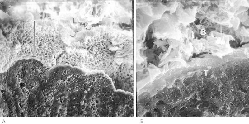
Figure 6-3. (A) Scanning electron micrograph of a cross-section of rat colonic mucosa.

Figure 6-3

(A) Scanning electron micrograph of a cross-section of rat colonic mucosa. The bar indicates the thick layer of bacteria between the mucosal surface and the lumen (L) (X 262,) (B) Higher magnification of the area indicated by the arrow in Fig. A, showing a mass of bacteria (B) immediately adjacent to colonized intestinal tissue (T), (X2,624.) (Figure from Davis CP: Preservation of bacteria and their microenvironmental association in the rat by freezing. Appl Environ Microbiol 31:310,1976, with permission.)

From: Chapter 6, Normal Flora

Cover of Medical Microbiology
Medical Microbiology. 4th edition.
Baron S, editor.
Copyright © 1996, The University of Texas Medical Branch at Galveston.

NCBI Bookshelf. A service of the National Library of Medicine, National Institutes of Health.