This is an open-access article distributed under the terms of the Creative Commons Attribution License, which permits unrestricted use, distribution, and reproduction in any medium, provided the original work is properly cited.
NCBI Bookshelf. A service of the National Library of Medicine, National Institutes of Health.
StemBook [Internet]. Cambridge (MA): Harvard Stem Cell Institute; 2008-. doi: 10.3824/stembook.1.67.1
Introduction
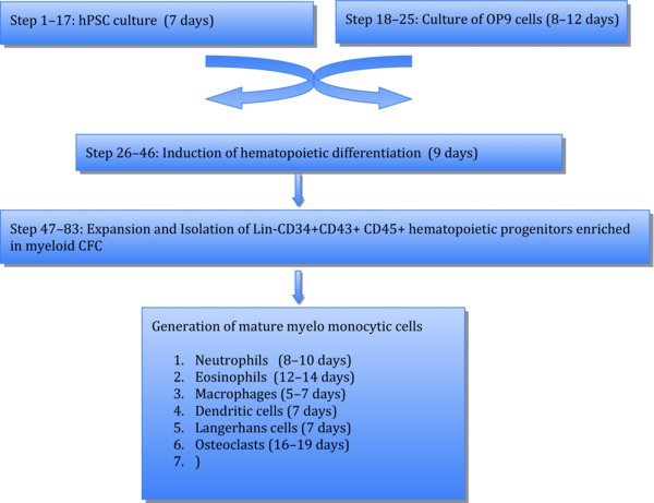
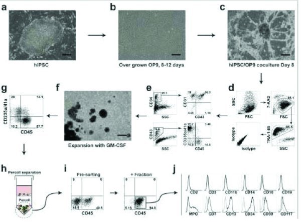
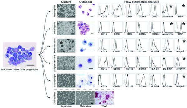
A protocol for hematopoietic differentiation of human pluripotent stem cells (hPSCs) and generation of mature myeloid cells from hPSCs through expansion and differentiation of hPSC-derived lin−CD34+CD43+CD45+ multipotent progenitors. The protocol comprises three major steps: (i) induction of hematopoietic differentiation by coculture of hPSCs with OP9 bone marrow stromal cells; (ii) short-term expansion of multipotent myeloid progenitors with a high dose of granulocyte-macrophage colony-stimulating factor; and (iii) directed differentiation of myeloid progenitors into neutrophils, eosinophils, dendritic cells, Langerhans cells, macrophages and osteoclasts. The generation of multipotent hematopoietic progenitors from hPSCs requires 9 d of culture and an additional 2 d to expand myeloid progenitors. Differentiation of myeloid progenitors into mature myeloid cells requires an additional 5–19 d of culture with cytokines, depending on the cell type.
Flow Chart
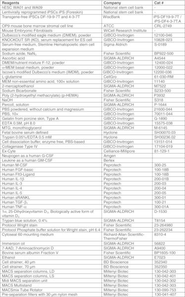
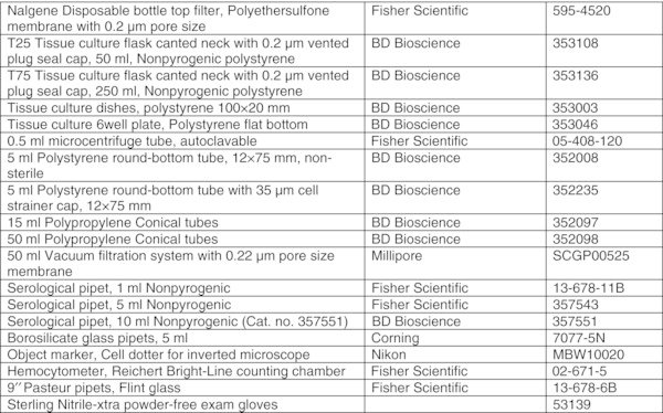
- hESC WA01 and WA09 (National KNOCKOUT SR (KO), serum replacement for ES cell (GIBCO-Invitrogen, Cat. no. 10828-028). CRITICAL: Each lot should be tested for its suitability for hPSC culture.
- Fetal bovine serum defined (Hyclone, Logan, UT, USA, Cat. no. SH30070.03). CRITICAL: FBS used in this protocol is defined FBS. Use FBS directly without the heat inactivation step for OP9 culture, hematopoietic differentiation, expansion of myeloid progenitors, and the generation of mature myelomonocytic cells from human pluripotent stem cells. Heat inactivation does not benefit culture but usually results in a higher adipogenic effect on OP9. CRITICAL: We have found that different lots of HyClone defined FBS provide relatively stable hematopoietic differentiation in hPSC/OP9 coculture and support efficient OP9 growth without significant adipogenesis. Results from other suppliers are more variable.
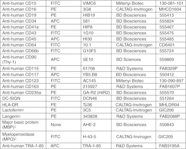
Antibody list

Antibodies used to analyze differentiation of hematopoietic progenitors and myeloid lineages from human pluripotent stem cells

Reagents setup
Collagenase solution (1 mg/ml): Add 50 mg of collagenase to 50 ml of DMEM/F-12 basal medium, and sterilize the solution by filtration using a 0.22 μm membrane filter. Keep solution at 2–8°C and use for up to one week.
0.1% Gelatin solution (w/v): Add 500 mg of gelatin to 500 ml of endotoxin-free reagent grade distilled water. Solubilize and sterilize by autoclaving for 20 min at 121°C. Store the solution at 4°C for up to 6 months. Keep sterile.
Magnetic cells sorting (MACS) buffer: MACS buffer contains 5% FBS (v/v) and 2 mM EDTA in PBS (Ca2+ and Mg2+ free). For 500 ml, add 25 ml of FBS and 2 ml of 0.5M EDTA (pH 8.0) into Ca2+ and Mg2+ free-PBS. Sterilize MACS buffer by filtration using a 0.22 μm membrane filter and keep at 2–8°C for up to 6 months. Optional: After filtration, close lid of filter unit and keep MACS buffer under vacuum for about 10–15 min for degassing.
Flow cytometry buffer: Flow cytometry buffer contains 2% FBS (v/v), 0.05% sodium azide (NaN3, w/v) and 2 mM EDTA in PBS (Ca2+ and Mg2+ free). For 500 ml, add 10 ml of FBS, 0.25 g of NaN3 and 2 ml of 0.5 M EDTA (pH 8.0) into Ca2+ and Mg2+ free-PBS. Filtrate the buffer using a 0.22 μm membrane filter and store at 2–8°C for up to 6 months.
10% pHEMA coating solution (w/v): Add 4 g of pHEMA to 40 ml of 95% ethanol containing 10 mM NaOH. Dissolve completely by continuously rotating at room temperature or 37°C overnight. Store at room temperature until needed for use. CAUTION: It is very hard to dissolve pHEMA crystals completely when they aggregate. Thus, shake the solution immediately after addition of pHEMA to prevent precipitation.
5× Percoll solution: Add 5 ml of 10× PBS (Ca2+ and Mg2+ free) to 45 ml of Percoll solution (90% Percoll and 10% 10× PBS). Store at 4°C for 6 months. For 1× Percoll working solution, dilute 10 ml of 5× Percoll solution in 40 ml of 1× PBS (1/5 dilution). Use fresh.
100× (100 mM) L-glutamine/2-mercaptoethanol solution. Add 146 mg of L-glutamine and 7 μl of 2-mercaptoethanol to 10 ml of PBS (Ca2+ and Mg2+ free). Sterilize the solution by filtration using a 0.22 μm membrane filter and store up to 2 weeks at 2–8°C.
1000× (100 mM) MTG solution: Add 87 μl of MTG to 10 ml of endotoxin-free reagent grade distilled water. Mix well and divide into 500 μl aliquots. Store up to 6 months at −20°C. CAUTION: MTG has high viscosity, thus pipet slowly to dispense MTG accurately.
1000× (50 mg/ml) ascorbic acid solution: Add 500 mg of ascorbic acid to 10 ml of endotoxin-free reagent grade distilled water. Dissolve completely, divide into 500 μl aliquots, and store up to 6 months at −20°C.
1 mM 1α, 25-Dihydroxyvitamin D3 stock solution: Dissolve 10 μg of 1α, 25-Dihydroxyvitamin D3 in 24 μl of 95% ethanol (Final concentration is 0.42 μg/μl in 95% EtOH). Store the stock solution at −20°C for up to 6 months.
50× 7-AAD solution: Dissolve 1 mg of 7-AAD in 50 μl of absolute methanol, then add 950 μl of 1× PBS. Final concentration is 1 mg/ml. Store the solution in an amber glass bottle or tube at 4°C protected from light. Solution can be stored for at least up to 6 months. For working solution, the stock solution (1 mg/ml) is diluted with flow cytometry buffer to 20 μg/ml concentration. Use 10 μl for staining.
0.1% BSA/PBS solution: Dissolve 25 mg of Bovine serum albumin Fraction V in 25 ml of PBS (Ca2+ and Mg2+ free). Sterilize the solution by filtration using a 0.22 μm membrane filter and store for up to 6 months at 2–8°C.
Reconstitution of cytokines: Centrifuge vials at maximum speed for 1 min to precipitate lyophilized pellet prior to opening vials. Reconstitute cytokines according to the product information provided by manufacturer. Dilute with 0.1% BSA/PBS solution for working concentration and store at −80°C until needed for use.
Gelatin-coated 10 cm culture dish and 6 well tissue-culture plate: Add 7–8 ml of autoclaved gelatin solution to a 10 cm culture dish or 2 ml to each well of a 6 well tissue-culture plate. Allow the gelatin solution to cover the entire plastic surface and incubate for at least 3 hrs at 37°C in an incubator. Dishes and 6-well plates containing gelatin solution can be stored for up to several days at 37°C in CO2 incubator. Do not allow the wells to dry. Before use, aspirate the gelatin solution from the dish.
pHEMA-coated culture flask: Add 5 ml of 10% pHEMA/ethanol solution to a T75 tissue culture flask or 2 ml of the solution to a T25 tissue culture flask. Rotate the flask gently to allow pHEMA solution to cover the entire surface of the flask. Make sure that the flask is completely covered with pHEMA solution. Tip the flask to remove excess pHEMA solution and save to reuse. After coating, dry the flask overnight in a sterile biosafety cabinet, close the cap and store under sterile conditions at room temperature until needed for use. CRITICAL Treatment with pHEMA should be done quickly to avoid irregular coating due to rapid ethanol evaporation.
Reconstitution of cytokines
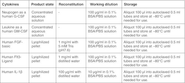
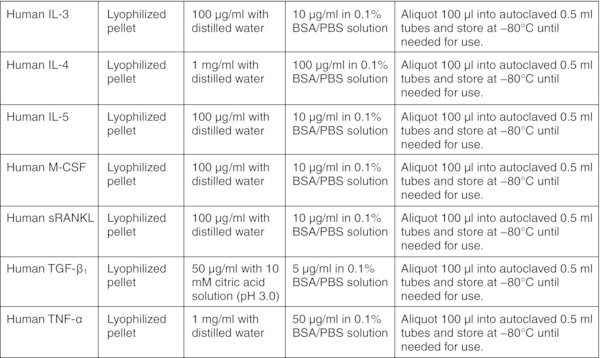
CRITICAL: Perform entire procedure in a sterile biosafety cabinet. Read the product information sheet carefully before preparation of working aliquots of all cytokines.
Medium composition
All basal medium, α-MEM, DMEM, DMEM/F-12, and IMDM should be prepared freshly from powder according to the manufacturer's instructions, sterilized by filtration using a 0.22 μm membrane filter and stored for up to 2 months at 2–8°C. Figures:


Eosinophil differentiation medium


MEFs preparation for Human ES/iPSC culture TIMING 24 hours
- Prepare MEFs according to WiCell protocol (http://www.wicell.org/index.php?option=com_content&task= category§ionid=7&id=246&Itemid=248).
- Inactivate MEFs with gamma irradiation at 8,000 rad
- Resuspend MEFs at 2×10⁵ cells/ml in prewarmed MEF growth.
- Add 2 mL/well of prewarmed MEF growth medium and then dispense MEF suspension on gelatin- coated 6-well plate (1 ml/well). CRITICAL STEP Distribute MEFs evenly with a back/forth and right/left movement twice. Irregular distribution of MEFs may cause death and unwanted differentiation of hPSC colonies during culture.
- Incubate MEF plates in a CO2 incubator at 37°C for at least 24 hrs before adding hPSCs. CRITICAL STEP MEFs should be used for hPSC passage within one week. Before plating hPSCs, aspirate MEF medium, add 2 ml of PBS, swirl once and aspirate PBS. Add 2 ml of prewarmed hPSC medium and place plate into CO2 incubator at 37°C. Now MEF feeders are ready for hPSC plating (step 15).hES/iPSC culture TIMING 7 days
- Aspirate hPSC growth medium from one well of the 6-well plate of hESCs or iPSCs. CRITICAL STEP: Note that cells will need to be split every 6–7 days.
- Wash cells with 2 ml/well of PBS (Ca2+ and Mg2+ free) stored at room temperature.
- Add 1 ml/well of collagenase IV solution (1 mg/ml) and incubate at 37°C in a CO2 incubator until the edges of the hPSC colonies begin to curl (approximately 7–10 minutes).
- Add 1 ml of hESC or hiPSC growth medium and break up the colonies into small cell aggregates by gently pipetting. CRITICAL STEP: After collagenase treatment, hPSC colonies are loosely attached and can be collected by gentle pipetting. Do not use excessive mechanical force or scraping which can provoke spontaneous differentiation.
- Transfer cells to a 15 ml conical tube.
- Centrifuge at 200×g at room temperature for 3–5 min.
- Aspirate the medium gently without disturbing the pellet.
- Resuspend cells in 3 ml of hESC or hiPSC growth medium, and wash cells by repeating steps 11 and 12.
- Resuspend cell pellet in 3 ml of hESC or hiPSC growth medium.
- Plate 0.5 ml/well of cell suspension onto MEF-grown 6-well plate from Step 5.Troubleshooting
- Feed hPSCs daily by replacing the old medium with 3 ml of prewarmed hESC or hiPSC medium.
- Passage undifferentiated hES/iPSCs (Fig. 1a) weekly at 1.2–1.5×10⁶ cells/well density on MEFs. CRITICAL STEP: If spontaneous differentiation of hESCs or hiPSCs occurs, differentiated hPSC colonies should be eliminated during the maintenance; observe hPSCs every day before changing medium. Mark differentiated colonies with an objective marker under the inverted microscope and aspirate marked areas using a glass Pasteur pipette while feeding hPSC with fresh medium. CRITICAL STEP: Alternatively, hPSCs can be maintained under feeder-free conditions.32 In OP9 coculture system, we did not observe significant differences in the efficiency of hematopoietic differentiation of hPSCs maintained on MEFs or in feeder-free cultures.
 Culture of mouse OP9 cells TIMING 8–12 days
 - Aspirate OP9 growth medium and wash cells twice with 10 ml of PBS. CRITICAL STEP: Note that cells will need to be split every 4 days.
- Add 5 ml of trypsin/EDTA(0.05%/0.5 mM) solution and incubate for 5 min at 37°C in a CO2 incubator. CRITICAL STEP: OP9 feeders consist of heterogeneous cell populations which include cells with at least adipogenic and osteogenic potential. To maintain a proper balance of cells following passage, OP9 feeders should be digested and detached completely by trypsin treatment. Inadequate washing of cells with PBS or using an old trypsin may results in partial detachment and enrichment in adipogenic cells.
- Add 5 ml of OP9 growth medium and collect cells by pipetting.
- Transfer cell suspension into a 15 ml conical tube and centrifuge for 5 min at 300×g at room temperature.
- Aspirate supernatant and resuspend cells in 1 ml of OP9 growth medium.
- Add 100 μl of cell suspension to 10 ml of OP9 growth medium and plate cells onto 10 cm gelatin-coated culture dishes. CRITICAL STEP: It is essential to culture OP9 on gelatin-coated plates to prevent spontaneous adipogenesis.Troubleshooting
- When cultures are confluent, split 1 dish for maintenance. This should occur after approximately 4 days of growth.
- To prepare overgrown OP9 for coculture with hPSCs, change half of the medium after 4 days of culture on gelatin-coated plates and incubate for an additional 4–8 days to achieve a dense OP9 monolayer. CRITICAL STEP: OP9 should be split every 4 days for maintenance/expansion. OP9 used for hPSC differentiation should be fed with fresh media at confluence (day 4) and incubated for an additional 4–8 days to form a dense monolayer embedded in extracellular matrix.Hematopoietic differentiation on OP9 TIMING 9 days
- Remove overgrown OP9 dishes prepared for coculture from the CO2 incubator.
- Aspirate OP9 growth medium.
- Add 10 ml of differentiation medium and keep at 37°C in a CO2 incubator.
- From one well of a 6-well hPSC plate from Step 17, aspirate hES/hiPSC growth medium. Add 1 ml of collagenase IV solution (1 mg/ml), and incubate cells for 10 min at 37°C.
- Add 1 ml/well of differentiation medium directly to the well and break up colonies into small cell aggregates by gentle pipetting. Transfer cells into a 15 ml conical tube. CRITICAL STEP: hPSCs for differentiation studies should be prepared as small aggregates. Single hPSCs will not survive on OP9.
- Centrifuge cells at 200×g for 3–5 min at room temperature.
- Aspirate the medium gently without disturbing the pellet.
- Resuspend the cell pellet with 1 mL of differentiation medium.
- Add 1 mL of hES/hiPSC suspension to 1 OP9 dish prepared in steps 26–28. CRITICAL STEP: Efficiency of hematopoietic differentiation is significantly affected by the density of hPSCs plated on OP9. In our experience, the optimal plating density of hPSCs is 1.0–1.5 × 10⁶ cells per 10 cm dish of OP9. To estimate the number of cells in a suspension consisting of small hPSC aggregates, one well of a 6-well plate can be used to prepare a single cell suspension by treatment with trypsin for cell counting. Alternatively, aliquots of hPSC aggregates can be collected, treated with trypsin, and counted.
- Distribute cells evenly with a back/forth and right/left movement twice.
- The following day (day 1), aspirate all of the media to waste and replace with 20 ml of prewarmed differentiation medium.
- On day 4, change half of the medium.
- On day 6, change half of the medium
- To collect cells on day 9, aspirate the supernatant and add 5 ml of prewarmed collagenase solution (1 mg/ml) to each dish of hPSC/OP9 coculture and incubate for 30 minutes at 37°C in a CO2 incubator.
- Remove the collagenase solution and keep it on ice in a 15 ml conical tube for subsequent collection of trypsin digested cells (cell collection tube). CRITICAL STEP: hPSC/OP9 coculture produces collagen-rich matrix and pretreatment with collagenase is essential to achieve efficient digestion of cells with trypsin. Because some cultures might form an excessive amount of extracellular matrix, the time of treatment with collagenase can be extended up to 40–50 minutes to achieve complete dissociation and maximize cell recovery.
- Add 5 ml of prewarmed Trypsin/EDTA solution (0.05%/0.5 mM) to the dish from Step 39 and incubate for 15–20 minutes at 37°C CO2 incubator.
- Add 2 ml/dish of MACS buffer, suspend coculture cells by pipetting and transfer to the collection tube from Step 40.
- Add an additional 5 ml/dish of MACS buffer to the coculture dish and collect the remaining cells into the collection tube.
- Centrifuge cell suspension at 300×g for 5 min at room temperature.
- Wash cells once by adding 5 ml of MACS buffer to cell pellet followed by pipetting and centrifugation at 300×g for 5 min at room temperature.
- Cells are ready to use in further applications such as flow cytometry, CFC assay, and further differentiation. CRITICAL STEP: The success of subsequent steps in this differentiation protocol largely depends on effective induction of hematopoietic differentiation and lin-CD34+CD43+CD45+ progenitors in coculture with OP9. Therefore analysis of CD43 expression and simultaneous detection of CD235a/CD41a+ and CD45+ cells within CD43+ population can be performed to confirm myeloid commitment28 (see Fig. 1). CRITICAL STEP: Because we observed a decline in the hematopoietic differentiation capacity of hESCs after passage 50, we do not recommend the use of hESC lines beyond this passage. OP9 cells should not be used beyond passage 60, because of significant decrease in hematopoiesis-inductive potential.TroubleshootingShort-term expansion of multipotent myeloid progenitors TIMING 2 days
- Wash out pHEMA-coated flasks with 20 ml (T75 flask) PBS.
- Resuspend differentiated hPSCs (from Step 46) in multipotent myeloid progenitor expansion medium at a concentration of ∼1×10⁶ cells/ml. CRITICAL STEP: Note that typically, 1.5–2×10⁷ cells are recovered from one 10 cm dish of hPSC/OP9 coculture. We usually culture cells collected from 2 dishes in one T75 flask. GM-CSF is a single key factor required for expansion of hPSC-derived myeloid progenitors. The addition of SCF and/or FLT3L to expansion cultures has little effect on the growth of myeloid precursors, but significantly increases the proportion of CD235a+ erythroid cells.
- Incubate 2 days at 37°C in a CO2 incubator. CRITICAL STEP: Differentiated hPSCs cultured in non-adherent conditions spontaneously reaggregate and form large floating cellular conglomerates with myeloid progenitors proliferating in suspension as single cells.Purification of multipotent myeloid progenitors TIMING 3 hrs
- Collect myeloid cultures (from Step 49) and filter through a 70 μm cell strainer into a 50 ml tube to remove cell aggregates.
- Pellet the cells by centrifugation at 250×g for 5 min at room temperature
- Resuspend cells with 5 ml of MACS buffer in a 15 ml tube.
- Underlay cell suspension with 1.0–1.5 ml of Percoll solution. Place a 1 ml plastic serological pipet filled with Percoll solution into the tube with cell suspension, so the pipet tip touches the bottom of the tube. Very carefully dispense Percoll solution to underlay cell suspension avoiding mixing.
- Centrifuge tube at 300×g for 10–15 min at room temperature.
- Aspirate supernatant and interface containing dead cells and debris.
- Resuspend cells with 5 ml of MACS buffer and centrifuge at 250×g for 5 min at room temperature. Take an aliquot of the resuspended cells before centrifugation to count the number of isolated cells.
- Aspirate supernatant and add 0.2 ml of MACS buffer to cell pellet.
- Add 1 μl of anti-human CD235a-PE, and 5 μl of anti-human CD41a-PE antibodies per 106 cells. CRITICAL STEP: Usually cells collected after Percoll separation are free of residual OP9 cells. If significant contamination of human cells with OP9 cells occurs, 10 μl of anti-mouse CD29-PE Ab can optionally be added to the cell pellet to deplete the mouse cells. The presence of contaminating OP9 cells in suspension can be evaluated by flow cytometry using mouse-specific CD29 antibodies.28
- Set up tube on the MACS mixer and incubate at the lowest rotation speed at 4°C for 15–20 minutes.
- Wash cells with ice-cold MACS buffer by adding 5 ml of MACS buffer to cell pellet followed by pipetting and centrifugation at 300×g for 5 min at 4°C, resuspend in 0.4 ml of MACS buffer, and add 10 μl of anti-PE magnetic beads.
- Repeat step 59.
- Wash cells with ice-cold MACS buffer as described in step 60 and resuspend in 1 ml of MACS buffer.
- Filter cells through a 30 μm pre-separation filter. CRITICAL STEP: It is important to filter cells before magnetic separation to remove large cell aggregates which may block the magnetic column.
- Assemble the MACS-LD separation column according to the manufacturer's instructions.
- Wash column with 2 ml of MACS buffer.
- Apply the cell suspension from Step 63 to the LD column allowing cells to pass completely through the column into 15 ml collection tube.
- Wash column with 2 ml of MACS buffer and collect in same collection tube.
- Recap and remove collection tube with unlabeled cells (CD235a-CD41a- human cells).
- Centrifuge cells at 300×g for 5 min at 4°C.
- Aspirate supernatant and add 80 μl of MACS buffer and 20 μl of anti-human CD45-FITC Ab.
- Place the tube on the MACS mixer and incubate at the lowest rotation speed at 4°C for 15–20 minutes.
- Wash cells with ice-cold MACS buffer as described in step 60, resuspend in 80 μl of MACS buffer, and add 20 μl of anti-FITC magnetic beads.
- Repeat step 69.
- Wash cells with ice-cold MACS buffer as described in step 60 and resuspend in 1 ml of MACS buffer.
- Filter cells through a 30 μm pre-separation filter.
- Assemble MACS-LS separation unit according to the manufacturer's instructions.
- Rinse column with 2 ml of MACS buffer.
- To purify CD235a/CD41a−CD45+ multipotent myeloid progenitors, apply the cell suspension from Step 75 to the LS column allowing cells to pass completely through the column into collection tube.
- Wash column with 2 ml of MACS buffer and collect in same collection tube then discard.
- Remove the column from the magnet and place in an empty 15 ml tube.
- Wash out CD45+ cells with 5 ml MACS buffer using the plunger supplied with column.
- Centrifuge cells at 300×g for 5 min at 4°C.
- Resuspend cells in 0.2 ml of MACS buffer and keep on ice. Cells are ready to use for further differentiation.TroubleshootingDifferentiation of hPSC-derived myelomonocytic cells
- Differentiate hPSC-derived lin−CD34+CD43+CD45+ progenitors using option A for neutrophils, option B for eosinophils, option C for macrophages, option D for DCs, option E for LCs, and option F for osteoclasts. Protocol for cytospin preparation and staining of differentiated cells is provided as option G.
(A) Neutrophil differentiation TIMING 8–10 days
- Prepare 6-well plates with semiconfluent OP9 monolayer.
- Add 5×10⁴ purified CD45+ cells in 2.5 ml of neutrophil differentiation medium per well of the 6-well plate.CRITICAL STEP: To obtain a pure population of neutrophils, it is essential to limit cytokine addition to G-CSF alone. Although IL-3, IL-6, and GM-CSF have been used to increase the output of neutrophils from somatic CD34+ cells, we found that addition of these cytokines to the neutrophil differentiation cultures resulted in production of a mixture of eosinophils and neutrophils, with eosinophils often predominating.
- On day 3, add 2.5 ml of neutrophil differentiation medium.
- On day 6, change half of the neutrophil differentiation medium.
- On day 8–10, collect differentiated cells from OP9 by gentle pipetting.CRITICAL STEP: The presence of mature neutrophils in cultures can be quickly evaluated using Wright-stained cytospins (see protocol G below). The mature neutrophils have segmented nucleus (usually 2–5 segments joined by thin filaments) and pale pink cytoplasm (see Fig. 2). Because the viability and functionality of neutrophils are decreased with the extension of culture time, the optimal time for cell collection should be determined using vital dye staining and functional analysis of collected cells.
(B) Eosinophil differentiation TIMING 12–14 days
- Prepare 6-well plates with semiconfluent OP9 monolayer.
- Add 5×10⁴ purified CD45+ cells in 2.5 ml of eosinophil differentiation medium per well of the 6-well plate.CRITICAL STEP: IL5 alone is sufficient to differentiate hPSC-derived myeloid progenitors into eosinophils, however the addition of IL3 significantly increases the total cell output.
- On day 3, add 2.5 ml of eosinophil differentiation medium.
- Change half of the eosinophil differentiation medium on days 6 and 9.
- On day 12–14, collect differentiated cells by gentle pipetting.CRITICAL STEP: The presence of mature eosinophils in cultures can be quickly evaluated using Wright-stained cytospins (see protocol G below). The mature eosinophils have classic bilobed nucleus and abundant eosinophilic cytoplasm filled by numerous coarse, orange-red granules of uniform size (see Fig. 2). In contrast, immature myeloid cells have round nucleus and less abundant basophilic or amphophilc cytoplasm. The optimal collection time for eosinophils should be determined based on predominance of viable mature cells in cultures.
(C) Macrophage differentiation TIMING 5–7 days
- Wash a pHEMA-coated T25 tissue culture flask with 10 ml of PBS.
- Add 1–5×10⁵ purified CD45+ cells in 5 mL of macrophage differentiation medium to the T25 flask.CRITICAL STEP: M-CSF and IL-1β are two critical cytokines for the differentiation of myeloid precursors into macrophages. We do not recommend using GM-CSF in macrophage differentiation cultures, because cells continue to proliferate and do not mature into macrophages in presence of GM-CSF.
- At day 3, add an additional 5 ml of macrophage differentiation medium.
- At day 5–7, collect differentiated cells.CRITICAL STEP: The presence of mature macrophages in cultures can be quickly evaluated using Wright-stained cytospins (see protocol G below). The macrophages have round nucleus and abundant foamy cytoplasm (see Fig. 2). Because the viability and functionality of macrophages are decreased with the extension of culture time, the optimal time for cell collection should be determined using vital dye staining and functional analysis of collected cells.
(D) Dendritic cell differentiation TIMING 7 days
- Wash a pHEMA-coated T25 tissue culture flask with 10 ml of PBS.
- Add 5×10⁵ isolated CD45+ cells in 5 ml of DC differentiation medium without TNF-α to the T25 flask.CRITICAL STEP: It is essential to avoid adding TNF-α during the first day of culture. We noted a significant decrease in cell viability if TNF-α was added to the freshly isolated cells.
- On day 2–3, add 5 ml of DC differentiation medium with TNF-α (2.5 ng/ml).
- On day 7, collect differentiated cells.CRITICAL STEP: On Wright-stained cytospins, cells of dendritic lineage can be recognized by the presence of cytoplasmic veils and dendritic projections (see Fig. 2). However, flow cytometric analysis20,28 is essential to identify myeloid DCs which have CD1a+HLA-DR+DC-SIGN+CD11b+Langerin− phenotype.
(E) Langerhans cell differentiation TIMING 7 days
- Wash a pHEMA-coated T25 tissue culture flask with 10 ml of PBS.
- Add 5×10⁵ cells in 5 ml LC differentiation medium without TNF-α and TGF-β1 to the T25 flask.CRITICAL STEP: It is essential to avoid adding TNF-α and TGF-β1 during the first day of culture. We noted a significant decrease in cell viability if these cytokines were added to the freshly isolated cells.
- On day 2–3, add 5 ml of complete LC differentiation medium with TNF-α and TGF-β1 (2.5 ng/ml).
- On day 7, collect differentiated cells.CRITICAL STEP: On Wright-stained cytospins, cells of dendritic lineage can be recognized by the presence of cytoplasmic veils and dendritic projections (see Fig. 2). However, flow cytometric analysis20,28 is essential to identify LCs which have CD1ahighHLA-DR+DC-SIGN−CD11blowLangerin+ phenotype.
(F) Osteoclast differentiation TIMING 16–19 days
- Expansion step for osteoclast precursors. Wash a pHEMA-coated T25 tissue culture flask with 10 ml of PBS.
- Add 5×10⁴ to 1×10⁵ cells in 5–10 ml of osteoclast progenitor expansion medium to the T25 flask.
- Change half of the medium on day 2–3.
- On day 4–5, collect cells; during the expansion step, osteoclast precursors adhere to the surface of the pHEMA coated-culture flask. Therefore, treat with 3 ml PBS (Ca2+ and Mg2+ free) or enzyme free cell dissociation buffer for 5–10 min and collect cells by gentle pipetting.
- Maturation step for osteoclasts. Plate 1–2×10⁵ cells/well of a 6-well plate in 2.5 ml of osteoclast maturation medium.
- Change half of the osteoclast maturation media every 3 days until the osteoclasts mature (12–14 days of culture). CRITICAL STEP: Large, adhesive and multinucleated cells are markers for mature osteoclasts. However, immature myeloid cells (small round and non-adherent cells) keep proliferating due to the presence of GM-CSF and these proliferating cells interfere with maturation of the osteoclasts. Therefore, eliminate non-adherent cells by aspiration during regular medium changes. We found that the addition of M-CSF had no effect on development of osteoclasts in our differentiation system. Moreover, the addition of M-CSF to osteoclast cultures shifted differentiation hiPSC-derived myeloid progenitors toward macrophages.
(G) Cytospin prepartion and Wright staining
- Use 2.5–3×10⁴ cells per slide for Cytospin preparation and Wright staining
- Cytospin preparation. Collect cells in 1.5 ml microcentrifuge tube and centrifuge at 400×g for 4 min.
- Wash cells once by adding ice cold 0.5 ml of flow cytometry buffer followed centrifugation at 400×g for 4 minutes. Discard supernate and resuspend cells with 250 μl of flow flow cytometry buffer.
- During centrifugation, label glass slide and assemble with Cytofuse filter concentrator unit according to manufacturer's instruction.
- Transfer cell suspension into sample chamber of assembled glass slide-Cytofuse filter concentrator unit.
- Spin samples at 700 rpm (27 ×g) for 4 min at room temperature.
- Dry slide completely at room temperature.
- Fix slide with absolute methanol for 30–60 seconds at room temperature.
- Wright staining. Cover cytospin area with 300 μl of Protocol Wright Stain and allow to stain for 3 minutes at room temperature. Make sure that stain covers the entire area with cells to ensure uniform staining of cells on cytospin.
- Add 450 μl of pH 6.4 Diluted Buffer to the Wright stain-covered slide.
- Mix the stain and buffer together by gently rocking slide for 1 minute and incubate the mixture for additional 4 minutes at room temperature.
- Rinse the slide with deionized water and dry completely in the air.
- Drop 2–3 droplets of Cytoseal 60 mounting medium on the stained cell area and cover with cover glass.
- Observe under the microscope.
References
- Choi K.-D, Vodyanik M, Slukvin I. I. Nature Protocols 6 296313 10.1038/nprot.2010.184, Published online 17 February 2011 2011 [PMC free article: PMC3066067] [PubMed: 21372811] [CrossRef]
Hematopoietic differentiation and production of mature myeloid cells from human pluripotent stem cells
Last revised March 28, 2012. Published June 10, 2012. This chapter should be cited as: Choi, K.-D., Vodyanik, M., and Slukvin, I. I., Hematopoietic differentiation (June 10, 2012), StemBook, ed. The Stem Cell Research Community, StemBook, doi/10.3824/stembook.1.67.1, http://www
.stembook.org.
- Hematopoietic differentiation - StemBookHematopoietic differentiation - StemBook
- Regulation of spermatogonia - StemBookRegulation of spermatogonia - StemBook
- PREDICTED: Homo sapiens TBC1 domain family member 5 (TBC1D5), transcript variant...PREDICTED: Homo sapiens TBC1 domain family member 5 (TBC1D5), transcript variant X12, mRNAgi|2217347134|ref|XM_047449289.1|Nucleotide
- Homo sapiens cDNA FLJ58392 complete cdsHomo sapiens cDNA FLJ58392 complete cdsgi|221045133|dbj|AK304712.1|Nucleotide
- Homo sapiens mucolipin 1, mRNA (cDNA clone MGC:3287 IMAGE:3507836), complete cdsHomo sapiens mucolipin 1, mRNA (cDNA clone MGC:3287 IMAGE:3507836), complete cdsgi|33869880|gb|BC005149.2|Nucleotide
Your browsing activity is empty.
Activity recording is turned off.
See more...
