ILLUSTRATIVE CASE
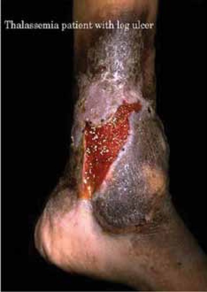
A 40-year-old man with β-thalassemia intermedia presented to his physician complaining of a painful lateral right ankle lesion. The lesion appeared a month ago and started progressively expanding and becoming more painful. He went to a primary care physician who prescribed topical antibiotics and advised continuous protection of lesion with a gauze, but the lesion did not improve. His past medical history included splenectomy at the age of 9 years and a deep vein thrombosis in his right arm at the age of 29 years. He was not taken any medications. On physical examination he had a 1 × 4 cm lesion on the lateral area of the right ankle, which was painful to palpation. The lesion did not look infected but the surrounding skin had a black hue. His laboratory work-up revealed a total hemoglobin level of 8.1 g/dl and a platelet count of 988 × 109/l. The patient was started on a once per month transfusion regimen. His leg ulcer progressively improved, and complete resolution was achieved by one year. His blood transfusions were later tapered and he was advised to start aspirin.
CONTEXT AND EVIDENCE
Leg ulcers are more common in NTDT compared with regularly-transfused β-thalassemia major patients [1-5]. The risk of leg ulcers in NTDT patients increases with advancing age [5-7]. The skin at the extremities of elderly patients can be thin due to reduced tissue oxygenation making the subcutaneous tissue fragile and increasing the risk of ulceration after minimal trauma. Severe anemia and ineffective erythropoiesis, as well as splenectomy and hypercoagulability levels have been described as risk factors for the development of leg ulcers [3, 8-9]. The hypercoagulable state and deformability of red blood cells in NTDT patients (see Chapter 6) has been incriminated in leg ulcer formation since this might cause ischemia to the skin and consequently friability and ulceration [10-11]. High venous pressure as a consequence in the subgroup of patients with right-heart failure and venous insufficiency may also be exacerbating factors [11-12]. Data on the role of fetal hemoglobin levels are conflicting. Although some propose that high fetal hemoglobin levels, by virtue of its oxygen retaining capacity, increase the risk of ulcers, other studies showed lower rates of leg ulcers in patients with high than low fetal hemoglobin levels [13]. Higher rates of leg ulcers have also been reported in NTDT patients with iron overload [7, 14-15]. Local iron overload is also thought to be a perpetuating factor causing chronicity of lesions especially when the heme from the degraded red blood cells accumulates locally and gives a dark hue [16].
Leg ulcers are often very painful and indolent. Observational studies indicate that blood transfusion or hydroxyurea therapy with or without erythropoietin may have a role [3, 12, 17]. The beneficial effects of hydroxyurea on leg ulcers in NTDT patients are not limited to fetal haemoglobin induction and improvement of anemia but also include improvement of red blood cell pathology, deformability, and hypercoagulability [18]. Pentoxifylline, which alters the rheological properties of the red blood cell, was also shown to accelerate the healing of leg ulcers [19]. The use of an oxygen chamber was also shown to provide moderate relief where tissue hypoxia may be an underlying cause of the ulceration [10]. The vasodilator dialzep (adenosine reuptake inhibitor) was shown to have some benefit in a trial of eight patients with haemoglobin E/β-thalassemia and chronic leg ulcers (three patients had total healing and four had improvement) [20]. Skin grafts have been tried by some plastic surgeons [10]. Both platelet derived wound healing factors and granulocyte macrophage colony-stimulating factor have been successfully used in some patients [21]. There is limited evidence on the benefit of anticoagulation for the management of leg ulcers in NTDT patients [11]. A recent trial has established benefit of sodium nitrite cream in patients with sickle cell disease and refractory leg ulcers [22].
PRACTICAL RECOMMENDATIONS
There is no sufficient evidence to recommend blood transfusion, iron chelation or hydroxyurea therapy for the prevention of leg ulcers in NTDT patients, although when used for different indications a beneficial effect may be observed
The skin of NTDT patients should always be inspected on routine physical examination
Patient with evidence of leg ulcers should be treated in close collaboration with a dermatologist and a plastic surgeon
Simple measures may be beneficial, such as keeping the patient’s legs and feet raised above the level of the heart for 1-2 hours during the day or sleeping with the end of the bed raised.
Topical antibiotics and occlusive dressing should be applied
Topical sodium nitrite cream may be considered
Blood transfusion should be considered as the first treatment option
The following treatment measures may also be considered in patients who have persistent leg ulcers, although no clinical trials to supporting their use exist:
REFERENCES
- 1.
Taher A, Isma’eel H, Cappellini MD. Thalassemia intermedia: revisited.
Blood Cells Mol Dis. 2006;37(1):12–20. [
PubMed: 16737833]
- 2.
- 3.
Taher AT, Musallam KM, Karimi M, El-Beshlawy A, Belhoul K, Daar S, Saned MS, El-Chafic AH, Fasulo MR, Cappellini MD. Overview on practices in thalassemia intermedia management aiming for lowering complication rates across a region of endemicity: the OPTIMAL CARE study.
Blood. 2010;115(10):1886–1892. [
PubMed: 20032507]
- 4.
Daneshmend TK, Peachey RD. Leg ulcers in alpha-thalassaemia (haemoglobin H disease).
Br J Dermatol. 1978;98(2):233–235. [
PubMed: 629879]
- 5.
Olivieri NF, Muraca GM, O’Donnell A, Premawardhena A, Fisher C, Weatherall DJ. Studies in haemoglobin E beta-thalassaemia.
Br J Haematol. 2008;141(3):388–397. [
PubMed: 18410572]
- 6.
Taher AT, Musallam KM, El-Beshlawy A, Karimi M, Daar S, Belhoul K, Saned MS, Graziadei G, Cappellini MD. Age-related complications in treatment-naive patients with thalassaemia intermedia.
Br J Haematol. 2010;150(4):486–489. [
PubMed: 20456362]
- 7.
Musallam KM, Cappellini MD, Daar S, Karimi M, El-Beshlawy A, Taher AT. Serum ferritin levels and morbidity in β-thalassemia intermedia: a 10-year cohort study [abstract] Blood. 2012;120(21):1021.
- 8.
Musallam KM, Taher AT, Duca L, Cesaretti C, Halawi R, Cappellini MD. Levels of growth differentiation factor-15 are high and correlate with clinical severity in transfusion-independent patients with beta thalassemia intermedia.
Blood Cells Mol Dis. 2011;47(4):232–234. [
PubMed: 21865063]
- 9.
- 10.
Gimmon Z, Wexler MR, Rachmilewitz EA. Juvenile leg ulceration in beta-thalassemia major and intermedia.
Plast Reconstr Surg. 1982;69(2):320–325. [
PubMed: 7034014]
- 11.
Levin C, Koren A. Healing of refractory leg ulcer in a patient with thalassemia intermedia and hypercoagulability after 14 years of unresponsive therapy.
Isr Med Assoc J. 2011;13(5):316–318. [
PubMed: 21845977]
- 12.
Gamberini MR, Fortini M, de Sanctis V. Healing of leg ulcers with hydroxyurea in thalassaemia intermedia patients with associated endocrine complications.
Pediatr Endocrinol Rev. 2004;2 Suppl 2:319–322. [
PubMed: 16462721]
- 13.
Musallam KM, Sankaran VG, Cappellini MD, Duca L, Nathan DG, Taher AT. Fetal hemoglobin levels and morbidity in untransfused patients with beta-thalassemia intermedia.
Blood. 2012;119(2):364–367. [
PubMed: 22096240]
- 14.
Musallam KM, Cappellini MD, Taher AT. Evaluation of the 5mg/g liver iron concentration threshold and its association with morbidity in patients with beta-thalassemia intermedia.
Blood Cells Mol Dis. 2013 [
PubMed: 23425967]
- 15.
Musallam KM, Cappellini MD, Wood JC, Motta I, Graziadei G, Tamim H, Taher AT. Elevated liver iron concentration is a marker of increased morbidity in patients with beta thalassemia intermedia.
Haematologica. 2011;96(11):1605–1612. [
PMC free article: PMC3208677] [
PubMed: 21791471]
- 16.
Ackerman Z. Local iron overload in chronic leg ulcers.
Isr Med Assoc J. 2011;13(10):647. [
PubMed: 22097241]
- 17.
al-Momen AK. Recombinant human erythropoietin induced rapid healing of a chronic leg ulcer in a patient with sickle cell disease.
Acta Haematol. 1991;86(1):46–48. [
PubMed: 1950363]
- 18.
Musallam KM, Taher AT, Cappellini MD, Sankaran VG. Clinical experience with fetal hemoglobin induction therapy in patients with beta-thalassemia.
Blood. 2013;121(12):2199–2212. [
PubMed: 23315167]
- 19.
Dettelbach HR, Aviado DM. Clinical pharmacology of pentoxifylline with special reference to its hemorrheologic effect for the treatment of intermittent claudication.
J Clin Pharmacol. 1985;25(1):8–26. [
PubMed: 3882773]
- 20.
Opartkiattikul N, Sukpanichnant S, Wanachiwanawin W, Fucharoen S, Funahara Y, Sumiyoshi A, Imai K, Sangtawesin W, Thientadakul P. A double-blind placebo control trial of dilazep in beta-thalassemia/hemoglobin E patients.
Southeast Asian J Trop Med Public Health. 1997;28 Suppl 3:167–171. [
PubMed: 9640622]
- 21.
Josifova D, Gatt G, Aquilina A, Serafimov V, Vella A, Felice A. Treatment of leg ulcers with platelet-derived wound healing factor (PDWHFS) in a patient with beta thalassaemia intermedia.
Br J Haematol. 2001;112(2):527–529. [
PubMed: 11167859]
- 22.
Minniti CP, Hon YY, Gorbach A, Delaney KM, Xu D, Malik N, Maivelett J, Novelli EM, Lanzkron S, Kato GJ. A phase 1, dose-escalation study of topical sodium nitrite in patients with sickle cell anemia and leg ulcers [abstract] Blood. 2012;120(21):86.