U.S. flag

An official website of the United States government

NCBI Bookshelf. A service of the National Library of Medicine, National Institutes of Health.

WHO guideline for screening and treatment of cervical pre-cancer lesions for cervical cancer prevention [Internet]. 2nd edition. Geneva: World Health Organization; 2021.

Cover of WHO guideline for screening and treatment of cervical pre-cancer lesions for cervical cancer prevention

WHO guideline for screening and treatment of cervical pre-cancer lesions for cervical cancer prevention [Internet]. 2nd edition.

Show details

ANNEX 5STANDARDIZED DEFINITIONS USED IN THIS GUIDELINE

5.1. Eligibility for ablative treatment

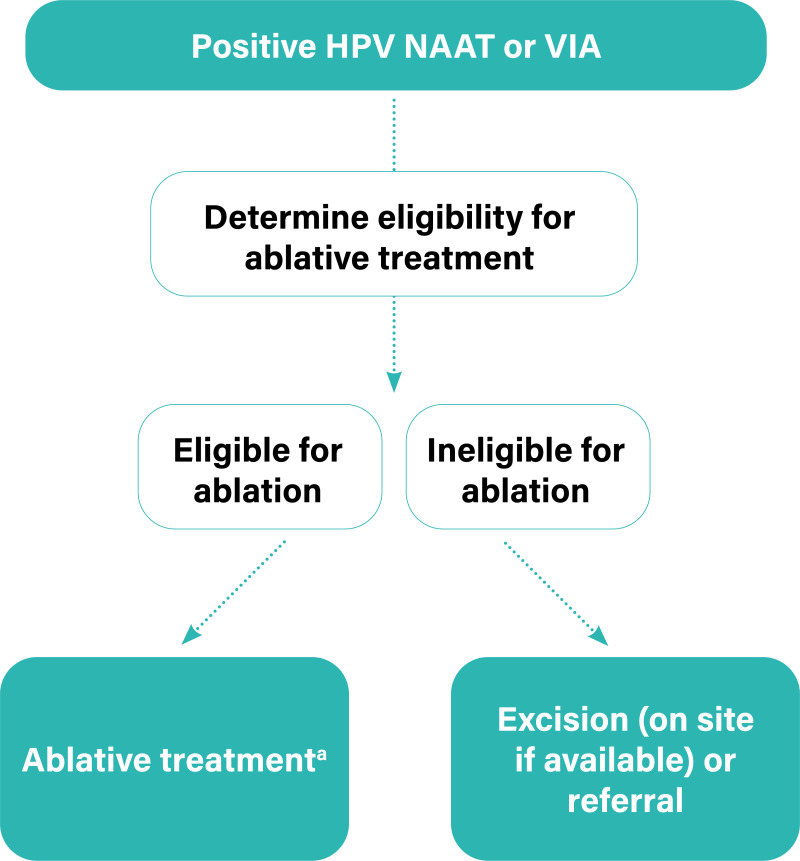
Women who screen positive with a human papillomavirus (HPV) nucleic acid amplification test (NAAT) are assessed for eligibility for ablative treatment so that women with or without a visible lesion (or lesions), who are eligible for ablation, can receive an ablative treatment (see section 5.1.2).

An assessment of eligibility for ablative treatment is done by identifying the transformation zone type (i.e. the visibility and position of the transformation zone) (see section 5.1.1), and the location and size of the lesion if the patient has a visible lesion. To assess eligibility, visualization of the cervix with magnified or naked-eye view after application of 3–5% acetic acid is required; histological diagnosis is not required.

Algorithm to determine eligibility for ablative treatment.

Algorithm to determine eligibility for ablative treatment

a Ablative treatments include cryotherapy and thermal ablation.

5.1.1. Types of transformation zone (section 5.5)1

On evaluation, the visibility and position of the transformation zone can be described as:

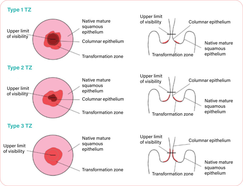
Type 1: The entire transformation zone is visible. The transformation zone is entirely visible and only ectocervical.

Type 2: The entire transformation zone is visible. The transformation zone is entirely visible and has an endocervical component.

Type 3: The transformation zone is not entirely visible. The transformation zone extends into the endocervical canal and is not fully visible.

5.1.2. Criteria for eligibility for ablative treatment1

There is no suspicion of invasive cancer or glandular disease (i.e. adenocarcinoma or adenocarcinoma in situ, AIS).

The transformation zone is fully visible, the whole lesion is visible, and it does not extend into the endocervix.

The lesion is type 1 transformation zone.

Interventions may be constrained by feasibility, training, programme quality-assurance and resources. In the absence of certain infrastructure, such as the availability of large-loop excision of the transformation zone (LLETZ) or of safe surgical settings, bridging strategies may be considered in carefully selected circumstances, to extend access to treatment rather than provide no treatment. For example, ablation may be an option for a carefully chosen, small type 2 transformation zone where the probe tip will achieve complete ablation of the squamocolumnar junction epithelium – that is, where it can reach the full extent, depth and upper limit of the transformation zone – and where adequate training for selection and follow-up is available.

5.1.3. Criteria for referral1

There is any suspicion of invasive cancer or glandular disease (i.e. adenocarcinoma or AIS).

The transformation zone is not fully visible because it is endocervical (type 2 or 3 transformation zone).

5.2. Outcome definitions

5.2.1. Persistent, residual or recurrent disease (histopathologically verified)

Definition: Persistent or residual disease is the detection of a histopathological high-grade lesion – including histological high-grade squamous intraepithelial lesions (HSIL) as per LAST terminology2 (HSIL-cervical intraepithelial neoplasia grade 2 [CIN2] and HSIL-CIN3); high-grade CIN as per CIN terminology (CIN2 and CIN3); HSIL not specified; and AIS – diagnosed from a punch biopsy or a subsequent surgical specimen (including hysterectomy, cold knife conization [CKC] and LLETZ) at any time interval after the initial treatment was performed with no intervening documented absence of disease. Persistent or residual disease excludes cancers that have been confirmed after diagnosis with surgical specimen.

Recurrent disease is defined as detection of a histopathological high-grade lesion (as described above) that was diagnosed from a biopsy or a subsequent surgical specimen (including hysterectomy, CKC and LLETZ) following a documented absence of high-grade lesions at any time interval after the initial treatment was performed.

Caveats: Studies have used the terms “persistent/residual disease” and “recurrence” to indicate the detection of disease post-treatment without making any distinction between them. Sub-analysis may be considered to distinguish between them. However, both conditions are treatment failures. Some studies use the terms “cure” or “treatment success” to indicate the absence of persistent/residual/recurrent disease following initial treatment and have estimated the rates. The specific location of the lesion on the cervix detected at follow-up is not important since the aim of the treatment of cervical pre-cancer is to treat the entire transformation zone and not any particular lesion. Any lesion detected at follow-up should be considered as representing treatment failure. The final diagnosis is the worst outcome found on histopathology.

Symptoms: Non-specific.

Consequences: Patient needs retreatment for high-grade lesions; adverse psychological impact; resource implications (including cost, human resources, hospital services).

Assessed by: Different studies may use different assessment protocols at follow-up. Some studies may take random punch biopsies from all the treated women undergoing follow-up. Other studies may have considered negative HPV test, cytology, VIA or normal colposcopy as evidence of absence or persistent/residual/recurrent disease. Such studies do biopsies only on selected patients who are positive on HPV test, cytology, VIA and/or colposcopy. These different methods of assessment should not exclude any studies, but a sensitivity analysis should then be conducted. Diagnosis of cancer at follow-up should ideally be excluded from verification of treatment outcome, as cancer should have been ruled out at baseline diagnosis.

Threshold: HSIL-CIN2 and HSIL-CIN3; high-grade CIN as per CIN terminology (CIN2 and CIN3); HSIL not specified; AIS. Some studies may consider low-grade squamous intraepithelial lesions (LSIL) or CIN1 as the threshold. Data that use LSIL+ or CIN1+ as the threshold should not be included but, if presented separately, data for HSIL-CIN2+ should be used.

Treatment: Treatment of recurrent or persistent HSIL/CIN2/CIN3 includes LLETZ or ablation (depending on the eligibility for ablation and the availability of treatment techniques). AIS is treated by LLETZ or CKC. Such treatment may be administered by a colposcopist, a general practitioner or a nurse.

Time horizon: In assessing research, the presence of disease should be measured beyond six months after treatment – and 1–3 years is preferred. A sensitivity analysis should exclude six months. Recurrence may be detected several years after treatment.

5.2.2. Persistent, residual or recurrent disease (not histopathologically verified)

Definition: Persistence or residual disease may be defined as a positive follow-up screening test (e.g. VIA, HPV test or cytology) at any time after the initial treatment. The women with positive tests were not further verified by histopathology. This is usually applicable in settings that use screen-and-treat approaches.

Recurrent disease may be defined as a positive follow-up screening test (e.g. VIA, HPV test or cytology) following a documented negative test at any time after the initial treatment in a screen-and-treat approach setting. There was no histopathological verification after a positive follow-up screening test.

Caveats: Studies have used the terms “persistent/residual disease” and “recurrence” to indicate the detection of disease post-treatment without making any distinction between them. Sub-analysis may be considered to distinguish between them. However, both conditions are treatment failures.

Symptoms: Non-specific.

Consequences: Patient needs retreatment, or verification with colposcopy and/or histopathology; adverse psychological impact; resource implications (including cost, human resources, hospital services).

Assessed by: Usually the same screening test at baseline is used to detect persistent/residual/recurrent disease at follow-up. HPV type-specific persistence/recurrence (detection of same HPV genotype at different time points) may be studied with an appropriate HPV detection test.

Threshold: The cytology threshold may vary between atypical squamous cells of uncertain significance (ASCUS), LSIL and HSIL. Ideally, HSIL should be used (LSIL could be used if identified). Other thresholds: VIA-positive and HPV-positive tests (any high-risk type detected by a validated HPV test).

Treatment: Treatment may include ablation or excision. Such treatment may be administered by a colposcopist, a general practitioner or a nurse.

Time horizon: In assessing research, the presence of disease should be measured beyond six months of treatment – and 1–3 years is preferred. There should be a sensitivity analysis to exclude six months. Recurrence may be detected several years after treatment.

5.2.3. Incidence of histopathological high-grade lesions and cancers

Definition: New (incident) cases of histopathological high-grade lesions and cancers (HSIL+ or CIN2+) detected following treatment. This includes histological HSIL as per LAST terminology (HSIL-CIN2 and HSIL-CIN3); high-grade CIN as per CIN terminology (CIN2 and CIN3); HSIL not specified; AIS, invasive squamous cell cancer, adenocarcinoma, carcinoma unspecified. All categories are combined together as HSIL+ or CIN2+.

Symptoms: Non-specific.

Consequences: Patient needs treatment for high-grade lesions or invasive cancer; adverse psychological consequences; resource implications (including cost, human resources, hospital services).

Assessed by: New cases are detected either through linkage with the screening registry or by active follow-up of the screening cohort. The treated women without any disease at first follow-up are advised to attend regular screening. Further evaluation and histopathology are performed if the screening test is positive (any screening tests at any follow-up visit).

Rates of HSIL+ or CIN2+ may be computed by each year since the index treatment on the basis of the number of women under active surveillance, with the number of women in active surveillance diagnosed with CIN2/3 in that year divided by the number in active surveillance screened in that year.

The cumulative rates of HSIL+ or CIN2+ or of invasive cancer alone may be estimated as number per 100 000 person-years of observation.

Threshold: The studies may have used HSIL+/CIN2+ or CIN3+ (HSIL-CIN3/CIN3, AIS, invasive cancer) or only invasive cancer as the end point. The studies should be analysed separately.

Treatment: The treatment of high-grade lesions requires ablation or excision depending on eligibility. AIS/microinvasive cancer requires excision (CKC or LLETZ). Such treatment may be administered by a colposcopist, a general practitioner or a nurse. Invasive cancer needs radical surgery or radiation therapy in a specialized setting. Additional testing for metastasis may be needed in patients with invasive cancers.

Time horizon: Incident CIN/cancer may be detected between six months and several years after treatment.

5.2.4. Incidence of cervical cancer (includes squamous cell carcinoma, adenocarcinoma or invasive cancer unspecified)

Definition: Newly diagnosed cases of invasive cervical cancer in the population under consideration within a defined time period, typically at least six months after screening and treatment.

Symptoms: Symptoms such as vaginal discharge, irregular bleeding, pain.

Consequences: Needs further investigations and treatment; adverse psychological consequences; disability; may be fatal, depending on stage at diagnosis; resource implications (including cost, human resources, hospital services).

Assessed by: The new cases of cancers are histopathologically diagnosed through active follow-up and/or linkage to the screening registry and/or population-based cancer registry. The incidence is usually documented as number per 100 000 person-years of observation.

Thresholds: Not applicable.

Treatment: Surgery or radiation therapy with concomitant chemotherapy, depending on stage.

Time horizon: Any time after initial treatment of premalignant lesions.

5.2.5. Mortality from cervical cancer (includes squamous cell carcinoma, adenocarcinoma or unspecified invasive cancer)

Definition: Deaths reported from cervical cancer in the population under consideration following treatment within a defined time period.

Symptoms: Not applicable.

Consequences: Not applicable.

Assessed by: Linkage with the population-based cancer registry and mortality databases, active follow-up.

Thresholds: Not applicable.

Treatment: Not applicable.

Time horizon: Any time following treatment.

5.2.6. Coverage of treatment

a. Treatment coverage

Definition: The proportion of eligible women treated in a population. The number of women completing treatment in a defined time frame divided by the total number of screen-positive women who were eligible to be treated in the same time frame and multiplied by 100; the time frame is not usually more than 12 months.

Symptoms: Not applicable.

Consequences: A low proportion of treatment will increase the risk of individual women detected with invasive cancer dying from the disease; low coverage will also prevent the screening programme from having the desired impact on cervical cancer incidence and mortality.

Assessed by: Data collected through an information system/screening registry, with linkage between screening and treatment services.

Thresholds: None; the higher the value, the better the impact.

Treatment: Not applicable.

Time horizon: 12 months.

b. Treatment rate

Definition: The proportion of eligible women treated in a study. The number of women who were treated divided by the number of women who were eligible to be treated.

Symptoms: Not applicable.

Consequences: Higher rates of treatment will increase the chances that the programme will have the desired impact on cervical cancer incidence and mortality.

Assessed by: Data will be recorded during the study.

Thresholds: Not applicable.

Treatment: Cryotherapy, thermal ablation, LLETZ, referral.

Time horizon: Duration of the study.

5.2.7. Same-day treatment rate

a. Population-level same-day treatment rate

Definition: Same-day treatment rate in a screen-and-treat approach setting is the number of eligible women treated on the same day of screening in a defined time frame divided by the total number of screen-positive women who are eligible for treatment on the same day during the same time frame and multiplied by 100.

Symptoms: Not applicable.

Consequences: Low compliance will increase the risk of being detected with invasive cancer, and of death from invasive cancer; low compliance will also prevent the screening programme from having the desired impact on cervical cancer incidence and mortality.

Assessed by: Systematic data collection; health information system.

Thresholds: Not applicable.

Treatment: Not applicable.

Time horizon: Same day or same visit.

b. Study-level same-day treatment rate

Definition: The proportion of eligible women treated in a study on the same day in a screen-and-treat approach setting. This is the number of women who were treated on the same day divided by the number of women who were eligible to be treated on the same day.

Symptoms: Not applicable.

Consequences: Higher rates of treatment will increase the chances that the programme will have the desired impact on cervical cancer incidence and mortality.

Assessed by: Data will be recorded during the study.

Thresholds: Not applicable.

Treatment: Cryotherapy, thermal ablation, LLETZ, referral.

Time horizon: Duration of the study.

5.2.8. Coverage of screening

a. Screening coverage

Definition: At the population level, the cervical cancer screening coverage is the number of screening-eligible women undergoing screening in a defined time frame divided by the total number of screening-eligible women during the same time frame and multiplied by 100; the time frame is equal to the defined screening interval or the reporting period.

Symptoms: Not applicable.

Consequences: Low screening coverage will increase the risk of individual women detected with invasive cancer, and dying from invasive cancer; low coverage will also prevent the screening programme from having the desired impact on cervical cancer incidence and mortality.

Assessed by: Data may be collected through an information system/screening registry or a survey. As population-based denominators are difficult to compare across studies, these data should be abstracted.

Thresholds: Not applicable.

Treatment: Not applicable.

Time horizon: Each programme has its own cycle of estimating coverage, ranging from one year to five years, but calculating annually is preferable.

b. Rate of participation in screening

Definition: At the study level, the rate of participation in screening is the number of screening-eligible women who were screened divided by the number of women who were invited to participate in a screening programme and multiplied by 100.

Symptoms: Not applicable.

Consequences: Higher rates of participation will increase the chances that a screening programme will have the desired impact on cervical cancer incidence and mortality.

Assessed by: Data will be recorded during the study.

Thresholds: Not applicable.

Treatment: Not applicable.

Time horizon: Duration of the study.

5.2.9. Treatment side-effects and complications

Definition: A complication is a condition or an event unfavourable to the patient’s health, causing irreversible damage or needing a change in therapeutic approach, including prolonged hospital stay. Side-effects are unwanted symptoms due to treatment that are mostly temporary and resolve spontaneously. These two terms are often used interchangeably.

Complications are graded as grade I (no pharmacological treatment or surgical intervention other than mild analgesics, antipyretics or antiemetics needed), grade II (needing pharmacological treatment, including intravenous fluid or blood transfusion), grade III (needing surgical intervention, e.g. cervical stiches), grade IV (life-threatening) and grade V (death). Most studies evaluating cervical pre-cancer treatment do not use such systematic classification or distinguish clearly between side-effects and complications. The most commonly evaluated significant treatment complications are described below. The proportion of treated women having one or several of these complications is assessed.

a. Major infections (grade II+)

Definition: Post-operative infection of the genital tract needing antibiotics and/or hospital admission.

Symptoms: Systemic: fever, nausea, vomiting, dehydration; local: foul-smelling discharge, pain.

Consequences: Hospitalization, antibiotic therapy, loss of workdays, psychological harm, long-term sequelae such as infertility, chronic pelvic inflammatory disease.

Assessed by: The diagnosis of major infection is mostly based on history and clinical assessment during follow-up. Acute pelvic inflammatory disease (presence of fever and/or lower abdominal pain and/or foul-smelling purulent vaginal discharge and/or adnexal tenderness and/or pain during cervical movement) is included in major infection. The diagnosis of major infection can also require blood tests, ultrasound and/or culture/sensitivity testing to adjust treatment.

Thresholds: Minor infections causing vaginal discharge and mild pain are often indistinguishable from side-effects and are not usually considered. Major infections can be distinguished from minor infections as they may include foul-smelling discharge plus other signs such as fever, tenderness on examination or severe pain.

Treatment: Hospitalization, antibiotics and rehydration, depending on the severity of the condition.

Time horizon: Usually 3–4 days after treatment and within one month; may take a few days to a few weeks to recover.

b. Major bleeding (grade II+)

Definition: Post-operative bleeding needing intervention (vaginal packing, cervical sutures), with or without hospitalization, with or without intravenous fluid or blood transfusion.

Symptoms: Vaginal bleeding, usually with the passage of clots, with or without pain. May have symptoms due to hypotension and shock (weakness, giddiness, vomiting, loss of consciousness).

Consequences: Shock, death in severe cases; may lead to anaemia; psychological harms; loss of workdays.

Assessed by: History and clinical examination, routine blood tests.

Thresholds: Major bleeding may be distinguished from minor bleeding as major bleeding may be accompanied by the passage of clots, a fall in blood pressure and/or significant drop in haemoglobin levels, or it may require intervention, such as vaginal packing or cervical sutures.

Treatment: Hospitalization followed by vaginal packing and antibiotics, and treatment of any shock; stitches may sometimes be needed under anaesthesia, and very rarely hysterectomy will be needed.

Time horizon: During the treatment and within one month of treatment. Recovery depends on severity – bleeding usually stops within a few days.

c. Procedure-associated pain

Symptoms: Pain, lower abdominal cramps during and/or immediately after the procedure.

Consequences: Medications; psychological harm; loss of workdays.

Assessed by: Feedback from the patient using a visual assessment scale; assessment by the treatment provider.

Thresholds: Exceeds pain that is mild and tolerable or the pain experienced during normal menstrual cycles.

Treatment: Analgesics advised depending on the severity of the pain.

Time horizon: During the procedure and up to one week after.

d. Cervical stenosis

Definition: Narrowing of the endocervical canal of the cervix to the extent that a cotton bud/swab or a uterine sound (diameter 3 mm) cannot be introduced.

Symptoms: May lead to dysmenorrhoea (rare) and/or haematocolpos.

Consequences: Dysmenorrhoea, decreased fertility (no consistent evidence), difficulty in obtaining endocervical samples during screening, difficulty in VIA or colposcopic assessment.

Assessed by: Clinical examination

Thresholds: Not applicable.

Treatment: May need dilatation of cervix.

Time horizon: Up to three years.

e. Spontaneous abortions

Definition: Clinically recognized spontaneous pregnancy loss before 20 weeks of gestation, or spontaneous expulsion of an embryo or fetus weighing 500 g or less. Also referred to as miscarriage.

Symptoms: In pregnant women, spotting, bleeding, pain, passage of fleshy mass through the vagina.

Consequences: Loss of pregnancy, hospitalization, psychological harm, loss of workdays.

Assessed by: History, clinical examination and ultrasound, or as reported by the patient, which may be less reliable.

Thresholds: Pregnancy may or may not be confirmed.

Treatment: Bed rest, hospitalization, tocolytic medications, evacuation in the case of incomplete abortion.

Time horizon: Not applicable.

f. Premature rupture of membranes

Definition: Rupture of membrane before the onset of labour at any gestational age (also known as pre-labour rupture of membranes).

Symptoms: Watery vaginal discharge in later gestational ages not associated with other signs of labour.

Consequences: Preterm labour, low birthweight, perinatal death, hospitalization, caesarean section, psychological harms.

Assessed by: Clinical examination, ultrasound, tests to detect amniotic fluid.

Thresholds: Not applicable.

Treatment: Bed rest, fetal monitoring, induction of labour, caesarean section.

Time horizon: Not applicable.

g. Preterm birth (early/late)

Definition: Delivery of fetus before 37 weeks of pregnancy. Early preterm birth is delivery before completion of 32 weeks of gestation.

Symptoms: Not applicable.

Consequences: Hospitalization, tocolytics, caesarean section, low birthweight, perinatal loss, psychological harms.

Assessed by: Gestational age is not always accurately determined; the best accuracy is obtained by ultrasound, but gestation could be clinically assessed or self-reported.

Thresholds: Not applicable.

Treatment: Vaginal delivery or caesarean section.

Time horizon: Not applicable.

h. Perinatal deaths

Definition: Death during the perinatal period between 22 completed weeks (154 days) of gestation and 7 days after birth.

Symptoms: Not applicable.

Consequences: Not applicable.

Assessed by: Standard definition as available from medical provider, but may be assessed by individual report, which is not confirmed by a medical provider.

Thresholds: Not applicable.

Treatment: Not applicable.

Time horizon: Not applicable.

i. Infertility

Definition: Women of reproductive age (15–49 years) at risk of becoming pregnant (not pregnant, sexually active, not using contraception and not lactating) who report trying unsuccessfully for a pregnancy for 12 months or more of regular unprotected sexual intercourse.

Symptoms: Not applicable.

Consequences: No pregnancy.

Assessed by: Self-reported.

Thresholds: Unsuccessful in becoming pregnant at 12 months.

Treatment: Various treatments can be provided to improve the chance of conception.

Time horizon: After treatment but before the first pregnancy following treatment.

j. Stigmatization

Definition: Stigma is a mark of disgrace associated with a particular circumstance or quality. Stereotypes, prejudice and discrimination are the main components of any type of stigma. Also, as defined by study authors.

Symptoms: Experience of fear, avoidance, depression, and so on.

Consequences: Stigma generally causes social and psychological harms, and may cause physical harm to the individual. The consequences include social isolation, intimate partner abuse, negative personal image, altered health-seeking behaviour.

Assessed by: There is no standardized tool. Assessment may be qualitative, by questionnaire surveys or the use of scales to measure stigma, which could be continuous or dichotomous.

Thresholds: To be determined.

Treatment: Community education and support for the individual experiencing stigma.

Time horizon: Any time after the receipt of screening results or after treatment.

k. Increased viral shedding in women living with HIV

Definition: Detection of HIV RNA in the genital tract of women living with HIV with or without detectable plasma viral loads.

Symptoms: Not applicable.

Consequences: Infection of non-infected partner.

Assessed by: mRNA-based viral load detection from vaginal fluid.

Thresholds: Vary from 40–200 copies per sample.

Treatment: None.

Time horizon: Usually estimated at baseline and then at 2 to 14 weeks at variable intervals.

5.2.10. Acceptability by women, providers or programme managers

Definition: The extent to which women receiving a screening test and/or treatment intervention consider it to be appropriate, based on anticipated or experienced cognitive and emotional responses to the intervention. This is a multicomponent construct based on an individual’s experience with side-effects, convenience, and perceived satisfaction with treatment and with services. It is also the extent to which providers and programme managers providing a screening test and/or treatment intervention consider it to be appropriate. Authors’ definitions of acceptability may also be used.

Symptoms: Not applicable.

Consequences: Better compliance with future screening and treatment, better community acceptance, better provision of screening and treatment interventions.

Assessed by: Questionnaires assessing the intensity of side-effects and complications, the level of satisfaction with the procedure and the services, and whether the treated person would recommend the services to others; visual scales are often used.

Thresholds: Scales may be dichotomized into acceptable or not acceptable.

Treatment: Not applicable.

Time horizon: May be immediately after treatment and/or during follow-up visits.

5.2.11. Screening test performance

a. Positive predictive value (PPV) to detect HSIL+, CIN2+ or CIN3+

Screen-and-treat studies usually report the outcomes only in the screen-positive women. Some of these studies may include cervical biopsy prior to administering ablative treatment. Such studies will allow an estimation of the PPV of the screening test to detect HSIL+, CIN2+ or CIN3+.

The PPV to detect HSIL+ or CIN2+ is the number of histopathological high-grade lesions – includes histological HSIL as per LAST terminology (HSIL-CIN2 and HSIL-CIN3); high-grade CIN as per CIN terminology (CIN2 and CIN3); HSIL not specified; AIS – divided by the total number of women who tested positive and had satisfactory histopathology results and multiplied by 100.

The PPV is directly related to overtreatment.

Overtreatment rate: The number of treated women without any CIN or AIS, on histopathology report made available after treatment, divided by the total number of women treated and with histopathology report available after treatment and multiplied by 100.

b. Sensitivity, specificity and negative predictive value (NPV) to detect HSIL+, CIN2+ or CIN3+

Screen-and-treat studies will rarely have these accuracy statistics because the screen-negative populations are not further evaluated, but it is ideal if sensitivity, specificity and the NPV are provided.

5.3. Outcome definitions: outcomes that required modelling

5.3.1. Modelled number of true positives, false positives, true negatives and false negatives to detect histopathologically verified HSIL+, CIN2+ or CIN3+

For example, screening with HPV test at a three-year interval compared with a five-year interval may detect few true positives, and at the cost of detecting a larger number of false positives.

5.3.2. Reduction in incidence of cervical cancer (squamous cell carcinoma or adenocarcinoma or cancer unspecified) over time

Definition: Reduction in the rate of newly diagnosed cases of cervical cancer in the population under consideration over a defined time period.

Symptoms: Not applicable.

Consequences: Not applicable.

Assessed by: The new cancer cases are histopathologically diagnosed through active follow-up and/or linkage to the screening registry and/or the population-based cancer registry.

Thresholds: Not applicable.

Treatment: Not applicable.

Time horizon: Reduction in incidence may be observed over 5–10 years.

5.3.3. Reduction in mortality from cervical cancer over time

Definition: Reduction in the rate of deaths from cervical cancer following treatment, within a defined time period, reported in the population under consideration.

Symptoms: Not applicable.

Consequences: Not applicable.

Assessed by/with: Linkage with the population-based cancer registry or the mortality databases, active follow-up.

Thresholds: Not applicable.

Treatment: Not applicable.

Time horizon: Reduction in mortality may be observed over 5–10 years.

5.3.4. Cervical cancer and death from the disease

The biggest negative impact of extending the interval between routine screening tests is cervical cancer and possible deaths from cervical cancer occurring in the interval. The most important consideration for defining an appropriate screening interval is to prevent such cancers and/or deaths. This kind of evidence can come only from modelling studies.

5.3.5. Cost and resource implications (load on services)

Every round of screening has its cost. Too-frequent screening will have a cost not only in the screening services, but also the associated false-positive diagnoses, overtreatment, and so on.

5.3.6. Number of screening tests provided

Likely calculated from modelling.

5.3.7. Coverage of screening

As above (see section 5.2.8a).

5.3.8. Coverage of treatment

As above (see section 5.2.6a).

5.3.9. Acceptability by the women, providers or programme managers

As above (see section 5.2.10).

5.3.10. Unnecessary interventions or overtreatment

Definition: Every round of screening is associated with unnecessary interventions such as colposcopy and biopsy (on women who received false-positive results) and overtreatment (treated but no CIN detected on histopathology; treated for CIN, many of which will not progress).

Symptoms: Not applicable.

Consequences: Side-effects and complications of biopsy and treatment, including psychological harms and resource implications.

Assessed by: To be estimated based on the false-positive rate of the screening test and colposcopy (since it is unlikely to be measured in studies); rate of progression of different grades of CIN are available from some of the natural history studies.

Threshold: Not applicable.

Treatment: Not applicable.

Time horizon: Depends on the interval being considered.

5.3.11. Consequential harms

See harms as above (see section 5.2.9).

5.4. Treatment definition

5.4.1. Treatment coverage

Definition: The proportion of eligible women treated in a population. The number of women completing treatment in a defined time frame divided by the total number of screen-positive women who were eligible to be treated in the same time frame and multiplied by 100; the time frame is not usually more than 12 months.

Symptoms: Not applicable.

Consequences: A low proportion of treatment will increase the risk of individual women being detected with invasive cancer, and of death from invasive cancer; low coverage will also prevent the screening programme from having the desired impact on cervical cancer incidence and mortality.

Assessed by: Data collected through an information system/screening registry with linkage between screening and treatment services.

Thresholds: None; the higher the value, the better the impact.

Treatment: Not applicable.

Time horizon: 12 months.

5.4.2. Treatment rate

Definition: The proportion of eligible women treated in a study. The number of women who were treated divided by the number of women who were eligible to be treated.

Symptoms: Not applicable.

Consequences: Higher rates of treatment will increase the chances that the programme will have the desired impact on cervical cancer incidence and mortality.

Assessed by: Data will be recorded during the study.

Thresholds: Not applicable.

Treatment: Cryotherapy, thermal ablation, LLETZ, referral.

Time horizon: Duration of the study.

5.4.3. Treatment intervention

Therapeutic intervention: A treatment for a diagnosed cervical lesion with the intention of completely removing the transformation zone either through ablation or excision. This is when there are no contraindications to ablation or local excision.

Diagnostic intervention: A procedure intended to obtain more tissue for analysis to guide further therapy.

5.4.4. Cervical anatomical description for treatment

Transformation zone: The region of the cervix where the columnar epithelium has been replaced and/or is being replaced by metaplastic squamous epithelium (squamous metaplasia). This corresponds to the area of the cervix bound by the original squamocolumnar junction, and proximally by the furthest extent that squamous metaplasia has occurred, as defined by the new squamocolumnar junction.

Types of transformation zone: On evaluation, the visibility and position of the transformation zone can be described as:

Type 1: The entire transformation zone is visible. The transformation zone is entirely visible and only ectocervical.

Type 2: The entire transformation zone is visible. The transformation zone is entirely visible and has an endocervical component.

Type 3: The transformation zone is not entirely visible. The transformation zone extends into the endocervical canal and is not fully visible.

Squamocolumnar junction (SCJ): the region of the cervix where the surface squamous epithelium is in continuity with the surface columnar epithelium. This varies over a woman’s lifetime. The zone between the original SCJ and the new SCJ undergoes squamous metaplasia, creating a transformation zone. The new SCJ defines the proximal border of the transformation zone.

Figure. Transformation zone (TZ) types.

FigureTransformation zone (TZ) types

Source: Colposcopy and treatment of cervical precancer. IARC Technical Publication No. 45. Lyon: International Agency for Research on Cancer; 2017. Annex 1, p. 157 (https://publications.iarc.fr/555) [PubMed: 33689255]. Shared with permission.

5.4.5. Methods of treatment for preinvasive cervical disease

Large-loop excision of the transformation zone (LLETZ): An excisional method for the treatment of CIN. A wire loop electrode powered by an electrosurgical unit (ESU) is used to resect the transformation zone along with the lesion. It removes the entire transformation zone (not just the lesion) along with an adequate extent of normal adjacent epithelium, to ensure there is a disease-free margin of at least 2–3 mm and the full depths of the crypts in the transformation zone have been removed. LLETZ is used for both diagnostic and therapeutic interventions.

Loop electrosurgical excision procedure (LEEP): See definition for LLETZ above. This terminology has commonly been replaced by the more specific and original term LLETZ for therapeutic interventions.

Small-loop electrosurgical biopsy: A small loop (3–5 mm diameter) excision as a directed diagnostic biopsy, as an alternative to punch biopsy, especially where cancer, microinvasive cancer or glandular disease is suspected.

Thermal ablation: Refers to the destruction of abnormal cervical tissue by extreme temperature, commonly used for hyperthermia (elevated tissue temperatures of at least 100 °C).

Cryotherapy: The application of extreme hypothermia to the cervix by applying a highly cooled metal disc (cryoprobe) to the cervix and freezing the abnormal areas (along with normal areas) covered by it. This is another form of ablative treatment.

Cold knife conization (CKC): The surgical removal of the central cervix, including portions of the outer (ectocervix) and inner cervix (endocervix) using a scalpel. It is usually performed with anaesthesia in a hospital. The amount of tissue removed will depend on the size and site of the transformation zone and the likelihood of finding invasive cancer. Excision treatments have been subdivided by type.3

Footnotes

1

WHO guidelines for the use of thermal ablation for cervical pre-cancer lesions. Geneva: World Health Organization; 2019 (https://apps​.who.int​/iris/bitstream/handle​/10665/329299/9789241550598-eng.pdf). [PubMed: 31661202]

2

Lower Anogenital Squamous Terminology (LAST). See: Waxman AG, Chelmow D, Darragh TM, Lawson H, Moscicki AB. Revised terminology for cervical histopathology and its implications for management of high-grade squamous intraepithelial lesions of the cervix. Obstet Gynecol. 2012;120(6):1465–71. doi:10.1097/aog.0b013e31827001d5. [PMC free article: PMC4054813] [PubMed: 23168774] [CrossRef]

3

Bornstein J, Bentley J, Bösze P, Girardi F, Haefner H, Menton M, et al. 2011 colposcopic terminology of the International Federation for Cervical Pathology and Colposcopy. Obstet Gynecol. 2012;120:166–72. [PubMed: 22914406]

© World Health Organization 2021.

Sales, rights and licensing. To purchase WHO publications, see http://apps.who.int/bookorders. To submit requests for commercial use and queries on rights and licensing, see http://www.who.int/about/licensing.

Third-party materials. If you wish to reuse material from this work that is attributed to a third party, such as tables, figures or images, it is your responsibility to determine whether permission is needed for that reuse and to obtain permission from the copyright holder. The risk of claims resulting from infringement of any third-party-owned component in the work rests solely with the user.

Some rights reserved. This work is available under the Creative Commons Attribution-NonCommercial-ShareAlike 3.0 IGO licence (CC BY-NC-SA 3.0 IGO; https://creativecommons.org/licenses/by-nc-sa/3.0/igo).

Under the terms of this licence, you may copy, redistribute and adapt the work for non-commercial purposes, provided the work is appropriately cited, as indicated below. In any use of this work, there should be no suggestion that WHO endorses any specific organization, products or services. The use of the WHO logo is not permitted. If you adapt the work, then you must license your work under the same or equivalent Creative Commons licence. If you create a translation of this work, you should add the following disclaimer along with the suggested citation: “This translation was not created by the World Health Organization (WHO). WHO is not responsible for the content or accuracy of this translation. The original English edition shall be the binding and authentic edition”.

Any mediation relating to disputes arising under the licence shall be conducted in accordance with the mediation rules of the World Intellectual Property Organization (http://www.wipo.int/amc/en/mediation/rules/).

Bookshelf ID: NBK572312

Views

Recent Activity

Your browsing activity is empty.

Activity recording is turned off.

Turn recording back on

See more...