From: The TRP Calcium Channel and Retinal Degeneration

NCBI Bookshelf. A service of the National Library of Medicine, National Institutes of Health.

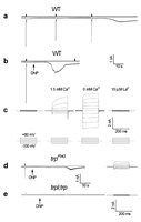
Single-cell functional analysis by whole-cell recordings from newly eclosed flies, showing activation of the TRP and TRPL channels in WT and trpP343 mutant, respectively, but not in the trpl;trpP343 double mutant. Membrane currents are usually elicited by light in the present of ATP and NAD in the pipette. Omission of these agents, combined with either few light pulses (A) or with application of DNP to the bath (B) induced constitutive activation of the light-sensitive channels in the dark as monitored by a sustained noisy inward current that had the characteristics of the TRP or TRPL-dependent current. None of these currents were observed in the double mutant trpl;trpP343 (E). A: Omission of ATP and NAD elicited a TRP-dependent current in the dark after few responses to light. Fig. 5A shows the typical LIC of a wild-type cell in response to orange lights (OG 590, attenuated by 1 log unit; left trace), followed by spontaneous inward current in the dark. The LM above all traces indicates the duration of the orange light stimulus. B, C: Application of DNP to the bath induced constitutive activation of the light-sensitive channels. Membrane currents were recorded 30 s after establishing the whole-cell configuration with physiological concentrations (1.5 mM) of Ca2+ in the bath. (B) Application of DNP (0.1 mM) induced spontaneous dark inward current that abolished the response to light. (C) Dark membrane currents were elicited in a DNP treated cell by stepping the holding voltage in the range of 100 and +80 mV, the currents were suppressed when 10 μM La3+ was applied to the bath. (C, right). Currents obtained with 0 mM Ca2+ in the bath reveal the typical inward and outward rectifications of the TRP dependent current. D, E: The trp mutation reduced, and the double mutation trpl;trp abolished the dark current induced by DNP. Fig. 5D shows an inward small dark current induced by DNP and families of current traces elicited by series of voltage steps from photoreceptors of trpP343 (D) and trpl;trpP343 (E) during recordings with pipettes in which ATP and NAD were omitted and DNP was applied to the external medium. For each experiment, a series of nine voltage steps was applied from a holding potential of 20 mV in 20 mV steps (bottom traces in C). (From Agam et al. 2000, Copyright 2000 by the Society for Neuroscience.)
From: The TRP Calcium Channel and Retinal Degeneration

NCBI Bookshelf. A service of the National Library of Medicine, National Institutes of Health.