Summary
Clinical characteristics.
Hypophosphatasia is characterized by defective mineralization of growing or remodeling bone, with or without root-intact tooth loss, in the presence of low activity of serum and bone alkaline phosphatase. Clinical features range from stillbirth without mineralized bone at the severe end to pathologic fractures of the lower extremities in later adulthood at the mild end. While the disease spectrum is a continuum, seven clinical forms of hypophosphatasia are usually recognized based on age at diagnosis and severity of features:
Perinatal (severe): Characterized by pulmonary insufficiency and hypercalcemia
Perinatal (benign): Prenatal skeletal manifestations that slowly resolve into one of the milder forms
Infantile: Onset between birth and age six months of clinical features of rickets without elevated serum alkaline phosphatase activity
Severe childhood (juvenile): Variable presenting features progressing to rickets
Mild childhood: Low bone mineral density for age, increased risk of fracture, and premature loss of primary teeth with intact roots
Adult: Characterized by stress fractures and pseudofractures of the lower extremities in middle age, sometimes associated with early loss of adult dentition
Odontohypophosphatasia: Characterized by premature exfoliation of primary teeth and/or severe dental caries without skeletal manifestations
Diagnosis/testing.
The clinical diagnosis of hypophosphatasia can be established in a proband with suggestive clinical, laboratory, and radiographic features by identification of reduced serum unfractionated alkaline phosphatase activity and/or identification on molecular genetic testing of biallelic loss-of-function ALPL variants or a heterozygous ALPL variant with dominant-negative effect.
Management.
Targeted therapy: Asfotase alfa (Strensiq®) enzyme replacement therapy (ERT) has been shown to improve pulmonary function, calcium homeostasis / bone health, and survival in individuals with the infantile and early childhood (juvenile) type of hypophosphatasia. There is growing experience with ERT in individuals with the perinatal (severe) type and emerging experience with ERT in treating osteomalacia in adults.
Supportive care: For the perinatal (severe) type: expectant management and family support; respiratory support; management of calcium homeostasis and bone health per endocrinologist and orthopedist; pain management; neurosurgical management of craniosynostosis; management of kidney disease per nephrologist; dental care. For the infantile and early childhood (juvenile) types: respiratory support; management of calcium homeostasis and bone health per endocrinologist and orthopedist; pain management; treatment of seizures with vitamin B6; neurosurgical management of craniosynostosis; management of kidney disease per nephrologist; dental care. For all other types: dental care starting at age one year; nonsteroidal anti-inflammatory drugs for osteoarthritis, bone pain, and osteomalacia; internal fixation for pseudofractures and stress fractures. In adult hypophosphatasia, there is limited experience in treating osteomalacia with teriparatide.
Surveillance: Monitor calcium homeostasis and bone health per endocrinologist, nephrologist, and orthopedist; physical medicine and rehabilitation, physical therapy, and occupational therapy evaluations as needed; monitor children with infantile type for increased intracranial pressure secondary to craniosynostosis; nephrology evaluations as needed for kidney disease; neurology evaluations as needed for seizures; dental visits twice yearly starting at age one year.
Agents/circumstances to avoid: Bisphosphonates and excess vitamin D; teriparatide is contraindicated in children.
Pregnancy management: The use of asfotase alfa (Strensiq®) ERT during human pregnancy has not been extensively studied; therefore, any potential risk to the fetus of a pregnant woman taking this therapy during pregnancy is unknown.
Genetic counseling.
Perinatal and infantile hypophosphatasia are typically inherited in an autosomal recessive manner. The milder forms, especially adult and odontohypophosphatasia, may be inherited in an autosomal recessive or autosomal dominant manner depending on the effect that the ALPL pathogenic variant has on TNSALP (alkaline phosphatase, tissue-nonspecific isozyme) activity.
Autosomal recessive hypophosphatasia: If both parents are known to be heterozygous for an ALPL pathogenic variant, each sib of an affected individual has at conception a 25% chance of inheriting biallelic pathogenic variants and being affected, a 50% chance of being heterozygous, and a 25% chance of inheriting neither of the familial pathogenic variants. Depending on the ALPL pathogenic variant, heterozygous sibs may be either clinically asymptomatic (manifesting only biochemical abnormality) or have milder clinical symptoms than the proband.
Autosomal dominant hypophosphatasia: All individuals reported to date with hypophosphatasia caused by a heterozygous ALPL variant with a dominant-negative effect inherited the ALPL pathogenic variant from a parent (who may or may not have clinical manifestations of hypophosphatasia). Unless an individual with autosomal dominant hypophosphatasia has children with an individual who has a heterozygous or biallelic ALPL pathogenic variant(s), offspring have a 50% chance of inheriting the ALPL pathogenic variant.
Once the ALPL pathogenic variant(s) have been identified in an affected family member, heterozygote testing for at-risk relatives, prenatal testing, and preimplantation genetic testing for hypophosphatasia are possible. Recurrence of perinatal and infantile hypophosphatasia may reliably be identified by prenatal ultrasound examination.
Diagnosis
No consensus clinical diagnostic criteria for hypophosphatasia have been published.
Suggestive Findings
Hypophosphatasia should be suspected in probands with the following clinical, laboratory, and radiographic features.
Clinical features
- Clinical features of infantile rickets: growth failure, craniotabes, craniosynostosis, blue sclerae, flail chest, costochondral enlargement ("rachitic rosary"), scoliosis, thickening of wrists, knees, and ankles, bowing of legs, lax ligaments, and hypotonia
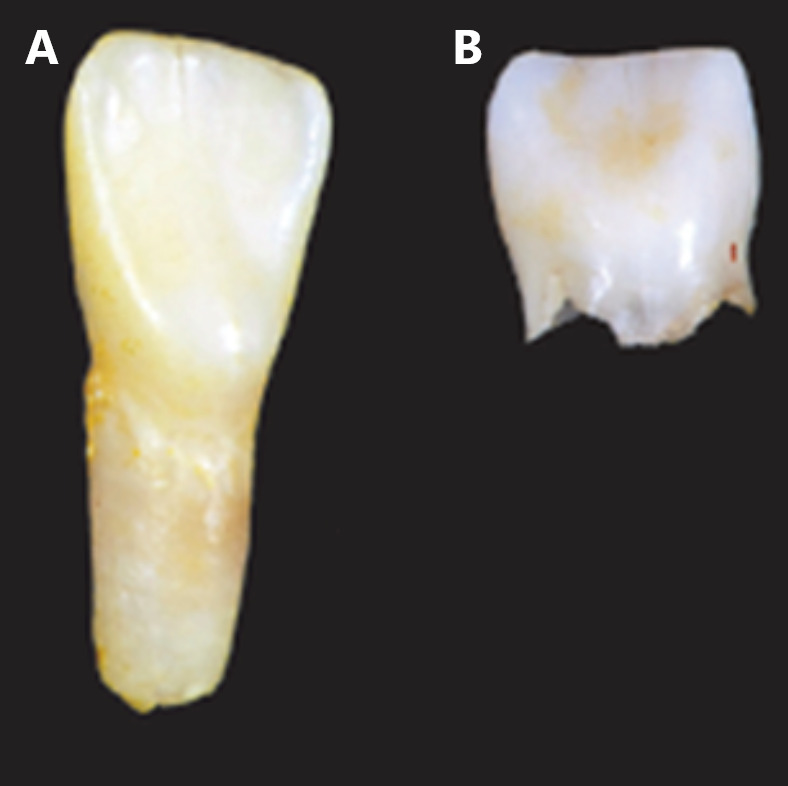
- Premature loss of deciduous teeth beginning with the incisors. Unusually and characteristically, the dental root remains attached to the lost tooth. Dental caries and early loss or extraction of adult teeth is also seen (see Figure 1).
- Vitamin B6 (pyridoxine)-responsive seizures
- Bone pain

Figure 1.
Lost incisors with and without hypophosphatasia A. Hypophosphatasia: root intact
Laboratory features
- Hypercalciuria particularly during the first year of life with or without hypercalcemia
- Typically normal serum calcium and ionized calcium. Note: May be elevated, particularly in the first year of life.
- Typically normal serum and urine inorganic phosphate. Note: May be elevated.
- Normal serum vitamin D (25-hydroxy and 1,25-dihydroxy) and parathyroid hormone
- Elevated plasma vitamin B6 without oral supplementation
- Elevated serum pyridoxal 5'-phosphate (PLP), a biologically active metabolite of vitamin B6. Note: (1) Reference laboratories may measure PLP and report as "vitamin B6." (2) Use of multivitamin or calcium supplements containing vitamin B6 within a week of assaying serum PLP may lead to false positive results.
- Elevated urine phosphoethanolamine (PEA) and proline on urine amino acid chromatogram. Note: (1) Urine PEA may be elevated with other metabolic bone diseases. (2) Urine PEA may be normal in affected individuals and can be elevated in asymptomatic heterozygotes.
- Elevated urine inorganic pyrophosphate (PPi). Note: (1) Assay is not available in North American clinical laboratories. (2) Asymptomatic heterozygotes can have elevated urine PPi.
- Reduced serum unfractionated alkaline phosphatase (ALP) activity. Note: (1) Transient increases in serum ALP activity can occur during pregnancy, with liver disease, and after acute fracture or surgery. Thus, serial measurements may be necessary in toddlers with unexplained fractures. Quantitation of the activity of the bone isoform of ALP in serum may be necessary in the setting of liver disease. The bone isoform is heat labile; the liver isoform is heat stable. (2) Asymptomatic heterozygotes can have reduced serum ALP activity.
Radiographic features
- Prenatal long bone bowing with osteochondral spurs
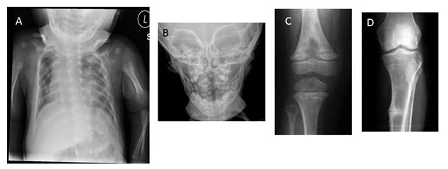
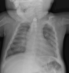
- Infantile rickets: undermineralized bones, widened-appearing sutures, brachycephaly, rachitic costochondral rib changes (see Figure 2A), flared metaphyses, poorly ossified epiphyses, and bowed long bones
- Focal bony defects of the metaphyses resembling radiolucent "tongues" (see Figure 2C) are fairly specific for childhood hypophosphatasia.
- Defective mineralization of growing/remodeling bone and/or teeth. Bone mineral content increases with age, and there may be improved mineralization during adolescence with decreased mineralization in middle age.
- Alveolar bone loss resulting in premature loss of deciduous teeth typically involving the anterior mandible, with the central incisors lost first. However, any tooth may be affected (see Figure 2B).
- Pathologic fractures. Growing children may have a predilection to metaphyseal fractures; however, epiphyseal and diaphyseal fractures are also seen. In adults, metatarsal stress fractures and femoral pseudofractures prevail.
- Osteomalacia with lateral pseudofractures ("Looser zones") in adult hypophosphatasia (see Figure 2D)

Figure 2.
Radiographic signs of hypophosphatasia A. Rachitic rib changes, flail chest, and metaphyseal dysplasia (proximal humerus) in infantile hypophosphatasia
Establishing the Diagnosis
The clinical diagnosis of hypophosphatasia can be established in a proband with suggestive clinical, radiographic, and laboratory features by identification of reduced serum unfractionated ALP activity.
The molecular diagnosis can be established in a proband with suggestive findings by identification of ONE of the following on molecular genetic testing (see Table 1):
- Biallelic loss-of-function ALPL variants
- A heterozygous ALPL variant with dominant-negative effect
Note: (1) Individuals with a heterozygous loss-of-function ALPL variant can have mild features of adult hypophosphatasia [Mornet et al 2021] (see Clinical Description, heterozygous loss-of-function variants). (2) Identification of a biallelic or heterozygous ALPL variant(s) of uncertain significance does not establish or rule out the diagnosis.
Molecular genetic testing approaches can include a combination of gene-targeted testing (single-gene testing, multigene panel) and comprehensive genomic testing (exome sequencing, genome sequencing) depending on the phenotype.
Gene-targeted testing requires that the clinician determine which gene(s) are likely involved, whereas genomic testing does not. Individuals with the distinctive findings described in Suggestive Findings are likely to be diagnosed using gene-targeted testing (see Option 1), whereas those with a phenotype indistinguishable from many other skeletal dysplasias are more likely to be diagnosed using genomic testing (see Option 2).
Option 1
Single-gene testing. Sequence analysis of ALPL is performed first to detect missense, nonsense, and splice site variants and small intragenic deletions/insertions. Note: Depending on the sequencing method used, single-exon, multiexon, or whole-gene deletions/duplications may not be detected. If only one or no variant is detected by the sequencing method used, the next step is to perform gene-targeted deletion/duplication analysis to detect exon and whole-gene deletions or duplications.
A multigene panel that includes ALPL and other genes of interest (see Differential Diagnosis) may be considered to identify the genetic cause of the condition while limiting identification of variants of uncertain significance and pathogenic variants in genes that do not explain the underlying phenotype. Note: (1) The genes included in the panel and the diagnostic sensitivity of the testing used for each gene vary by laboratory and are likely to change over time. (2) Some multigene panels may include genes not associated with the condition discussed in this GeneReview. (3) In some laboratories, panel options may include a custom laboratory-designed panel and/or custom phenotype-focused exome analysis that includes genes specified by the clinician. (4) Methods used in a panel may include sequence analysis, deletion/duplication analysis, and/or other non-sequencing-based tests.
For an introduction to multigene panels click here. More detailed information for clinicians ordering genetic tests can be found here.
Option 2
When the phenotype is indistinguishable from many other skeletal dysplasias, comprehensive genomic testing (which does not require the clinician to determine which gene is likely involved) is likely the best option. Exome sequencing is most commonly used; genome sequencing is also possible.
For an introduction to comprehensive genomic testing click here. More detailed information for clinicians ordering genomic testing can be found here.
Table 1.
Molecular Genetic Testing Used in Hypophosphatasia
Clinical Characteristics
Clinical Description
Hypophosphatasia is characterized by defective mineralization of bone and/or teeth and reduced serum alkaline phosphatase (ALP). The phenotypic spectrum ranges from stillbirth without mineralized bone at the severe end to pathologic stress fractures of the lower extremities in older adults at the mild end (Table 2). Intrafamilial clinical variability is common, particularly when some affected family members have a heterozygous ALPL pathogenic variant and other affected family members have biallelic pathogenic variants. Sibs with compound heterozygous variants tend to display less clinical variability at the severe end of the spectrum and more variability at the milder end of the spectrum.
Table 2.
Select Clinical, Radiographic, and Laboratory Features of Hypophosphatasia by Type
Perinatal (severe) hypophosphatasia is typically identified by prenatal ultrasound examination. Pregnancies may end in stillbirth. Small thoracic cavity and short, bowed limbs are seen in both stillborn and live-born infants. A flail chest may be present (see Figure 2A). Infants with perinatal hypophosphatasia may experience pulmonary insufficiency; restrictive lung disease is the most frequent cause of death. Hypercalcemia is common and may be associated with apnea or seizures. In those treated with asfotase alfa enzyme replacement therapy (ERT), a new phenotype of "treated perinatal and infantile hypophosphatasia" is emerging. However, even when the diagnosis is made expediently, unfavorable outcomes with ERT are possible [Duffus et al 2018]. Infants with perinatal (severe) hypophosphatasia started on ERT between age one day and age 78 months showed improvement in pulmonary function and survival. The effect of ERT on fractures remains unclear [Whyte et al 2019]. In the past, individuals with severe phenotypes died before dental eruption; emerging data suggest the possibility of dental features in infants treated with ERT.
Perinatal (benign) hypophosphatasia is typically identified by prenatal ultrasound examination showing short and bowed long bones but normal or slightly decreased mineralization. Postnatally, skeletal manifestations slowly resolve with a less severe hypophosphatasia phenotype [Wenkert et al 2011].
Infantile hypophosphatasia. There may be no clinical features apparent at birth. Clinical signs may be recognized between birth and age six months and resemble rickets (see Figure 2A). Clinical severity depends on the degree of pulmonary insufficiency; the infantile phenotype has high mortality. Prior to the availability of ERT, 50% of individuals succumbed to respiratory failure caused by undermineralization of the ribs. Other complications include hypercalcemia, irritability, poor feeding, failure to thrive, hypotonia, and more rarely vitamin B6-responsive seizures (see Management). Open fontanels and wide sutures may be deceptive, in that the hypomineralized bone causing this radiographic appearance is prone to premature fusion. Craniosynostosis and intracranial hypertension are potential complications. Older children may have kidney damage. Clinical trials with ERT have shown improvement in developmental milestones and pulmonary function (see Figure 3) [Whyte et al 2019].
Severe childhood (juvenile) hypophosphatasia displays wide variability in initial clinical presentation but often progresses to rickets. More severely affected toddlers have short stature and delay in walking, developing a waddling myopathic gait. Bone and joint pain are typical. Diaphyseal and metaphyseal fractures may occur. Gait, six-minute walk test, and step length improved in individuals treated with ERT. To date, data are insufficient to assess the effect of ERT on fractures in juvenile hypophosphatasia [Whyte et al 2016].
Mild childhood hypophosphatasia is characterized by low bone mineral density for age with unexplained fractures. Children may have premature loss of deciduous teeth (prior to age 5 years), usually beginning with incisors, with the dental root characteristically remaining attached to the lost tooth. Bone and joint pain are atypical.
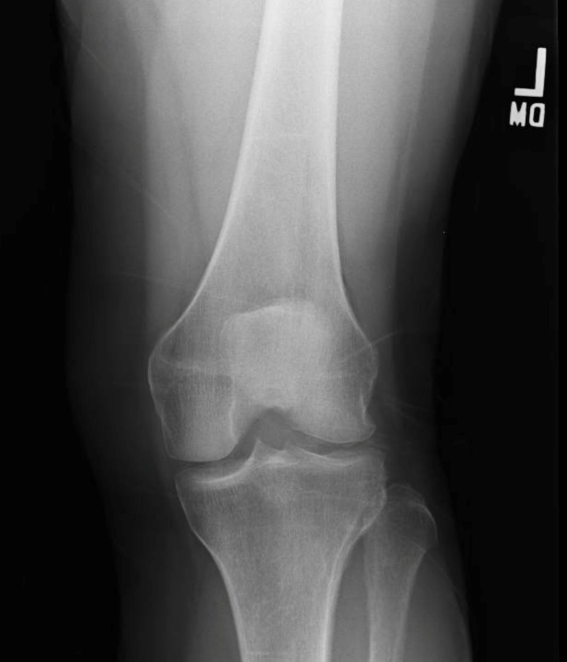
Adult hypophosphatasia is sometimes associated with a history of transient rickets in childhood and/or premature loss of deciduous teeth. Early loss of adult dentition is common. Other dental problems in adolescents and adults with hypophosphatasia are more poorly characterized, although enamel hypoplasia and tooth mobility have been described. Adult hypophosphatasia is usually recognized in middle age, the cardinal features being stress fractures and pseudofractures of the lower extremities. Foot pain and slow-to-heal stress fractures of the metatarsals are common. Thigh and hip pain may reflect pseudofractures ("Looser zones") in the lateral cortex of the femoral diaphysis (see Figure 2D). Chondrocalcinosis and osteoarthropathy may develop with age (see Figure 4). Osteomalacia distinguishes adult hypophosphatasia from odontohypophosphatasia.
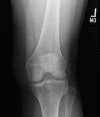
Figure 4.
Radiograph of treated adult hypophosphatasia: linear sclerosis in remodeling distal femur and proximal tibia, osteophytes mid-proximal tibia, and chondrocalcinosis medial lateral compartment
Odontohypophosphatasia can be seen as an isolated finding without additional abnormalities of the skeletal system or can be variably seen in the above forms of hypophosphatasia. Caution should be exercised in citing extradental manifestations of other forms of hypophosphatasia in individuals with odontohypophosphatasia, in that such features may be common and multifactorial (e.g., low bone density for age). Premature exfoliation of primary teeth and/or severe dental caries may be seen, with the incisors most frequently lost.
Phenotype in those with heterozygous loss-of-function variants. Heterozygous loss-of-function ALPL variants have been identified in adults with osteoporosis, musculoskeletal pain, and an increased risk of fractures [Mornet et al 2021]. These individuals are ascertained by low serum ALP and tend to have additional biochemical evidence of hypophosphatasia (elevated serum pyridoxal 5'-phosphate [PLP] or urine phosphoethanolamine [PEA]). Those ascertained as an incidental finding on molecular testing have lower ALP activity but may not display additional biochemical evidence. In this latter circumstance, elevated serum PLP or urine PEA may predict disease potential.
Histopathology
- Bone histology reveals rachitic abnormalities of the growth plate. Histochemical testing of osteoclasts reveals lack of membrane-associated ALP activity. Osteoclasts and osteoblasts otherwise appear normal.
- Tooth histology reveals a decrease in cementum, which varies with the severity of the disease.
Genotype-Phenotype Correlations
Most individuals with hypophosphatasia have unique ALPL variants, preventing the identification of genotype-phenotype correlations. However, site-directed mutagenesis experiments have identified variants producing significant residual enzymatic activity and variants with a dominant-negative effect (see Molecular Genetics).
Less severe phenotypes have been observed in individuals with biallelic loss-of-function variants that allow residual enzymatic activity or heterozygous variants exhibiting a dominant-negative effect [Fauvert et al 2009, Mornet et al 2021]. Clinical features of individuals with reported variants, as well as residual enzyme activity for some of those variants, can be found in the ALPL Variants Database.
Penetrance
While some argue that penetrance is complete, reduced penetrance is possible in autosomal dominant hypophosphatasia due to ALPL variants manifesting a dominant-negative effect.
Nomenclature
Hypophosphatasia takes its name from low activity of the enzyme ALP, rather than reflecting serum concentration of phosphorus.
In classifications of genetic conditions, hypophosphatasia may be considered a metabolic bone disease, a skeletal dysplasia, a metaphyseal dysplasia, a dental disorder, or a disorder of membrane-bound ectoenzyme activity in the extracellular matrix.
Prevalence
Based on pediatric hospital records in Ontario, Canada, the birth prevalence of (autosomal recessive) perinatal and infantile hypophosphatasia was estimated at 1:100,000 [Fraser 1957]. Applying the Hardy-Weinberg equation to this estimate, the frequency of heterozygotes for ALPL pathogenic variants in Ontario, Canada, is about 1:150.
In the Canadian Mennonite population, the prevalence of the perinatal (severe) form is 1:2,500 (carrier frequency 1:25) due to founder the variant p.Gly334Asp [Triggs-Raine et al 2016].
On the basis of molecular diagnosis in France and elsewhere in Europe, the prevalence of severe forms has been estimated at 1:300,000. For mild forms (perinatal benign, mild childhood, adult, and odontohypophosphatasia), the prevalence is expected to be as high as 1:6,300 [Mornet et al 2011] because heterozygotes may express the disease with low selective pressure. Applying the Hardy-Weinberg equation to this estimate for severe forms, the frequency of heterozygotes for ALPL pathogenic variants in France is about 1:275.
In Japan, the birth prevalence of severe hypophosphatasia may be estimated at 1:150,000 on the basis on the frequency of individuals homozygous for the pathogenic variant c.1559delT (1:900,000 [Watanabe et al 2011]) and on the proportion of this pathogenic variant in affected individuals of Japanese ancestry (45.4% [Michigami et al 2020]).
In China, some pathogenic variants have been reported [Wei et al 2010, Zhang et al 2012, Yang et al 2013] but the birth prevalence is unknown.
In Africa, no individuals with hypophosphatasia have been reported in the medical literature outside of North Africa and South Africa; however, clinical ascertainment bias is significant. African American individuals with hypophosphatasia are rare; it is assumed that pathogenic variants in this population represent European admixture.
Genetically Related (Allelic) Disorders
No phenotypes other than those discussed in this GeneReview are known to be associated with germline pathogenic variants in ALPL.
Differential Diagnosis
The differential diagnosis of hypophosphatasia depends on the age at which the diagnosis is considered. Clinical features that help differentiate hypophosphatasia from other conditions include bone hypomineralization prenatally and immediately postnatally; elevated serum concentrations of calcium and phosphorus postnatally; and persistently low serum alkaline phosphatase (ALP) enzyme activity.
In Utero
Early prenatal ultrasound examination may lead to a consideration of osteogenesis imperfecta (OI) type II, campomelic dysplasia, and chondrodysplasias with defects in bone mineralization, as well as hypophosphatasia. Experienced sonographers usually have little difficulty in distinguishing among these disorders. Fetal radiographs are sometimes helpful in recognizing the undermineralization of bone that is more typical of perinatal hypophosphatasia than of the other disorders considered in the differential diagnosis.
At Birth
Outwardly difficult to distinguish, OI type II, thanatophoric dysplasia, campomelic dysplasia, and chondrodysplasias with bone mineralization defects are readily distinguished from hypophosphatasia by radiograph. In individuals in which the diagnosis is in doubt, analysis of serum ALP activity, pyridoxal 5'-phosphate (PLP) or vitamin B6, and urine phosphoethanolamine (PEA) can suggest the diagnosis pending confirmation with molecular genetic testing.
Infancy and Childhood
Irritability, poor feeding, failure to thrive, hypotonia, and seizures place the infantile type in a broad differential diagnosis that includes inborn errors of energy metabolism, organic acidemia, primary and secondary rickets, neglect, and non-accidental trauma. Infantile hypophosphatasia is suspected with low serum ALP enzyme activity, making the argument for routine screening of serum ALP enzyme activity in infants and children with failure to thrive, unexplained seizures, and suspected non-accidental skeletal injury.
Table 3.
Acquired Disorders and Disorders of Unknown Cause in the Differential Diagnosis of Infantile and Childhood-Onset Hypophosphatasia
Table 4.
Hereditary Disorders in the Differential Diagnosis of Infantile and Childhood-Onset Hypophosphatasia
Management
Evaluations Following Initial Diagnosis
To establish the extent of disease and needs in an individual diagnosed with hypophosphatasia, the evaluations summarized in Table 7 and Table 8 (if not performed as part of the evaluation that led to the diagnosis) are recommended.
Table 7.
Recommended Evaluations Following Initial Diagnosis in Infants with Perinatal Hypophosphatasia
Table 8.
Recommended Evaluations Following Initial Diagnosis in Older Individuals with Hypophosphatasia
Treatment of Manifestations
There is no cure for hypophosphatasia. Targeted therapy in the form of enzyme replacement therapy (ERT) is available, and supportive care by specialists is recommended.
Targeted Therapy
In GeneReviews, a targeted therapy is one that addresses the specific underlying mechanism of disease causation (regardless of whether the therapy is significantly efficacious for one or more manifestation of the genetic condition); would otherwise not be considered without knowledge of the underlying genetic cause of the condition; or could lead to a cure. —ED
Asfotase alfa (Strensiq®) ERT has been shown to improve pulmonary function, calcium homeostasis / bone health, and survival in individuals with the infantile and early childhood (juvenile) type of hypophosphatasia. There is growing experience with ERT in individuals with the perinatal (severe) type and emerging experience with ERT in treating osteomalacia in adults.
Table 9.
Targeted Treatment of Hypophosphatasia
Supportive Care
At all ages, supportive care to improve quality of life, maximize function, and reduce complications is recommended. This ideally involves multidisciplinary care by specialists in relevant fields (see Table 10).
Table 10.
Treatment of Manifestations in Individuals with Hypophosphatasia
Surveillance
Table 11.
Recommended Surveillance for Individuals with Hypophosphatasia
Agents/Circumstances to Avoid
Bisphosphonates are relatively contraindicated in hypophosphatasia. Although adverse outcomes have not been identified in children with the infantile type [Deeb et al 2000], theoretic concern has long been raised based on the structure of bisphosphonates. The phosphate motifs in bisphosphonates have a similar conformation to inorganic pyrophosphate (PPi), the natural substrate of TNSALP (alkaline phosphatase, tissue-nonspecific isozyme); thus, treatment with bisphosphonates is thought to be analogous to "adding fuel to the fire." In adults with hypophosphatasia and osteomalacia treated with bisphosphonates, lateral subtrochanteric femoral pseudofractures have been described [Whyte 2009]. As the prevalence of adult hypophosphatasia is not known and many undiagnosed adults undoubtedly are treated with bisphosphonates, the frequency of this unusual complication is not known.
Excess vitamin D can exacerbate hypercalcemia/hypercalciuria in children with infantile hypophosphatasia who have hypercalcemia.
Teriparatide (recombinant human parathyroid hormone fragment, amino acids 1-34) at high doses induces osteosarcoma in rats and may increase the risk of radiation-induced osteosarcoma (a pediatric growth plate tumor) in humans. Thus, it is contraindicated in children with hypophosphatasia.
Evaluation of Relatives at Risk
It is appropriate to clarify the genetic status of apparently asymptomatic older and younger at-risk relatives of an affected individual in order to identify as early as possible those who would benefit from prompt initiation of treatment and preventive measures.
See Genetic Counseling for issues related to testing of at-risk relatives for genetic counseling purposes.
Pregnancy Management
The use of asfotase alfa (Strensiq®) ERT during human pregnancy has not been extensively studied; therefore, any potential risk to the fetus of a pregnant woman taking this therapy during pregnancy is unknown.
See MotherToBaby for further information on medication use during pregnancy.
Therapies Under Investigation
Osteoblast enhancement by anti-sclerostin antibodies. Teriparatide enhances osteoblast production of TNSALP, and sclerostin inhibits osteoblast differentiation. Anti-sclerostin therapies have emerged for metabolic bone diseases. A specific Phase II clinical open-label trial for eight adults with hypophosphatasia (mean age 47.8 years) using anti-sclerostin monoclonal antibodies (BPS804) showed early improvement in bone density and markers of bone turnover in seven individuals completing the 16-week study period. Hypophosphatasia-specific biomarkers other than serum alkaline phosphatase were not reported, and functional assessments were beyond the scope of a Phase II study [Seefried et al 2017].
Bone marrow transplantation (hematopoietic cell transplantation) was used to treat an eight-month-old girl with severe hypophosphatasia with prolonged, significant clinical and radiologic improvement [Whyte et al 2003]. Seven years after transplantation, she was reported to be active and growing, and to have the clinical phenotype of the childhood (juvenile) form of hypophosphatasia [Cahill et al 2007]. In another trial, both bone marrow and allogenic mesenchymal stem cells were implanted in an eight-month-old infant, resulting in improvement of respiratory conditions [Tadokoro et al 2009]. However, the infant developed therapy-related leukemia [Taketani et al 2013]. Transplantation of ex vivo expanded mesenchymal stem cells for individuals who had previously undergone bone marrow transplantation improved bone mineralization, muscle mass, respiratory function, intellectual development, and survival [Taketani et al 2015].
Search ClinicalTrials.gov in the US and EU Clinical Trials Register in Europe for access to information on clinical studies for a wide range of diseases and conditions.
Genetic Counseling
Genetic counseling is the process of providing individuals and families with information on the nature, mode(s) of inheritance, and implications of genetic disorders to help them make informed medical and personal decisions. The following section deals with genetic risk assessment and the use of family history and genetic testing to clarify genetic status for family members; it is not meant to address all personal, cultural, or ethical issues that may arise or to substitute for consultation with a genetics professional. —ED.
Mode of Inheritance
Perinatal and infantile hypophosphatasia are typically inherited in an autosomal recessive manner.
Milder forms of hypophosphatasia, especially adult and odontohypophosphatasia, may be inherited in an autosomal recessive or autosomal dominant manner depending on the effect of the ALPL pathogenic variant on TNSALP (alkaline phosphatase, tissue-nonspecific isozyme) activity [Mornet et al 2021]. ALPL variants with a dominant-negative effect are associated with autosomal dominant inheritance.
Intrafamilial clinical variability is common, particularly when some affected family members have a heterozygous ALPL pathogenic variant and other affected family members have biallelic pathogenic variants. Individuals with severe perinatal, childhood, and adult forms of hypophosphatasia may be seen in families segregating two ALPL pathogenic variants.
Reliable assessment of recurrence risk requires identification of the causative pathogenic variant(s) in the proband and molecular genetic testing of the proband's parents to confirm their genetic status.
Autosomal Recessive Inheritance (Proband with Biallelic Pathogenic Variants) – Risk to Family Members
Parents of a proband
- The parents of a child with biallelic ALPL pathogenic variants are typically heterozygous for one ALPL pathogenic variant.
- Molecular genetic testing is recommended for the parents of a proband to confirm that both parents are heterozygous for an ALPL pathogenic variant and to allow reliable recurrence risk assessment.
- If a pathogenic variant is detected in only one parent and parental identity testing has confirmed biological maternity and paternity, it is possible that one of the pathogenic variants identified in the proband occurred as a de novo event in the proband [Taillandier et al 2005, Zhang et al 2012] or as a postzygotic de novo event in a mosaic parent. If the proband appears to have homozygous pathogenic variants (i.e., the same two pathogenic variants), additional possibilities to consider include:
- A single- or multiexon deletion in the proband that was not detected by sequence analysis and that resulted in the artifactual appearance of homozygosity;
- Uniparental isodisomy for the parental chromosome with the pathogenic variant that resulted in homozygosity for the pathogenic variant in the proband [Watanabe et al 2014, Hancarova et al 2015].
- Depending on the ALPL pathogenic variant, heterozygous parents are either clinically asymptomatic (manifesting only biochemical abnormality) or have milder clinical symptoms than their child (see Molecular Pathogenesis).
Sibs of a proband
- If both parents are known to be heterozygous for an ALPL pathogenic variant, each sib of an affected individual has at conception a 25% chance of inheriting biallelic pathogenic variants, a 50% chance of being heterozygous, and a 25% chance of inheriting neither of the familial pathogenic variants.
- Sibs who inherit biallelic pathogenic variants tend to have similar disease severity; however, growth differences, nutrition, activity level, and earlier age of diagnosis all may influence phenotype. Sibs with compound heterozygous variants tend to display less intrafamilial clinical variability at the severe end of the spectrum and more variability at the milder end of the spectrum.
- Depending on the ALPL pathogenic variant, heterozygous sibs may be either clinically asymptomatic (manifesting only biochemical abnormality) or have milder clinical symptoms than the proband (see Molecular Pathogenesis).
Offspring of a proband. Unless an individual with autosomal recessive hypophosphatasia has children with an affected individual or a heterozygote, offspring will be obligate heterozygotes for a pathogenic variant in ALPL. Note: In the Canadian Mennonite population, the prevalence of the perinatal (severe) form is 1:2,500, with a carrier frequency of 1:25, due to a founder variant (see Prevalence).
Other family members. Each sib of the proband's parents is at a 50% risk of being heterozygous for a pathogenic variant in ALPL.
Heterozygote Detection
Heterozygote testing for at-risk relatives requires prior identification of the ALPL pathogenic variants in the family.
Autosomal Dominant Inheritance (Proband with a Heterozygous ALPL Variant with a Dominant-Negative Effect) – Risk to Family Members
Parents of a proband
- All individuals reported to date with hypophosphatasia caused by a heterozygous ALPL variant with a dominant-negative effect inherited the ALPL pathogenic variant from a parent (who may or may not have clinical manifestations of hypophosphatasia).
- Recommendations for the evaluation of parents of a proband include review of clinical history and laboratory evaluations for signs of hypophosphatasia. Molecular genetic testing is recommended for the parents of the proband to confirm their genetic status and to allow reliable recurrence risk counseling.
- If the pathogenic variant identified in the proband is not identified in either parent and parental identity testing has confirmed biological maternity and paternity, the following possibilities should be considered:
- The proband has a de novo pathogenic variant.
- The proband inherited a pathogenic variant from a parent with germline (or somatic and germline) mosaicism. Note: Testing of parental leukocyte DNA may not detect all instances of somatic mosaicism and will not detect a pathogenic variant that is present in the germ cells only.
- Evaluation of parents may determine that a parent is affected but has escaped previous diagnosis because of failure by health care professionals to recognize the disorder, reduced penetrance, and/or a milder phenotypic presentation. Therefore, an apparently negative family history cannot be confirmed unless molecular genetic testing has demonstrated that neither parent is heterozygous for the ALPL pathogenic variant identified in the proband.
Sibs of a proband. The risk to the sibs of the proband depends on the genetic status of the proband's parents:
- If a parent of the proband is known to have the pathogenic variant identified in the proband, the risk to the sibs of inheriting the pathogenic variant is 50%.
- Clinical severity is often similar in affected family members but cannot be reliably predicted by family history or molecular genetic testing due to reduced penetrance and variable expressivity.
- If the ALPL pathogenic variant identified in the proband cannot be detected in the leukocyte DNA of either parent, the recurrence risk to sibs is estimated to be 1% because of the theoretic possibility of parental germline mosaicism [Rahbari et al 2016].
Offspring of a proband. Each child of an individual with a heterozygous ALPL pathogenic variant has a 50% chance of inheriting the pathogenic variant.
Other family members. The risk to other family members depends on the status of the proband's parents: if a parent has an ALPL pathogenic variant, the parent's family members may be at risk.
Related Genetic Counseling Issues
See Management, Evaluation of Relatives at Risk for information on evaluating at-risk relatives for the purpose of early diagnosis and treatment.
Family planning
- The optimal time for determination of genetic risk and discussion of the availability of prenatal/preimplantation genetic testing is before pregnancy.
- It is appropriate to offer genetic counseling (including discussion of potential risks to offspring and reproductive options) to young adults who are affected, are heterozygous, or are at risk of being heterozygous.
DNA banking. Because it is likely that testing methodology and our understanding of genes, pathogenic mechanisms, and diseases will improve in the future, consideration should be given to banking DNA from probands in whom a molecular diagnosis has not been confirmed (i.e., the causative pathogenic mechanism is unknown). For more information, see Huang et al [2022].
Prenatal Testing and Preimplantation Genetic Testing
Pregnancy with high a priori risk (pregnancy known to be at increased risk based on family history)
- Molecular genetic testing. Once the ALPL pathogenic variant(s) have been identified in an affected family member, prenatal testing and preimplantation genetic testing for hypophosphatasia are possible.
- Fetal ultrasonography. Recurrence of perinatal hypophosphatasia may reliably be identified by prenatal ultrasound examination. Undermineralization, small thoracic cavity, shortened long bones, and bowing are typical features of autosomal recessive and severe hypophosphatasia. Long bone bowing has been reported prenatally in affected sibs and in children of individuals with childhood (juvenile) or adult hypophosphatasia, but the finding is not diagnostic of perinatal severe hypophosphatasia, since it may also be seen in perinatal benign hypophosphatasia, a clinical form that can improve during later stages of pregnancy and result in nonlethal hypophosphatasia [Wenkert et al 2011]. Established information on the functional effect of some ALPL pathogenic variants can assist in distinguishing lethal and nonlethal hypophosphatasia prenatally [Sperelakis-Beedham et al 2021].
- Biochemical testing. Concentration of alkaline phosphatase in amniotic fluid, amniocytes, and chorionic villous samples is prone to misinterpretation (particularly in distinguishing unaffected heterozygotes); molecular genetic testing is the preferred method in confirming prenatal diagnosis [Sperelakis-Beedham et al 2021].
Pregnancy with low a priori risk (pregnancy not known to be at risk)
- Fetal ultrasonography. Although perinatal hypophosphatasia may be distinguished from other skeletal dysplasias by prenatal ultrasonography, care must be taken in the interpretation of bowed long bones. Undermineralization, small thoracic cavity, shortened long bones, and bowing are typical features of autosomal recessive and severe hypophosphatasia. However, prognosis is difficult to predict based on ultrasound findings alone: bowed and shortened long bones have been observed on prenatal ultrasound in individuals who ultimately were shown to have – variably – perinatal (benign), childhood (juvenile), or adult hypophosphatasia. The bowing resolves postnatally. In 50% of individuals, when ALPL molecular testing has been performed, a single pathogenic variant in ALPL has been identified, confirming the benign nature of the phenotype and excluding perinatal (severe) hypophosphatasia.
Differences in perspective may exist among medical professionals and within families regarding the use of prenatal testing. While most centers would consider use of prenatal testing to be a personal decision, discussion of these issues may be helpful.
Resources
GeneReviews staff has selected the following disease-specific and/or umbrella support organizations and/or registries for the benefit of individuals with this disorder and their families. GeneReviews is not responsible for the information provided by other organizations. For information on selection criteria, click here.
- Hypophosphatasie EuropeFranceEmail: contact@hypophosphatasie.com
- MAGIC Foundation6645 West North AvenueOak Park IL 60302Phone: 800-362-4423; 708-383-0808Fax: 708-383-0899Email: ContactUs@magicfoundation.org
- Soft Bones CanadaPO Box 882Winkler Manitoba R6W 4A9CanadaPhone: 204-202-3211Email: contactus@softbonescanada.ca
- Soft Bones, Inc.121 Hawkins Place #267Boonton NJ 07005Phone: 866-827-9937
- National Organization for Rare Disorders (NORD)Phone: 800-999-6673
- UCLA International Skeletal Dysplasia Registry (ISDR)Phone: 310-825-8998
Molecular Genetics
Information in the Molecular Genetics and OMIM tables may differ from that elsewhere in the GeneReview: tables may contain more recent information. —ED.
Table A.
Hypophosphatasia: Genes and Databases
Table B.
OMIM Entries for Hypophosphatasia (View All in OMIM)
Molecular Pathogenesis
ALPL encodes alkaline phosphatase, tissue-nonspecific isozyme (TNSALP), the isozyme present in liver, kidney, and bone. It is functional as a homodimer. The enzyme acts as a (lipid) membrane-bound ectophosphatase with inorganic pyrophosphate (PPi), pyridoxal 5'-phosphate (PLP), and phosphoethanolamine (PEA) as natural substrates.
ALPL pathogenic variants are distributed throughout the 12 exons of the gene. Pathogenic missense variants account for 74.6% of variants; the remainder comprise microdeletions/insertions (13.3%), pathogenic splice site variants (6.0%), pathogenic nonsense variants (3.7%), gross deletions (1.3%), and a nucleotide substitution affecting the major transcription initiation site. This variety of pathogenic variants results in highly variable clinical expression and in a great number of compound heterozygous genotypes.
Genotype-phenotype correlations have been studied using site-directed mutagenesis and 3D enzyme modeling. These studies have allowed the characterization of severe and moderate alleles (alleles producing significant residual enzymatic activity) and alleles with a dominant-negative effect responsible for dominant inheritance [Fukushi et al 1998, Shibata et al 1998, Zurutuza et al 1999, Mornet et al 2001, Watanabe et al 2002, Nasu et al 2006, Brun-Heath et al 2007, Fauvert et al 2009, Mornet et al 2021]. However, such tools do not always predict the severity of pathogenic variants.
Mechanism of disease causation. Pathogenic variants may result in various consequences, sometimes cumulative: decrease or abolition of the catalytic activity, inability to form homodimers, and sequestration of mutated proteins in cell compartments resulting in an inability to reach the cell membrane [Cai et al 1998, Fukushi et al 1998, Shibata et al 1998, Watanabe et al 2002, Brun-Heath et al 2007, Sultana et al 2013, Numa-Kinjoh et al 2015].
Table 12.
Notable ALPL Pathogenic Variants
Chapter Notes
Acknowledgments
Michael Whyte, MD is the foremost authority on hypophosphatasia in all its clinical forms, and his mentorship inspires this chapter. Etienne Mornet, PhD is the foremost authority on ALPL variants; he co-authored the first edition of this chapter in 2007, and his continued work in the field informs this current edition. Jose Luis Millan, PhD literally wrote the book on alkaline phosphatase, and his groundbreaking basic science has transformed hypophosphatasia from a manageable to a treatable disorder.
Author History
Etienne Mornet, PhD; Centre Hospitalier de Versailles (2007-2022)
Mark E Nunes, MD (2007-present)
Revision History
- 30 March 2023 (ma) Revision: asfotase alfa (Strensiq®) enzyme replacement therapy added as a targeted therapy
- 7 April 2022 (sw) Comprehensive update posted live
- 4 February 2016 (ha) Comprehensive update posted live
- 10 November 2011 (cd) Revision: deletion/duplication analysis of ALPL available clinically
- 5 August 2010 (me) Comprehensive update posted live
- 20 November 2007 (me) Review posted live
- 18 December 2006 (men) Original submission
References
Literature Cited
- Brun-Heath I, Lia-Baldini AS, Maillard S, Taillandier A, Utsch B, Nunes ME, Serre JL, Mornet E. Delayed transport of tissue-nonspecific alkaline phosphatase with missense mutations causing hypophosphatasia. Eur J Med Genet. 2007;50:367–78. [PubMed: 17719863]
- Cahill RA, Wenkert D, Perlman SA, Steele A, Coburn SP, McAlister WH, Mumm S, Whyte MP. Infantile hypophosphatasia: transplantation therapy trial using bone fragments and cultured osteoblasts. J Clin Endocrinol Metab. 2007;92:2923–30. [PubMed: 17519318]
- Cai G, Michigami T, Yamamoto T, Yasui N, Satomura K, Yamagata M, Shima M, Nakajima S, Mushiake S, Okada S, Ozono K. Analysis of localization of mutated tissue-nonspecific alkaline phosphatase proteins associated with neonatal hypophosphatasia using green fluorescent protein chimeras. J Clin Endocrinol Metab. 1998;83:3936–42. [PubMed: 9814472]
- Camacho PM, Mazhari AM, Wilczynski C, Kadanoff R, Mumm S, Whyte MP. Adult hypophosphatasia treated with teriparatide: report of 2 patients and review of the literature. Endocr Pract. 2016;22:941–50. [PubMed: 27042741]
- Deeb AA, Bruce SN, Morris AA, Cheetham TD. Infantile hypophosphatasia: disappointing results of treatment. Acta Paediatr. 2000;89:730–3. [PubMed: 10914973]
- Duffus S, Thrasher B, Calikoglu AS. Brief clinical report: hypophosphatasia-diagnostic considerations and treatment outcomes in an infant. Case Rep Pediatr. 2018;2018:5719761. [PMC free article: PMC5901473] [PubMed: 29808151]
- Fauvert D, Brun-Heath I, Lia-Baldini AS, Bellazi L, Taillandier A, Serre JL, de Mazancourt P, Mornet E. Mild forms of hypophosphatasia mostly result from dominant negative effect of severe alleles or from compound heterozygosity for severe and moderate alleles. BMC Med Genet. 2009;10:51. [PMC free article: PMC2702372] [PubMed: 19500388]
- Fraser D. Hypophosphatasia. Am J Med. 1957;22:730–46. [PubMed: 13410963]
- Fukushi M, Amizuka N, Hoshi K, Ozawa H, Kumagai H, Omura S, Misumi Y, Ikehara Y, Oda K. Intracellular retention and degradation of tissue-nonspecific alkaline phosphatase with a Gly317-->Asp substitution associated with lethal hypophosphatasia. Biochem Biophys Res Commun. 1998;246:613–8. [PubMed: 9618260]
- Greenberg CR, Taylor CL, Haworth JC, Seargeant LE, Philipps S, Triggs-Raine B, Chodirker BN. A homoallelic Gly317-->Asp mutation in ALPL causes the perinatal (lethal) form of hypophosphatasia in Canadian mennonites. Genomics. 1993;17:215–7. [PubMed: 8406453]
- Hacıhamdioğlu B, Özgürhan G, Pereira C, Tepeli E, Acar G, Cömert S. A case of the perinatal form hypophosphatasia caused by a novel large duplication of the ALPL gene and report of one year follow-up with enzyme replacement therapy. J Clin Res Pediatr Endocrinol. 2019;11:306–10. [PMC free article: PMC6745457] [PubMed: 30468149]
- Hancarova M, Krepelova A, Puchmajerova A, Soucek O, Prchalova D, Sumnik Z, Sedlacek Z. Hypophosphatasia due to uniparental disomy. Bone. 2015;81:765–6. [PubMed: 25937451]
- Huang SJ, Amendola LM, Sternen DL. Variation among DNA banking consent forms: points for clinicians to bank on. J Community Genet. 2022;13:389–97. [PMC free article: PMC9314484] [PubMed: 35834113]
- Huggins E, Ong R, Rockman-Greenberg C, Flueckinger LB, Dahir KM, Kishnani PS. Multigenerational case examples of hypophosphatasia: Challenges in genetic counseling and disease management. Mol Genet Metab Rep. 2020;25:100661. [PMC free article: PMC7578550] [PubMed: 33101980]
- Lia-Baldini AS, Muller F, Taillandier A, Gibrat JF, Mouchard M, Robin B, Simon-Bouy B, Serre JL, Aylsworth AS, Bieth E, Delanote S, Freisinger P, Hu JC, Krohn HP, Nunes ME, Mornet E. A molecular approach to dominance in hypophosphatasia. Hum Genet. 2001;109:99–108. [PubMed: 11479741]
- Michigami T, Tachikawa K, Yamazaki M, Kawai M, Kubota T, Ozono K. Hypophosphatasia in Japan: ALPL mutation analysis in 98 unrelated patients. Calcif Tissue Int. 2020;106:221–31. [PubMed: 31707452]
- Mornet E, Taillandier A, Domingues C, Dufour A, Benaloun E, Lavaud N, Wallon F, Rousseau N, Charle C, Guberto M, Muti C, Simon-Bouy B. Hypophosphatasia: a genetic-based nosology and new insights in genotype-phenotype correlation. Eur J Hum Genet. 2021;29:289–99. [PMC free article: PMC7868366] [PubMed: 32973344]
- Mornet E, Stura E, Lia-Baldini AS, Stigbrand T, Menez A, Le Du MH. Structural evidence for a functional role of human tissue nonspecific alkaline phosphatase in bone mineralization. J Biol Chem. 2001;276:31171–8. [PubMed: 11395499]
- Mornet E, Yvard A, Taillandier A, Fauvert D, Simon-Bouy B. A molecular-based estimation of the prevalence of hypophosphatasia in the European population. Ann Hum Genet. 2011;75:439–45. [PubMed: 21488855]
- Nasu M, Ito M, Ishida Y, Numa N, Komaru K, Nomura S, Oda K. Aberrant interchain disulfide bridge of tissue-nonspecific alkaline phosphatase with an Arg433-->Cys substitution associated with severe hypophosphatasia. FEBS J. 2006;273:5612–24. [PubMed: 17212778]
- Numa-Kinjoh N, Komaru K, Ishida Y, Sohda M, Oda K. Molecular phenotype of tissue-nonspecific alkaline phosphatase with a proline (108) to leucine substitution associated with dominant odontohypophosphatasia. Mol Genet Metab. 2015;115:180–5. [PubMed: 25982064]
- Rahbari R, Wuster A, Lindsay SJ, Hardwick RJ, Alexandrov LB, Turki SA, Dominiczak A, Morris A, Porteous D, Smith B, Stratton MR, Hurles ME, et al. Timing, rates and spectra of human germline mutation. Nat Genet. 2016;48:126–33. [PMC free article: PMC4731925] [PubMed: 26656846]
- Schalin-Jäntti C, Mornet E, Lamminen A, Välimäki MJ. Parathyroid hormone treatment improves pain and fracture healing in adult hypophosphatasia. J Clin Endocrinol Metab. 2010;95:5174–9. [PubMed: 20739387]
- Seefried L, Baumann J, Hemsley S, Hofmann C, Kunstmann E, Kiese B, Huang Y, Chivers S, Valentin MA, Borah B, Roubenoff R, Junker U, Jakob F. Efficacy of anti-sclerostin monoclonal antibody BPS804 in adult patients with hypophosphatasia. J Clin Invest. 2017;127:2148–58. [PMC free article: PMC5451251] [PubMed: 28436937]
- Shibata H, Fukushi M, Igarashi A, Misumi Y, Ikehara Y, Ohashi Y, Oda K. Defective intracellular transport of tissue-nonspecific alkaline phosphatase with an Ala162-->Thr mutation associated with lethal hypophosphatasia. J Biochem (Tokyo). 1998;123:968–77. [PubMed: 9562633]
- Spentchian M, Brun-Heath I, Taillandier A, Fauvert D, Serre JL, Simon-Bouy B, Carvalho F, Grochova I, Mehta SG, Müller G, Oberstein SL, Ogur G, Sharif S, Mornet E. Characterization of missense mutations and large deletions in the ALPL gene by sequencing and quantitative multiplex PCR of short fragments. Genet Test. 2006;10:252–7. [PubMed: 17253930]
- Sperelakis-Beedham B, Taillandier A, Domingues C, Guberto M, Colin E, Porquet-Bordes V, Rothenbuhler A, Salles JP, Wenkert D, Zankl A, Muti C, Bacrot S, Simon-Bouy B, Mornet E. Utility of genetic testing for prenatal presentations of hypophosphatasia. Mol Genet Metab. 2021;132:198–203. [PubMed: 33549410]
- Sultana S, Al-Shawafi HA, Makita S, Sohda M, Amizuka N, Takagi R, Oda K. An asparagine at position 417 of tissue-nonspecific alkaline phosphatase is essential for its structure and function as revealed by analysis of the N417S mutation associated with severe hypophosphatasia. Mol Genet Metab. 2013;109:282–8. [PubMed: 23688511]
- Tadokoro M, Kanai R, Taketani T, Uchio Y, Yamaguchi S, Ohgushi H. New bone formation by allogeneic mesenchymal stem cell transplantation in a patient with perinatal hypophosphatasia. J Pediatr. 2009;154:924–30. [PubMed: 19446101]
- Taillandier A, Sallinen SL, Brun-Heath I, De Mazancourt P, Serre JL, Mornet E. Childhood hypophosphatasia due to a de novo missense mutation in the tissue-nonspecific alkaline phosphatase gene. J Clin Endocrinol Metab. 2005;90:2436–9. [PubMed: 15671102]
- Taketani T, Kanai R, Abe M, Mishima S, Tadokoro M, Katsube Y, Yuba S, Ogushi H, Fukuda S, Yamaguchi S. Therapy-related Ph+ leukemia after both bone marrow and mesenchymal stem cell transplantation for hypophosphatasia. Pediatr Int. 2013;55:e52–5. [PubMed: 23782379]
- Taketani T, Oyama C, Mihara A, Tanabe Y, Abe M, Hirade T, Yamamoto S, Bo R, Kanai R, Tadenuma T, Michibata Y, Yamamoto S, Hattori M, Katsube Y, Ohnishi H, Sasao M, Oda Y, Hattori K, Yuba S, Ohgushi H, Yamaguchi S. Ex vivo expanded allogeneic mesenchymal stem cells with bone marrow transplantation improved osteogenesis in infants with severe hypophosphatasia. Cell Transplant. 2015;24:1931–43. [PubMed: 25396326]
- Triggs-Raine B, Dyck T, Boycott KM, Innes AM, Ober C, Parboosingh JS, Botkin A, Greenberg CR, Spriggs EL. Development of a diagnostic DNA chip to screen for 30 autosomal recessive disorders in the Hutterite population. Mol Genet Genomic Med. 2016;4:312–21. [PMC free article: PMC4867565] [PubMed: 27247959]
- Watanabe H, Goseki-Sone M, Orimo H, Hamatani R, Takinami H, Ishikawa I. Function of mutant (G1144A) tissue-nonspecific ALP gene from hypophosphatasia. J Bone Miner Res. 2002;17:1945–8. [PubMed: 12412800]
- Watanabe A, Karasugi T, Sawai H, Naing BT, Ikegawa S, Orimo H, Shimada T. Prevalence of c.1559delT in ALPL, a common mutation resulting in the perinatal (lethal) form of hypophosphatasia in Japanese and effects of the mutation on heterozygous carriers. J Hum Genet. 2011;56:166–8. [PubMed: 21179104]
- Watanabe A, Satoh S, Fujita A, Naing BT, Orimo H, Shimada T. Perinatal hypophosphatasia caused by uniparental isodisomy. Bone. 2014;60:93–7. [PubMed: 24334170]
- Wei KW, Xuan K, Liu YL, Fang J, Ji K, Wang X, Jin Y, Watanabe S, Watanabe K, Ojihara T. Clinical, pathological and genetic evaluations of Chinese patients with autosomal-dominant hypophosphatasia. Arch Oral Biol. 2010;55:1017–23. [PubMed: 20828673]
- Wenkert D, McAlister WH, Coburn SP, Zerega JA, Ryan LM, Ericson KL, Hersh JH, Mumm S, Whyte MP. Hypophosphatasia: nonlethal disease despite skeletal presentation in utero (17 new cases and literature review). J Bone Miner Res. 2011;26:2389–98. [PubMed: 21713987]
- Whyte MP. Atypical femoral fractures, bisphosphonates, and adult hypophosphatasia. J Bone Miner Res. 2009;24:1132–4. [PubMed: 19113923]
- Whyte MP, Kurtzberg J, McAlister WH, Mumm S, Podgornik MN, Coburn SP, Ryan LM, Miller CR, Gottesman GS, Smith AK, Douville J, Waters-Pick B, Armstrong RD, Martin PL. Marrow cell transplantation for infantile hypophosphatasia. J Bone Miner Res. 2003;18:624–36. [PubMed: 12674323]
- Whyte MP, Rockman-Greenberg C, Ozono K, Riese R, Moseley S, Melian A, Thompson DD, Bishop N, Hofmann C. Asfotase alfa treatment improves survival for perinatal and infantile hypophosphatasia. J Clin Endocrinol Metab. 2016;101:334–42. [PMC free article: PMC4701846] [PubMed: 26529632]
- Whyte MP, Simmons JH, Moseley S, Fujita KP, Bishop N, Salman NJ, Taylor J, Phillips D, McGinn M, McAlister WH. Asfotase alfa for infants and young children with hypophosphatasia: 7 year outcomes of a single-arm, open-label, phase 2 extension trial. Lancet Diabetes Endocrinol. 2019;7:93–105. [PubMed: 30558909]
- Yang H, Wang L, Geng J, Yu T, Yao RE, Shen Y, Yin L, Ying D, Huang R, Zhou Y, Chen H, Liu L, Mo X, Shen Y, Fu Q, Yu Y. Characterization of six missense mutations in the tissue-nonspecific alkaline phosphatase (TNSALP) gene in Chinese children with hypophosphatasia. Cell Physiol Biochem. 2013;32:635–44. [PubMed: 24022022]
- Zhang H, Ke YH, Wang C, Yue H, Hu WW, Gu JM, Zhang ZL. Identification of the mutations in the tissue-nonspecific alkaline phosphatase gene in two Chinese families with hypophosphatasia. Arch Med Res. 2012;43:21–30. [PubMed: 22300680]
- Zurutuza L, Muller F, Gibrat JF, Taillandier A, Simon-Bouy B, Serre JL, Mornet E. Correlations of genotype and phenotype in hypophosphatasia. Hum Mol Genet. 1999;8:1039–46. [PubMed: 10332035]
Publication Details
Author Information and Affiliations
Valley Children's Hospital
Madera, California
Publication History
Initial Posting: November 20, 2007; Last Revision: March 30, 2023.
Copyright
GeneReviews® chapters are owned by the University of Washington. Permission is hereby granted to reproduce, distribute, and translate copies of content materials for noncommercial research purposes only, provided that (i) credit for source (http://www.genereviews.org/) and copyright (© 1993-2024 University of Washington) are included with each copy; (ii) a link to the original material is provided whenever the material is published elsewhere on the Web; and (iii) reproducers, distributors, and/or translators comply with the GeneReviews® Copyright Notice and Usage Disclaimer. No further modifications are allowed. For clarity, excerpts of GeneReviews chapters for use in lab reports and clinic notes are a permitted use.
For more information, see the GeneReviews® Copyright Notice and Usage Disclaimer.
For questions regarding permissions or whether a specified use is allowed, contact: ude.wu@tssamda.
Publisher
University of Washington, Seattle, Seattle (WA)
NLM Citation
Nunes ME. Hypophosphatasia. 2007 Nov 20 [Updated 2023 Mar 30]. In: Adam MP, Feldman J, Mirzaa GM, et al., editors. GeneReviews® [Internet]. Seattle (WA): University of Washington, Seattle; 1993-2024.