Summary
Clinical characteristics.
Hypomyelination and congenital cataract (HCC) is usually characterized by bilateral congenital cataracts and normal psychomotor or only mildly delayed development in the first year of life, followed by slowly progressive neurologic impairment manifest as ataxia, spasticity (brisk tendon reflexes and bilateral extensor plantar responses), and mild-to-moderate cognitive impairment. Dysarthria and truncal hypotonia are observed. Cerebellar signs (truncal titubation and intention tremor) and peripheral neuropathy (muscle weakness and wasting of the legs) are present in the majority of affected individuals. Seizures can occur. Cataracts may be absent in some individuals.
Diagnosis/testing.
The diagnosis of HCC can be established in individuals with typical clinical findings, characteristic abnormalities on brain MRI, and biallelic pathogenic variants in HYCC1 (formerly FAM126A) identified by molecular genetic testing.
Management.
Treatment of manifestations: Cataract extraction usually in the first months of life. Therapy support for developmental delays; special education; physical medicine and rehabilitation for spasticity and ataxia. Consider pharmacologic agents for spasticity; anti-seizure medication as needed. Treatment for scoliosis and contractures per orthopedist; feeding therapy and or gastrostomy tube as needed.
Surveillance: Eye examinations if cataracts were not identified in neonatal period. Developmental, neurologic, and musculoskeletal assessments at each visit. Growth measurement, nutrition assessment, and assessment of family need for social work support and care coordination at each visit.
Genetic counseling.
HCC is inherited in an autosomal recessive manner. If both parents are known to be heterozygous for a HYCC1 pathogenic variant, each sib of an affected individual has at conception a 25% chance of being affected, a 50% chance of being an asymptomatic carrier, and a 25% chance of being unaffected and not a carrier. Once the HYCC1 pathogenic variants have been identified in an affected family member, prenatal testing for a pregnancy at increased risk and preimplantation genetic testing are possible.
Diagnosis
Suggestive Findings
Hypomyelination and congenital cataract (HCC) should be suspected in individuals with the following clinical findings [Biancheri et al 2007] and characteristic abnormalities on brain MRI [Rossi et al 2008].
Clinical findings
- Bilateral congenital cataracts. One individual had juvenile cataract [Ugur & Tolun 2008]; one individual had only a mild lens opacity, noted at age three years [Biancheri et al 2011].
- Nystagmus present from the first few weeks of life
- Classic presentation shows normal or mildly delayed psychomotor development in the first year of life, followed by slowly progressive neurologic impairment manifest as:
- Ataxia
- Spasticity
- Loss of the ability to walk
- Mild-to-moderate cognitive impairment
- Uncommon presentations [Biancheri et al 2011]
- Early-onset severe variant. Hypotonia and feeding difficulties in the neonatal period, developmental delay in the first months of life, and wheelchair dependency in early childhood
- Late-onset mild variant. Normal development in the first two years of life with subsequent sudden motor regression
MRI findings
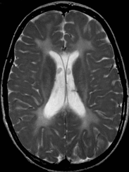
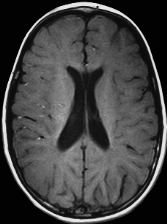
- Diffusely abnormal supratentorial white matter in all individuals
- Abnormal white matter signal behavior consistent with hypomyelination:
- Areas of higher T2-weighted signal intensity with corresponding low-signal intensity on T1-weighted images consistent with areas of increased white matter water content of variable extension in some individuals (Figure 3)
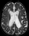
- White matter bulk loss in older individuals (Figure 4)
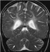
- Medullary centers of the cerebellar hemispheres showing mildly increased T2-weighted signal intensity, paralleling that of the adjacent cortical gray matter and resulting in a "blurred" gray-white matter interface in some individuals (Figure 5)
- Sparing of the cortical and deep gray matter structures

Figure 1.
Axial T2-weighted image shows diffuse hyperintensity of supratentorial white matter consistent with hypomyelination.

Figure 2.
Axial T1-weighted image shows diffusely isointense white matter with poor demarcation from adjacent gray matter, consistent with hypomyelination.

Figure 3.
Axial T1-weighted image (panel A) and axial T2-weighted image (panel B) show areas of increased water content involving the deep frontal white matter.

Figure 5.
Coronal T2-weighted image shows poor gray-white matter demarcation at the level of the medullary centers of the cerebellum, suggesting abnormal myelination of the cerebellar white matter.
Establishing the Diagnosis
The diagnosis of HCC is established in a proband with suggestive findings and biallelic pathogenic (or likely pathogenic) variants in HYCC1 (formerly FAM126A) identified by molecular genetic testing (see Table 1).
Note: (1) Per ACMG/AMP variant interpretation guidelines, the terms "pathogenic variant" and "likely pathogenic variant" are synonymous in a clinical setting, meaning that both are considered diagnostic and can be used for clinical decision making [Richards et al 2015]. Reference to "pathogenic variants" in this GeneReview is understood to include any likely pathogenic variants. (2) Identification of biallelic HYCC1 variants of uncertain significance (or of one known HYCC1 pathogenic variant and one HYCC1 variant of uncertain significance) does not establish or rule out the diagnosis.
Molecular testing approaches can include a combination of gene-targeted testing (single-gene testing, multigene panel) and comprehensive genomic testing (exome sequencing, genome sequencing) depending on the phenotype.
Gene-targeted testing requires that the clinician determine which gene(s) are likely involved, whereas genomic testing does not. Individuals with the distinctive findings described in Suggestive Findings are likely to be diagnosed using gene-targeted testing (see Option 1), whereas those with a phenotype indistinguishable from many other inherited disorders with cataracts and/or leukodystrophy are more likely to be diagnosed using genomic testing (see Option 2).
Option 1
Single-gene testing. Sequence analysis of HYCC1 is performed first to detect missense, nonsense, and splice site variants and small intragenic deletions/insertions. Note: Depending on the sequencing method used, single-exon, multiexon, or whole-gene deletions/duplications may not be detected. If only one or no variant is detected by the sequencing method used, the next step is to perform gene-targeted deletion/duplication analysis to detect exon and whole-gene deletions or duplications.
A multigene panel that includes HYCC1 and other genes of interest (see Differential Diagnosis) may also be considered. Note: (1) The genes included in the panel and the diagnostic sensitivity of the testing used for each gene vary by laboratory and are likely to change over time. (2) Some multigene panels may include genes not associated with the condition discussed in this GeneReview; thus, clinicians need to determine which multigene panel is most likely to identify the genetic cause of the condition while limiting identification of variants of uncertain significance and pathogenic variants in genes that do not explain the underlying phenotype. (3) In some laboratories, panel options may include a custom laboratory-designed panel and/or custom phenotype-focused exome analysis that includes genes specified by the clinician. (4) Methods used in a panel may include sequence analysis, deletion/duplication analysis, and/or other non-sequencing-based tests.
For an introduction to multigene panels click here. More detailed information for clinicians ordering genetic tests can be found here.
Option 2
When the phenotype is indistinguishable from many other inherited disorders characterized by cataracts and/or leukodystrophy, comprehensive genomic testing (which does not require the clinician to determine which gene is likely involved) is the best option. Exome sequencing is most commonly used; genome sequencing is also possible.
For an introduction to comprehensive genomic testing click here. More detailed information for clinicians ordering genomic testing can be found here.
Table 1.
Molecular Genetic Testing Used in Hypomyelination and Congenital Cataract
Clinical Characteristics
Clinical Description
Hypomyelination and congenital cataract (HCC) phenotype is quite consistent in the affected individuals described to date.
Table 2.
Hypomyelination and Congenital Cataract: Frequency of Select Features
Prenatal/perinatal. All affected individuals have normal prenatal and perinatal histories.
Ophthalmologic. Bilateral congenital cataracts identified at birth or within the first month of life are the first clinical sign. All children underwent ocular surgery in the first months of life with the exception of the one child who had adolescent-onset cataracts [Ugur & Tolun 2008].
Psychomotor development is normal up until the end of the first year of life, when developmental delays appear [Biancheri et al 2007]. The ability to walk with support is achieved between ages 12 and 24 months. Independent walking is not achieved in all individuals. Slowly progressive neurologic impairment then becomes apparent with gradual loss of the ability to walk. Most individuals become wheelchair bound between ages eight and nine years [Biancheri et al 2007].
Feeding issues occur as a result of neurologic impairment. Swallowing may become difficult, and growth may be affected by suboptimal intake.
Cognitive skills. All individuals have mild-to-moderate intellectual disability without deterioration in cognitive ability over time.
Neurologic findings. Clinical examination reveals the following from the onset of the disease course:
- Dysarthria
- Truncal hypotonia
- Pyramidal signs and spasticity. Tendon reflexes may be decreased or lost as a result of peripheral neuropathy.
- Cerebellar signs/ataxia (including truncal titubation and intention tremor)
- Peripheral neuropathy, present in most individuals, manifest as muscle weakness, wasting of the legs and ataxia. Peripheral neuropathy is absent in individuals with a milder form of the disorder (see Genotype-Phenotype Correlations).
Seizures including those triggered by fever may occur, but are not a predominant clinical feature.
Neurophysiologic investigations show the following from the onset of the disease course:
- Motor nerve conduction velocity. Slightly to markedly slowed in most individuals, with lower values in older persons
- Compound muscle action potentials. Reduced amplitude
- Electromyography. Signs of denervation in the absence of spontaneous activity
- Waking EEG. Irregular background activity; multifocal epileptiform discharges may be recorded.
- Brain stem auditory evoked potentials. Increased I-V interpeak conduction time in individuals older than age two years
- Electroretinogram. Normal
Neuropathologic findings
- Sural nerve biopsy of individuals with peripheral neuropathy shows a slight-to-severe reduction in density of myelinated fibers, with several axons surrounded by a thin myelin sheath or devoid of myelin.
- Uncompaction of the myelin sheath, which in some fibers appears redundant and irregularly folded, is occasionally seen.
- Electron microscopy confirms the presence of axons devoid of myelin, together with thinly myelinated fibers, sometimes surrounded by few Schwann cells processes, forming small onion bulbs.
Orthopedic issues. A slowly progressive scoliosis appears concurrently with the loss of the ability to walk [Biancheri et al 2007].
Life expectancy is unknown; the oldest living affected individual is age 34 years.
Genotype-Phenotype Correlations
Pathogenic variants leading to the complete absence of HYCC1 (formerly FAM126A) protein expression are associated with the full phenotype of bilateral cataract, central nervous system hypomyelination, and peripheral nerve hypomyelination.
Pathogenic variants leading to a partial protein deficiency are associated with the milder form without peripheral nervous system involvement.
An individual with deletion of exons 8 and 9 did not have congenital cataracts; cataracts developed at age nine years. A second individual had congenital unilateral cataract. However, of the four children in this family who survived beyond age two years, none was able to walk even with support after age six years [Ugur & Tolun 2008].
Because of the limited number of individuals with HCC described so far, these correlations should be further confirmed.
Penetrance
Penetrance is complete.
Prevalence
HCC is likely a rare disorder. No epidemiologic studies are available.
Genetically Related (Allelic) Disorders
No phenotypes other than those discussed in this GeneReview are known to be associated with germline pathogenic variants in HYCC1 (formerly FAM126A).
Differential Diagnosis
The association of congenital cataract and CNS hypomyelination is typical of hypomyelination and congenital cataract (HCC). However, the differential diagnosis with other hypomyelinating disorders should include the disorders summarized in Table 3. MRI usually shows areas with an even higher T2-weighted signal in HCC, whereas the white matter signal is homogeneously hyperintense in other hypomyelinating disorders.
Table 3.
Hypomyelinating Disorders of Interest in the Differential Diagnosis of Hypomyelination and Congenital Cataract
Management
Evaluations Following Initial Diagnosis
To establish the extent of disease and needs in an individual diagnosed with hypomyelination and congenital cataract (HCC), the evaluations summarized in Table 4 (if not performed as part of the evaluation that led to the diagnosis) are recommended.
Table 4.
Recommended Evaluations Following Initial Diagnosis in Individuals with Hypomyelination and Congenital Cataract
Treatment of Manifestations
Table 5.
Treatment of Manifestations in Individuals with Hypomyelination and Congenital Cataract
Surveillance
Table 6.
Recommended Surveillance for Individuals with Hypomyelination and Congenital Cataract
Agents/Circumstances to Avoid
None are known. Some individuals are prone to febrile seizures.
Evaluation of Relatives at Risk
See Genetic Counseling for issues related to testing of at-risk relatives for genetic counseling purposes.
Therapies Under Investigation
Search ClinicalTrials.gov in the US and EU Clinical Trials Register in Europe for access to information on clinical studies for a wide range of diseases and conditions. Note: There may not be clinical trials for this disorder.
Genetic Counseling
Genetic counseling is the process of providing individuals and families with information on the nature, mode(s) of inheritance, and implications of genetic disorders to help them make informed medical and personal decisions. The following section deals with genetic risk assessment and the use of family history and genetic testing to clarify genetic status for family members; it is not meant to address all personal, cultural, or ethical issues that may arise or to substitute for consultation with a genetics professional. —ED.
Mode of Inheritance
Hypomyelination and congenital cataract (HCC) is inherited in an autosomal recessive manner.
Risk to Family Members
Parents of a proband
- The parents of an affected child are obligate heterozygotes (i.e., presumed to be carriers of one HYCC1 [formerly FAM126A] pathogenic variant based on family history).
- Molecular genetic testing is recommended for the parents of a proband to confirm that both parents are heterozygous for a HYCC1 pathogenic variant and to allow reliable recurrence risk assessment. (De novo variants are known to occur at a low but appreciable rate in autosomal recessive disorders [Jónsson et al 2017].)
- Heterozygotes (carriers) are asymptomatic and are not at risk of developing the disorder.
Sibs of a proband
- If both parents are known to be heterozygous for a HYCC1 pathogenic variant, each sib of an affected individual has at conception a 25% chance of being affected, a 50% chance of being an asymptomatic carrier, and a 25% chance of being unaffected and not a carrier.
- Heterozygotes (carriers) are asymptomatic and are not at risk of developing the disorder.
Offspring of a proband. Unless an affected individual's reproductive partner also has HCC or is a carrier, offspring will be obligate heterozygotes (carriers) for a pathogenic variant in HYCC1.
Other family members. Each sib of the proband's parents is at a 50% risk of being a carrier of a HYCC1 pathogenic variant.
Carrier Detection
Carrier testing for at-risk relatives requires prior identification of the HYCC1 pathogenic variants in the family.
Related Genetic Counseling Issues
Family planning
- The optimal time for determination of genetic risk and discussion of the availability of prenatal/preimplantation genetic testing is before pregnancy.
- It is appropriate to offer genetic counseling (including discussion of potential risks to offspring and reproductive options) to young adults who are carriers or are at risk of being carriers.
Prenatal Testing and Preimplantation Genetic Testing
Once the HYCC1 pathogenic variants have been identified in an affected family member, prenatal testing for a pregnancy at increased risk and preimplantation genetic testing are possible.
Differences in perspective may exist among medical professionals and within families regarding the use of prenatal testing. While most centers would consider use of prenatal testing to be a personal decision, discussion of these issues may be helpful.
Resources
GeneReviews staff has selected the following disease-specific and/or umbrella support organizations and/or registries for the benefit of individuals with this disorder and their families. GeneReviews is not responsible for the information provided by other organizations. For information on selection criteria, click here.
- National Library of Medicine Genetics Home Reference
- European Leukodystrophy Association (ELA)Phone: 03 83 30 93 34
- National Eye Institute31 Center DriveMSC 2510Bethesda MD 20892-2510
- Prevent Blindness America211 West Wacker DriveSuite 1700Chicago IL 60606Phone: 800-331-2020 (toll-free)Email: info@preventblindness.org
- Myelin Disorders Bioregistry ProjectPhone: 215-590-1719Email: sherbinio@chop.edu
Molecular Genetics
Information in the Molecular Genetics and OMIM tables may differ from that elsewhere in the GeneReview: tables may contain more recent information. —ED.
Table A.
Hypomyelination and Congenital Cataract: Genes and Databases
Table B.
OMIM Entries for Hypomyelination and Congenital Cataract (View All in OMIM)
Molecular Pathogenesis
HYCC1 (formerly FAM126A) encodes the membrane protein hyccin [Zara et al 2006]. This protein belongs to a complex needed for the synthesis of phosphatidylinositol 4-phosphate, essential for the growth of the myelin sheaths [Baskin et al 2016].
Splicing variants (c.414+1G>T and c.51+1G>A) lead to the premature truncation of protein. Missense variant c.158T>C does not alter mRNA expression but leads to severe protein deficit through unknown cellular pathways. The genomic deletion 531-439_743+348del is expected to result in a 308-amino acid deletion. The effect of the latter variant was not investigated by immunoblot analysis.
Mechanism of disease causation. Loss of function
Table 7.
Notable HYCC1 Pathogenic Variants
Chapter Notes
Acknowledgments
We thank the Cell Line and DNA Bank from Patients Affected by Genetic Diseases collection, supported by the Italian Telethon, for allowing us to obtain samples.
Revision History
- 14 January 2021 (sw) Comprehensive update posted live
- 4 June 2015 (me) Comprehensive update posted live
- 27 October 2011 (cd) Revision: mutation scanning and deletion/duplication analysis no longer available clinically; sequence analysis now available clinically
- 27 January 2011 (cd) Revision: prenatal testing available clinically
- 16 November 2010 (me) Comprehensive update posted live
- 14 October 2008 (me) Review posted live
- 14 May 2008 (rb) Original submission
References
Literature Cited
- Baskin JM, Wu X, Christiano R, Oh MS, Schauder CM, Gazzerro E, Messa M, Baldassari S, Assereto S, Biancheri R, Zara F, Minetti C, Raimondi A, Simons M, Walther TC, Reinisch KM, De Camilli P. The leukodystrophy protein FAM126A (hyccin) regulates PtdIns(4)P synthesis at the plasma membrane. Nat Cell Biol. 2016;18:132–8. [PMC free article: PMC4689616] [PubMed: 26571211]
- Biancheri R, Zara F, Bruno C, Rossi A, Bordo L, Gazzerro E, Sotgia F, Pedemonte M, Scapolan S, Bado M, Uziel G, Bugiani M, Lamba LD, Costa V, Schenone A, Rozemuller AJ, Tortori-Donati P, Lisanti MP, van der Knaap MS, Minetti C. Phenotypic characterization of hypomyelination and congenital cataract. Ann Neurol. 2007;62:121–7. [PubMed: 17683097]
- Biancheri R, Zara F, Rossi A, Mathot M, Nassogne MC, Yalcinkaya C, Erturk O, Tuysuz B, Di Rocco M, Gazzerro E, Bugiani M, van Spaendonk R, Sistermans EA, Minetti C, van der Knaap MS, Wolf NI. Hypomyelination and congenital cataract: broadening the clinical phenotype. Arch Neurol. 2011;68:1191–4. [PubMed: 21911699]
- Jónsson H, Sulem P, Kehr B, Kristmundsdottir S, Zink F, Hjartarson E, Hardarson MT, Hjorleifsson KE, Eggertsson HP, Gudjonsson SA, Ward LD, Arnadottir GA, Helgason EA, Helgason H, Gylfason A, Jonasdottir A, Jonasdottir A, Rafnar T, Frigge M, Stacey SN, Th Magnusson O, Thorsteinsdottir U, Masson G, Kong A, Halldorsson BV, Helgason A, Gudbjartsson DF, Stefansson K. Parental influence on human germline de novo mutations in 1,548 trios from Iceland. Nature. 2017;549:519–22. [PubMed: 28959963]
- Richards S, Aziz N, Bale S, Bick D, Das S, Gastier-Foster J, Grody WW, Hegde M, Lyon E, Spector E, Voelkerding K, Rehm HL, et al. Standards and guidelines for the interpretation of sequence variants: a joint consensus recommendation of the American College of Medical Genetics and Genomics and the Association for Molecular Pathology. Genet Med. 2015;17:405–24. [PMC free article: PMC4544753] [PubMed: 25741868]
- Rossi A, Biancheri R, Zara F, Bruno C, Uziel G, van der Knaap MS, Minetti C, Tortori-Donati P. Hypomyelination and congenital cataract: neuroimaging features of a novel inherited white matter disorder. AJNR Am J Neuroradiol. 2008;29:301–5. [PMC free article: PMC8118974] [PubMed: 17974614]
- Traverso M, Assereto S, Gazzerro E, Savasta S, Abdalla EM, Rossi A, Baldassari S, Fruscione F, Ruffinazzi G, Fassad MR, El Beheiry A, Minetti C, Zara F, Biancheri R. Novel FAM126A mutations in hypomyelination and congenital cataract disease. Biochem Biophys Res Commun. 2013a;439:369–72. [PubMed: 23998934]
- Traverso M, Yuregir OO, Mimouni-Bloch A, Rossi A, Aslan H, Gazzerro E, Baldassari S, Fruscione F, Minetti C, Zara F, Biancheri R. Hypomyelination and congenital cataract: identification of novel mutations in two unrelated families. Eur J Paediatr Neurol. 2013b;17:108–11. [PubMed: 22749724]
- Ugur SA, Tolun A. A deletion in DRCTNNB1A associated with hypomyelination and juvenile onset cataract. Eur J Hum Genet. 2008;16:261–4. [PubMed: 17928815]
- Zara F, Biancheri R, Bruno C, Bordo L, Assereto S, Gazzerro E, Sotgia F, Wang XB, Gianotti S, Stringara S, Pedemonte M, Uziel G, Rossi A, Schenone A, Tortori-Donati P, van der Knaap MS, Lisanti MP, Minetti C. Deficiency of hyccin, a newly identified membrane protein, causes hypomyelination and congenital cataract. Nat Genet. 2006;38:1111–3. [PubMed: 16951682]
Publication Details
Author Information and Affiliations
Amsterdam Leukodystrophy Center
Amsterdam University Medical Centers; Amsterdam Neuroscience, Vrije Universiteit
Amsterdam, the Netherlands
Department of Neuroscience
Great Ormond Street Hospital
London, United Kingdom
G Gaslini Pediatric Institute and University of Genova
Genova, Italy
G Gaslini Pediatric Institute and University of Genova
Genova, Italy
G Gaslini Pediatric Institute and University of Genova
Genova, Italy
G Gaslini Pediatric Institute and University of Genova
Genova, Italy
Vrije Universiteit Medical Center
Amsterdam, the Netherlands
G Gaslini Pediatric Institute and University of Genova
Genova, Italy
Publication History
Initial Posting: October 14, 2008; Last Update: January 14, 2021.
Copyright
GeneReviews® chapters are owned by the University of Washington. Permission is hereby granted to reproduce, distribute, and translate copies of content materials for noncommercial research purposes only, provided that (i) credit for source (http://www.genereviews.org/) and copyright (© 1993-2024 University of Washington) are included with each copy; (ii) a link to the original material is provided whenever the material is published elsewhere on the Web; and (iii) reproducers, distributors, and/or translators comply with the GeneReviews® Copyright Notice and Usage Disclaimer. No further modifications are allowed. For clarity, excerpts of GeneReviews chapters for use in lab reports and clinic notes are a permitted use.
For more information, see the GeneReviews® Copyright Notice and Usage Disclaimer.
For questions regarding permissions or whether a specified use is allowed, contact: ude.wu@tssamda.
Publisher
University of Washington, Seattle, Seattle (WA)
NLM Citation
Wolf NI, Biancheri R, Zara F, et al. Hypomyelination and Congenital Cataract. 2008 Oct 14 [Updated 2021 Jan 14]. In: Adam MP, Feldman J, Mirzaa GM, et al., editors. GeneReviews® [Internet]. Seattle (WA): University of Washington, Seattle; 1993-2024.


