Summary
Clinical characteristics.
Koolen-de Vries syndrome (KdVS) is characterized by congenital malformations, developmental delay / intellectual disability, neonatal/childhood hypotonia, epilepsy, dysmorphisms, and behavioral features. Psychomotor developmental delay is noted in all individuals from an early age. The majority of individuals with KdVS function in the mild-to-moderate range of intellectual disability. Other findings include speech and language delay (100%), epilepsy (~33%), congenital heart defects (25%-50%), renal and urologic anomalies (25%-50%), and cryptorchidism. Behavior in most is described as friendly, amiable, and cooperative.
Diagnosis/testing.
The diagnosis of KdVS is established in a proband who has either a heterozygous 500- to 650-kb deletion at chromosome 17q21.31 that includes KANSL1 or a heterozygous intragenic pathogenic variant in KANSL1. Note: The 17q21.31 deletion cannot be identified by analysis of G-banded chromosomes or other cytogenetic banding techniques.
Management.
Treatment of manifestations: Supportive care, ideally through a multidisciplinary team of specialists, to improve quality of life, maximize function, and reduce complications is recommended. Speech therapy to support early feeding challenges and communication development; physiotherapy for gross and fine motor delays; educational programs directed to specific disabilities identified. Growth hormone therapy is indicated for those with growth hormone deficiency. Routine treatment of vision issues / strabismus; hearing loss; cardiac, renal, and urologic issues; epilepsy; scoliosis, hip dislocation, and positional deformities of the feet; multiple nevi.
Surveillance: At each visit: measurement of growth parameters; evaluation of nutritional status and safety of oral intake; monitoring of developmental progress and educational needs; assessment for new manifestations, such as seizures and changes in tone; behavioral assessment; assessment of mobility and self-help skills. Annual full skin examination in those with lighter skin tones or skin types who are at greater risk for developing melanoma. Ophthalmology and hearing evaluations annually or as clinically indicated.
Genetic counseling.
KdVS, caused by a heterozygous deletion at chromosome 17q21.31 or a heterozygous intragenic KANSL1 pathogenic variant, is an autosomal dominant disorder. Almost all affected individuals represent simplex cases (i.e., a single affected individual in the family).The recurrence risk for future pregnancies is slightly greater than that of the general population because of the possibility of germline mosaicism in one of the parents. Once the KdVS-related genetic alteration has been identified in an affected family member, prenatal and preimplantation genetic testing are possible.
Diagnosis
No consensus clinical diagnostic criteria for Koolen-de Vries syndrome (KdVS) have been published.
Suggestive Findings
KdVS should be considered in probands with the following clinical and family history findings.
Clinical findings
- Mild-to-moderate developmental delay or intellectual disability in which speech and language development is particularly affected
AND
- Neonatal/childhood hypotonia and feeding difficulties
- Epilepsy
- Dysmorphic facial features (See Clinical Description and Figure 1.)
- Hypermetropia
- Congenital heart anomalies
- Congenital renal/urologic anomalies
- Hypermobility of the joints and/or joint dislocation/dysplasia
- Deformities of the spine and/or feet
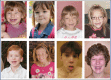
Figure 1.
Photographs of eight individuals with a 17q21.31 deletion
Family history. Because KdVS is typically caused by a de novo pathogenic variant, most probands represent a simplex case (i.e., a single occurrence in a family).
Establishing the Diagnosis
The diagnosis of KdVS is established in a proband with typical clinical findings and of any of the following (see Table 1):
- A heterozygous deletion at chromosome 17q21.31 that includes KANSL1 (~60% of affected individuals). The 17q21.31 deletion is typically 500 to 650 kb in size (hg19: chr17:43700000-44250000) and is flanked by segmental duplications.
- A heterozygous intragenic pathogenic (or likely pathogenic) variant in KANSL1 (~40% of affected individuals)
- Haploinsufficiency of KANSL1 due to chromosome rearrangements [Moreno-Igoa et al 2015]
Note: (1) Per ACMG/AMP variant interpretation guidelines, the terms "pathogenic variant" and "likely pathogenic variant" are synonymous in a clinical setting, meaning that both are considered diagnostic and can be used for clinical decision making [Richards et al 2015]. Reference to "pathogenic variants" in this GeneReview is understood to include any likely pathogenic variants. (2) Identification of a heterozygous KANSL1 variant of uncertain significance does not establish or rule out the diagnosis. (3) A characteristic epigenetic signature for KdVS has been established and may aid in the determination of the clinical significance of uncertain variants (see Further Testing to Consider).
Molecular genetic testing approaches can include a combination of gene-targeted testing (chromosomal microarray analysis, single-gene testing, multigene panel) and comprehensive genomic testing (exome sequencing, genome sequencing) depending on the phenotype.
Gene-targeted testing requires that the clinician determine which gene(s) are likely involved, whereas genomic testing does not. Individuals with the distinctive findings described in Suggestive Findings are likely to be diagnosed using gene-targeted testing (see Option 1), whereas those in whom the diagnosis of KdVS has not been considered are more likely to be diagnosed using genomic testing (see Option 2).
Option 1
When the phenotypic findings suggest the diagnosis of KdVS, molecular genetic testing approaches can include chromosomal microarray analysis (CMA), single-gene testing, or use of a multigene panel.
- CMA uses oligonucleotide or SNP arrays to detect genome-wide large deletions/duplications (including KANSL1) that cannot be detected by sequence analysis. If no DNA copy variant is detected by CMA, the next step is to perform either single-gene testing or a multigene panel.
- Single-gene testing. Sequence analysis of KANSL1 is performed to detect missense, nonsense, and splice site variants and small intragenic deletions/insertions. Note: Depending on the sequencing method used, single-exon, multiexon, or whole-gene deletions/duplications may not be detected. If no variant is detected by the sequencing method used, gene-targeted deletion/duplication analysis to detect exon-level deletions or duplications that could have been missed on CMA could be considered.
- An intellectual disability multigene panel that includes KANSL1 and other genes of interest (see Differential Diagnosis) may also be considered to identify the genetic cause of this condition while limiting identification of variants of uncertain significance and pathogenic variants in genes that do not explain the underlying phenotype. Note: (1) The genes included in the panel and the diagnostic sensitivity of the testing used for each gene vary by laboratory and are likely to change over time. (2) Some multigene panels may include genes not associated with the condition discussed in this GeneReview. (3) In some laboratories, panel options may include a custom laboratory-designed panel and/or custom phenotype-focused exome analysis that includes genes specified by the clinician. (4) Methods used in a panel may include sequence analysis, deletion/duplication analysis, and/or other non-sequencing-based tests.
Option 2
When the diagnosis of KdVS is not considered because an individual has atypical phenotypic features, comprehensive genomic testing (which does not require the clinician to determine which gene[s] are likely involved) is the best option. Exome sequencing is most commonly used; genome sequencing is also possible.
Note: Some exome sequencing platforms also provide information on DNA copy number variants (CNVs); genome sequencing frequently includes information on DNA CNVs. As such, exome sequencing with CNV analysis or genome sequencing could be considered as a first-line test for KdVS.
For an introduction to comprehensive genomic testing click here. More detailed information for clinicians ordering genomic testing can be found here.
Further Testing to Consider
Epigenetic signature analysis / methylation array. A distinctive epigenetic signature (disorder-specific genome-wide changes in DNA methylation profiles) in peripheral blood leukocytes has been identified in individuals with KdVS [Aref-Eshghi et al 2020, Levy et al 2021]. Epigenetic signature analysis of a peripheral blood sample or DNA banked from a blood sample can therefore clarify the diagnosis in individuals with: (1) convincing findings of KdVS but in whom no 17q21.31 deletion or pathogenic variant in KANSL1 has been identified via sequence analysis or genomic testing; or (2) suggestive findings of KdVS and a KANSL1 variant of uncertain clinical significance identified by molecular genetic testing. The data from the episignature analysis must be interpreted carefully. A positive result on the methylation array is insufficient for diagnosis without clinical findings consistent with KdVS. For an introduction to epigenetic signature analysis click here.
Karyotype. If a 17q21.31 deletion is not identified on CMA and an intragenic pathogenic variant in KANSL1 has not been identified on either a multigene panel or comprehensive genomic testing (exome or genome sequencing), additional options for testing include karyotype. A chromosome translocation with a 17q21.31 breakpoint that disrupted KANSL1 has been observed in one case report [Moreno-Igoa et al 2015].
Table 1.
Molecular Genetic Testing Used in Koolen-de Vries Syndrome
Clinical Characteristics
Clinical Description
Koolen-de Vries syndrome (KdVS) has a clinically recognizable phenotype that includes neonatal/childhood hypotonia, developmental delay / intellectual givdisability, dysmorphisms (see Figure 1), speech and language delays, congenital malformations, and behavioral features. To date, more than 200 individuals have been identified with KdVS [Koolen et al 2006, Sharp et al 2006, Koolen et al 2008, Grisart et al 2009, Tan et al 2009, Koolen et al 2012b, Zollino et al 2012, Zollino et al 2015, Koolen et al 2016, Morgan et al 2018a, Myers et al 2017, Amenta et al 2022, St John et al 2023]. The following description of the phenotypic features associated with this condition is based on these reports.
Table 2.
Koolen-de Vries Syndrome: Frequency of Select Features
Dysmorphic craniofacial features that may suggest KdVS include:
- Upslanted palpebral fissures
- Blepharophimosis
- Epicanthus
- Ptosis
- Pear-shaped nose
- Bulbous nose
- Large/protruding ears
The nose can have a high nasal bridge, a broad nasal root, long columella, and underdeveloped and/or thick alae nasi. The facial characteristics change with age. In infancy the facial gestalt is mostly characterized by hypotonia with an "open mouth" appearance. With increasing age there is usually elongation of the face and broadening of the chin, and the "tubular'' or "pear'' shape of the nose may become more apparent.
Developmental delay / intellectual disability. Psychomotor delay is noted in all affected individuals from an early age. The level of developmental delay varies significantly. The majority of individuals with KdVS function in the mild-to-moderate range of intellectual disability.
- Communication disorder is a core feature of KdVS, with a common speech and language phenotype seen. This includes an overriding "double hit" of oral hypotonia and apraxia in infancy and preschool, associated with severely delayed speech development [Morgan et al 2018a]. St John et al [2023] defined speech, language, and functional/adaptive behavior in 81 individuals with KdVS.
- First words occur on average between ages 2.5 and 3.5 years.
- Childhood apraxia of speech (CAS) is common in the preschool years, and speech development is effortful even when supported with intensive therapy.Augmentative (e.g., sign language) or alternative (e.g., communication devices) communication may alleviate frustration for the child and promote communication development.Overall, however, speech prognosis is positive, with CAS improving markedly around age eight to 12 years. At this time, the dysarthric element of speech is more apparent with a slow rate and monotone presentation.
- Stuttering has been described in 76.6% of verbal individuals and follows a unique trajectory of late onset and fluctuating presence [St John et al 2023].
- Receptive and expressive language abilities are commensurate, but literacy skills remain a relative weakness.
- Social competence, successful behavioral/emotional control, and coping skills are areas of relative strength, while communication difficulties affect daily living skills as an area of comparative difficulty.
Hypotonia with poor sucking and slow feeding can be evident in the neonatal period and during childhood. Feeding difficulties may require hospitalization and/or nasogastric tube feeding in some neonates. Beyond infancy and into the preschool years, many children experience issues chewing difficult, lumpy, or solid textures [Morgan et al 2018a].
Epilepsy, including generalized seizures and unilateral clonic seizures, is noted in approximately 33% of affected individuals. The epilepsy phenotypic spectrum in KdVS is broad; however, most individuals have focal seizures, with some having a phenotype resembling the self-limited focal epilepsies of childhood [Myers et al 2017].
- The typical epilepsy phenotype of KdVS involves childhood-onset focal seizures that are prolonged and have prominent autonomic features.
- Multifocal epileptiform discharges are the typical EEG pattern.
Neurobehavioral/psychiatric manifestations. In many affected individuals, behavior is described as friendly, amiable, and cooperative, with or without frequent laughing. However, behavioral findings including attention-deficit/hyperactivity disorder have been reported [Koolen et al 2008, Tan et al 2009, Koolen et al 2016]. A subset of affected individuals have autism and/or anxiety.
Growth. Short stature is not one of the most common clinical features of the syndrome. However, El Chehadeh-Djebbar et al [2011] reported a child with a 17q21.31 deletion, short stature (4 SD below the mean), complete growth hormone deficiency, and gonadotropic deficiency [El Chehadeh-Djebbar et al 2011]. Brain MRI showed partial pituitary stalk interruption, expanding the phenotypic spectrum of the syndrome.
Ophthalmologic involvement in individuals with this condition include hypermetropia, strabismus, congenital cataract, and optic atrophy.
Hearing impairment. A minority of affected individuals experience recurrent otitis media.
Neuroimaging/other neurodevelopmental features
- Brain MRI. Structural brain abnormalities may be universal, including signs of abnormal neuroblast migration and abnormal axonal guidance. Affected individuals have been described as having:
- Ventriculomegaly
- Aplasia/hypoplasia of the corpus callosum
- Hydrocephalus
- Arnold-Chiari malformation
- Intraventricular hemorrhage
- Infrequent findings (present in fewer than 10% of reported individuals) may include the following:
- Sacral dimple
- Dural ectasia
- Spina bifida
- Pineal cyst
- Cervical spinal canal stenosis
Musculoskeletal. Joint hypermobility is common. Affected individuals may also experience joint dislocations. Other findings can include:
- Long, slender fingers
- Persistence of the fetal fingertip pads
- Hypoplasia of the hand muscles
- Pes planus
- Pes cavus
- Calcaneovalgus deformity
- Congenital hip dislocation
- Scoliosis/kyphosis
- Pectus anomalies, including pectus excavatum or pectus carinatum
- Slender build
- Spondylolisthesis (infrequently)
- Craniosynostosis (infrequently), most commonly sagittal, but metopic has also been observed
Congenital heart defects mainly include septal heart defects; however, cardiac valve disease, aortic root dilatation, and pulmonary stenosis have also been described.
Renal and urologic anomalies include vesicoureteral reflux, hydronephrosis, pyelectasis, and duplex renal system. Cryptorchidism has been reported in the majority of males. A minority of affected individuals experience recurrent urinary tract infection.
Respiratory. Some affected individuals experience recurrent respiratory infections. There is no known immune deficiency described in affected individuals that can explain this finding. Tracheo-/laryngomalacia has also been reported in a relatively small number of affected individuals.
Other associated features reported infrequently (fewer than 10% of known affected individuals)
- Endocrinology. In addition to at least one affected individual with growth hormone deficiency, other reported hormonal issues include hypothyroidism, precocious puberty, and primary adrenal insufficiency.
- Integument. A myriad of skin findings have been described, typically in a few individuals each, including [Wright et al 2011, Zollino et al 2015, Koolen et al 2016]:
- Multiple nevi
- Other pigmentary skin abnormalities, such as vitiligo and café au lait macules
- Hemangioma
- Eczema
- Ichthyosis/hyperkeratosis
- Hair abnormalities, such as fair hair and/or alopecia
- Neoplasia. It is unclear if individuals with this condition have an increased risk above the general population risk of developing a malignancy. Infrequent malignancies in affected individuals have included melanoma and testicular neoplasms. Individuals with KdVS who have lighter skin tones or skin types who are at greater risk for developing melanoma should be evaluated annually to assess ectodermal findings and cutaneous changes (see Management). Currently, there is no consensus tumor screening protocols that have been proposed or published for individuals with KdVS.
Life span. Longitudinal data are insufficient to determine life expectancy, although survival into adulthood is typical. One reported individual is alive at age 63 years [Farnè et al 2022].
Genotype-Phenotype Correlations
Genotype-phenotype correlations in KdVS have not been demonstrated. Notably, the clinical features of affected individuals with atypical deletions and those with pathogenic variants in KANSL1 are in keeping with the phenotype seen in individuals with a classic 17q21.31 deletion [Zollino et al 2015, Koolen et al 2016].
Penetrance
Penetrance is 100%. Clinical features of KdVS are apparent in all individuals with a deletion of or a pathogenic variant in KANSL1, although the extent and severity of clinical findings vary among individuals.
Nomenclature
The disorder was first recognized following chromosomal microarray analysis among large cohorts of unselected individuals with intellectual disability [Koolen et al 2006, Sharp et al 2006, Shaw-Smith et al 2006]. The identification of individuals with a similar phenotype and a de novo KANSL1 pathogenic variant [Koolen et al 2012b, Zollino et al 2012] led OMIM to assign the name "Koolen-de Vries syndrome" to the condition.
Prevalence
The prevalence of KdVS is unknown. The authors estimate the prevalence of the 17q21.31 deletion to be 1:55,000 individuals [Koolen et al 2016]. The prevalence of individuals with a pathogenic sequence variant in KANSL1 cannot be determined with precision owing to the limited number of such affected individuals identified thus far. Preliminary data suggest that pathogenic KANSL1 sequence variants may be as frequent as deletions, but more studies are needed to determine an unbiased prevalence.
Genetically Related (Allelic) Disorders
No phenotypes other than those discussed in this GeneReview are known to be associated with deletion of the genes located within the 17q21.31 chromosome locus or with pathogenic variants in KANSL1. Besides the recurrent classic 17q21.31 microdeletion, several atypical 17q21.31 deletions have been described in children with clinical features typically associated with the classic 17q21.31 microdeletion [Cooper et al 2011, Dubourg et al 2011, Kitsiou-Tzeli et al 2012, Koolen et al 2012b]. All these atypical deletions encompass at least KANSL1.
Duplication of 17q21.31 (OMIM 613533). Persons with a reciprocal duplication of the region deleted in Koolen-de Vries syndrome differ phenotypically from those with the 17q21.31 deletion. The reciprocal duplication has been found in a female with severe psychomotor developmental delay, microcephaly, facial dysmorphisms, abnormal digits, and hirsutism [Kirchhoff et al 2007] and in four individuals with mild psychomotor developmental delay and behavioral findings [Grisart et al 2009].
MAPT. Pathogenic gain-of-function variants in MAPT, the gene encoding microtubule-associated protein tau, have been identified in individuals diagnosed with frontotemporal dementia with parkinsonism-17 (FTDP-17). These variants result in pathogenic deposits of hyperphosphorylated tau. This is in contrast to the haploinsufficiency of MAPT in Koolen-de Vries syndrome due to a deletion of 17q21.31 that includes KANSL1 and MAPT. Therefore, individuals who have the 17q21.31 deletion are not at an increased risk for FTDP-17 or related tauopathies.
Differential Diagnosis
The most common findings in Koolen-de Vries syndrome (KdVS) – developmental delay and childhood hypotonia – are common and relatively nonspecific indications for molecular cytogenetic analysis. However, the concurrent finding of characteristic facial dysmorphic features, epilepsy, hypermetropia, congenital heart defects, renal or urologic anomalies, cryptorchidism, and/or distinctive friendly/amiable behavior may prompt specific consideration of the diagnosis of KdVS. See Table 3 for other diagnoses that may be considered in individuals with developmental delay, childhood hypotonia, and additional findings overlapping those observed in KdVS.
Table 3.
Selected Disorders with Developmental Delay, Childhood Hypotonia, and Concurrent Findings Similar to Koolen-de Vries Syndrome
Management
Evaluations Following Initial Diagnosis
To establish the clinical consequences in an individual diagnosed with Koolen-de Vries syndrome (KdVS), the evaluations in Table 4 (if not performed as part of the evaluation that led to diagnosis) are recommended.
Table 4.
Recommended Evaluations Following Initial Diagnosis of Koolen-de Vries Syndrome
Treatment of Manifestations
There is no cure for Koolen-de Vries syndrome.
Supportive care to improve quality of life, maximize function, and reduce complications is recommended. This ideally involves multidisciplinary care by specialists in relevant fields (see Table 5).
Table 5.
Treatment of Manifestations in Individuals with Koolen-de Vries Syndrome
Developmental Delay / Intellectual Disability Management Issues
Children with KdVS require early, intensive speech motor and language therapy, with targeted literacy and social language interventions as developmentally appropriate.
The following information represents typical management recommendations for individuals with developmental delay / intellectual disability in the United States (US); standard recommendations may vary from country to country.
Ages 0-3 years. Referral to an early intervention program is recommended for access to occupational, physical, speech, and feeding therapy. In the US, early intervention is a federally funded program available in all states.
Ages 3-5 years. In the US, developmental preschool through the local public school district is recommended. Before placement, an evaluation is made to determine needed services and therapies and an individualized education plan (IEP) is developed.
Ages 5-21 years
- In the US, an IEP based on the individual’s level of function should be developed by the local public school district. Affected children are permitted to remain in the public school district until age 21.
- Discussion about transition plans including financial, vocation/employment, and medical arrangements should begin at age 12 years. Developmental pediatricians can provide assistance with transition to adulthood.
All ages. Consultation with a developmental pediatrician is recommended to ensure the involvement of appropriate community, state, and educational agencies and to support parents in maximizing quality of life.
Consideration of private supportive therapies based on the affected individual's needs is recommended. Specific recommendations regarding type of therapy can be made by a developmental pediatrician.
In the US:
- Developmental Disabilities Administration (DDA) enrollment is recommended. DDA is a public agency that provides services and support to qualified individuals. Eligibility differs by state but is typically determined by diagnosis and/or associated cognitive/adaptive disabilities.
- Families with limited income and resources may also qualify for supplemental security income (SSI) for their child with a disability.
Motor Dysfunction
Gross motor dysfunction
- Physical therapy is recommended to maximize mobility and to reduce the risk for later-onset orthopedic complications (e.g., contractures, scoliosis, hip dislocation).
- Consider use of durable medical equipment as needed (e.g., walkers, bath chairs, orthotics, adaptive strollers).
Fine motor dysfunction. Occupational therapy is recommended for difficulty with fine motor skills that affect adaptive function such as feeding, grooming, dressing, and writing.
Oral motor dysfunction should be assessed at each visit and clinical feeding evaluations and/or radiographic swallowing studies should be obtained for choking/gagging during feeds, poor weight gain, frequent respiratory illnesses, or feeding refusal that is not otherwise explained. Assuming that the child is safe to eat by mouth, feeding therapy (typically from an occupational or speech therapist) is recommended to help improve coordination or sensory-related feeding issues. Feeds can be thickened or chilled for safety. When feeding dysfunction is severe, an NG-tube or G-tube may be necessary.
Communication issues. Consider evaluation for alternative means of communication (e.g., augmentative and alternative communication [AAC]) used alongside verbal therapies for individuals who have expressive language difficulties. Intensive verbal speech therapy approaches for childhood apraxia of speech are recommended in the early years [Morgan et al 2018b, St John et al 2023], and literacy, dysarthria, and social skill therapies are required in the school years.
Social/Behavioral Concerns
Children may qualify for and benefit from interventions used in treatment of autism spectrum disorder, including applied behavior analysis (ABA). ABA therapy is targeted to the individual child's behavioral, social, and adaptive strengths and weaknesses and is typically performed one on one with a board-certified behavior analyst.
Consultation with a developmental pediatrician may be helpful in guiding parents through appropriate behavior management strategies or providing prescription medications when necessary.
Surveillance
To monitor existing manifestations, the individual's response to supportive care, and the emergence of new manifestations, the evaluations in Table 6 are recommended.
Table 6.
Recommended Surveillance for Individuals with Koolen-de Vries Syndrome
Evaluation of Relatives at Risk
See Genetic Counseling for issues related to testing of at-risk relatives for genetic counseling purposes.
Therapies Under Investigation
Search ClinicalTrials.gov in the US and EU Clinical Trials Register in Europe for access to information on clinical studies for a wide range of diseases and conditions. Note: There may not be clinical trials for this disorder.
Genetic Counseling
Genetic counseling is the process of providing individuals and families with information on the nature, mode(s) of inheritance, and implications of genetic disorders to help them make informed medical and personal decisions. The following section deals with genetic risk assessment and the use of family history and genetic testing to clarify genetic status for family members; it is not meant to address all personal, cultural, or ethical issues that may arise or to substitute for consultation with a genetics professional. —ED.
Mode of Inheritance
Koolen-de Vries syndrome (KdVS), caused by a heterozygous deletion at chromosome 17q21.31 or a heterozygous intragenic KANSL1 pathogenic variant, is an autosomal dominant disorder. Almost all affected individuals represent simplex cases (i.e., a single affected individual in the family).
Risk to Family Members
Parents of a proband
- To date, all reported intragenic KANSL1 pathogenic variants and almost all reported 17q21.31 deletions have been de novo in the proband.
- Evaluation of the parents by testing that will detect the 17q21.31 deletion or intragenic KANSL1 pathogenic variant present in the proband is recommended to confirm their genetic status and to allow reliable recurrence risk counseling. FISH analysis in the parents to evaluate for a balanced insertion and/or translocation may also be considered.
- All unaffected parents tested to date from whom a deleted chromosome 17 originated have shown a 900-kb inversion involving chromosome 17q21.31. This inversion (also referred to as the H2 lineage) is enriched in Europeans, and carriers are predisposed to the 17q21.31 deletion (see Molecular Genetics).Note: Testing for the 17q21.31 inversion polymorphism in parents is not recommended for recurrence risk assessment because it does not provide additional information that is of clinical use. The inversion is common in northern European populations, and although it seems to be a necessary factor for the deletion to occur, many other factors are important given the fact that the 17q21.31 deletion is relatively rare.
- If the 17q21.31 deletion or intragenic KANSL1 pathogenic variant identified in the proband is not identified in either parent and parental identity testing has confirmed biological maternity and paternity, the following possibilities should be considered:
- The proband has a de novo genetic alteration.
- The proband inherited a genetic alteration from a parent with germline (or somatic and germline) mosaicism. Somatic and (presumed) germline mosaicism for a 17q21.31 deletion has been identified in at least two parents [Koolen et al 2012a].Note: Testing of parental leukocyte DNA may not detect all instances of somatic mosaicism and will not detect a genetic alteration that is present in the germ cells only.
- Theoretically, a parent could have a balanced chromosome rearrangement involving 17q21.31 resulting in a 17q21.31 deletion in an affected child; balanced chromosome rearrangements in parents involving 17q21.31 have not been reported to date.
Sibs of a proband. The risk to the sibs of the proband depends on the genetic status of the proband's parents:
- If the parents are clinically unaffected and the 17q21.31 deletion or intragenic KANSL1 pathogenic variant detected in the proband cannot be detected in the leukocyte DNA of either parent, the recurrence risk to sibs is slightly greater than that of the general population because of the possibility of:
- Parental germline mosaicism [Koolen et al 2012a];
- A balanced chromosome rearrangement involving 17q21.31 (not reported, but theoretically possible).
Offspring of a proband
- Individuals who have the 17q21.31 deletion or an intragenic KANSL1 pathogenic variant have a 50% chance of transmitting the genetic alteration to each child.
- To date, one individual diagnosed with KdVS has been known to reproduce [Author, personal observation].
Other family members. The risk to other family members depends on the status of the proband's parents: if a parent has a KdVS-related genetic alteration or, theoretically, a balanced chromosomal rearrangement, the parent's family members may be at risk.
Related Genetic Counseling Issues
Family planning
- The optimal time for determination of genetic risk and discussion of the availability of prenatal/preimplantation genetic testing is before pregnancy.
- It is appropriate to offer genetic counseling (including discussion of potential risks to offspring and reproductive options) to parents of a child with KdVS.
Prenatal Testing and Preimplantation Genetic Testing
Once the KdVS-related genetic alteration has been identified in an affected family member, prenatal and preimplantation genetic testing are possible.
Differences in perspective may exist among medical professionals and within families regarding the use of prenatal testing. While most centers would consider use of prenatal testing to be a personal decision, discussion of these issues may be helpful.
Resources
GeneReviews staff has selected the following disease-specific and/or umbrella support organizations and/or registries for the benefit of individuals with this disorder and their families. GeneReviews is not responsible for the information provided by other organizations. For information on selection criteria, click here.
- Kool Kid Alliance
- Koolen-de Vries Syndrome FoundationEnriching lives through education, awareness and research.Phone: 833-721-KDVS
- Chromosome Disorder Outreach Inc.Phone: 561-395-4252Email: info@chromodisorder.org
- MedlinePlus
- Unique: Understanding Rare Chromosome and Gene DisordersUnited KingdomPhone: +44 (0) 1883 723356Email: info@rarechromo.org
- GenIDA Registry: Genetically Determined Intellectual Disabilities and Autism Spectrum DisordersA website for Patients, Families and ProfessionalsFrance
- Human Disease Gene Website Series - RegistryEmail: info@humandiseasegenes.com
Molecular Genetics
Information in the Molecular Genetics and OMIM tables may differ from that elsewhere in the GeneReview: tables may contain more recent information. —ED.
Table A.
Koolen-de Vries Syndrome: Genes and Databases
Table B.
OMIM Entries for Koolen-de Vries Syndrome (View All in OMIM)
Molecular Pathogenesis
De novo pathogenic variants in KANSL1 were identified in children with clinical features that are in keeping with the phenotype seen in individuals with a classic 17q21.31 deletion, demonstrating that KANSL1 is the primary gene involved in this deletion syndrome [Koolen et al 2012b, Zollino et al 2012].
KANSL1 encodes KAT8 regulatory NSL complex subunit 1 (KANSL1), the longer isoform of which (NP_001180395.1) has 1,105 amino acids. KANSL1 is a scaffold protein of the nonspecific lethal complex that contains the histone acetyltransferase MOF, which acetylates histone H4 on lysine 16 (H4K16ac) to facilitate transcriptional activation [Mendjan et al 2006].
H4K16ac activates the expression of a broad set of genes including several autophagy-related genes [Füllgrabe et al 2013]. Autophagy is a catabolic process important for the clearance of protein aggregates and damaged organelles within the cell, which is essential for cell homeostasis and survival. Autophagy is essential in neurons, not only for cell homeostasis but also for regulation of development and function [Shehata et al 2012, Tang et al 2014].
Studies in mice have shown that heterozygous loss of Kansl1 leads to changes in gene expression related to synaptic transmission and to a decrease in basal synaptic transmission and plasticity [Arbogast et al 2017], but the underlying cellular mechanisms remain unknown.
Linda et al [2022] reported that KANSL1 deficiency leads to increased oxidative stress and autophagosome formation in iPSCs and iNeurons. In neurons, increased reactive oxygen species (ROS)-activated autophagy reduced neuronal synaptic connectivity and activity. The observed neuronal phenotype could be rescued by treatment with apocynin, an antioxidant that reduced oxidative stress and autophagosome accumulation. These findings were supported by the study of Li et al [2022], in which KANSL1 was identified as an essential gene for autophagy using siRNA screening. Kansl1+/- mice exhibit impairment in the autophagic clearance of damaged mitochondria and accumulation of reactive oxygen species, thereby resulting in defective neuronal and cardiac function.
Laboratory technical considerations. Genetic testing of the 17q21.31 genomic region is challenging. The mapping and interpretation of the deletion breakpoints are confounded by the structural complexity and genomic variation of the 17q21.31 locus [Koolen et al 2016]. Two haplotypes exist, in direct (H1) and inverted (H2) orientation [Stefansson et al 2005]. The H2 haplotype is enriched in Europeans, and those with this haplotype are predisposed to the 17q21.31 deletion [Koolen et al 2006, Sharp et al 2006, Koolen et al 2008, Zody et al 2008]. However, the frequency of de novo 17q21.31 deletions in those with the H2 inversion is low, and other as yet poorly understood factors are likely to be important in the generation of the deletion.
The 17q21.31 inversion polymorphism (H2 haplotype) and the copy number polymorphism clusters encompassing exons 1-3 of KANSL1 contribute to difficulties in single nucleotide variant calling, such as loss-of-function variant "artifacts" in KANSL1 [Koolen et al 2016]. The detection of a truncating variant in exons 1-3 of KANSL1 is not sufficient to make a diagnosis of KdVS. In these cases, a compatible clinical phenotype and variant analysis of parental samples is of the utmost importance to verify that the possibly pathogenic variant occurred de novo.
Mechanism of disease causation. Loss of function. The 17q21.31 deletion is typically 500 to 650 kb in size (hg19: chr17:43700000-44250000) and is flanked by segmental duplications that mediate nonallelic homologous recombination [Itsara et al 2012].
Chapter Notes
Author Notes
Radboudumc Center of Expertise: rare congenital developmental disorders
Acknowledgments
The authors gratefully acknowledge the KdVS Foundation, other support groups and the parents/caregivers for their participation in research and for their generous sharing of information.
Revision History
- 2 February 2023 (ma) Comprehensive update posted live
- 13 June 2019 (ma) Comprehensive update posted live
- 20 November 2012 (me) Comprehensive update posted live
- 26 January 2010 (me) Review posted live
- 28 August 2009 (dak) Original submission
References
Literature Cited
- Amenta S, Frangella S, Marangi G, Lattante S, Ricciardi S, Doronzio PN, Orteschi D, Veredice C, Contaldo I, Zampino G, Gentile M, Scarano E, Graziano C, Zollino M. Adult phenotype in Koolen-de Vries/KANSL1 haploinsufficiency syndrome. J Med Genet. 2022;59:189-95. [PubMed: 33361104]
- Arbogast T, Iacono G, Chevalier C, Afinowi NO, Houbaert X, van Eede MC, Laliberte C, Birling MC, Linda K, Meziane H, Selloum M, Sorg T, Nadif Kasri N, Koolen DA, Stunnenberg HG, Henkelman RM, Kopanitsa M, Humeau Y, De Vries BBA, Herault Y. Mouse models of 17q21.31 microdeletion and microduplication syndromes highlight the importance of Kansl1 for cognition. PLoS Genet. 2017;13:e1006886. [PMC free article: PMC5531616] [PubMed: 28704368]
- Aref-Eshghi E, Kerkhof J, Pedro VP, Groupe DI. France, Barat-Houari M, Ruiz-Pallares N, Andrau JC, Lacombe D, Van-Gils J, Fergelot P, Dubourg C, Cormier-Daire V, Rondeau S, Lecoquierre F, Saugier-Veber P, Nicolas G, Lesca G, Chatron N, Sanlaville D, Vitobello A, Faivre L, Thauvin-Robinet C, Laumonnier F, Raynaud M, Alders M, Mannens M, Henneman P, Hennekam RC, Velasco G, Francastel C, Ulveling D, Ciolfi A, Pizzi S, Tartaglia M, Heide S, Héron D, Mignot C, Keren B, Whalen S, Afenjar A, Bienvenu T, Campeau PM, Rousseau J, Levy MA, Brick L, Kozenko M, Balci TB, Siu VM, Stuart A, Kadour M, Masters J, Takano K, Kleefstra T, de Leeuw N, Field M, Shaw M, Gecz J, Ainsworth PJ, Lin H, Rodenhiser DI, Friez MJ, Tedder M, Lee JA, DuPont BR, Stevenson RE, Skinner SA, Schwartz CE, Genevieve D, Sadikovic B. Evaluation of DNA methylation episignatures for diagnosis and phenotype correlations in 42 mendelian neurodevelopmental disorders. Am J Hum Genet. 2020;106:356-70.
- Cooper GM, Coe BP, Girirajan S, Rosenfeld JA, Vu TH, Baker C, Williams C, Stalker H, Hamid R, Hannig V, Abdel-Hamid H, Bader P, McCracken E, Niyazov D, Leppig K, Thiese H, Hummel M, Alexander N, Gorski J, Kussmann J, Shashi V, Johnson K, Rehder C, Ballif BC, Shaffer LG, Eichler EE. A copy number variation morbidity map of developmental delay. Nat Genet. 2011;43:838-46. [PMC free article: PMC3171215] [PubMed: 21841781]
- Dubourg C, Sanlaville D, Doco-Fenzy M, Le Caignec C, Missirian C, Jaillard S, Schluth-Bolard C, Landais E, Boute O, Philip N, Toutain A, David A, Edery P, Moncla A, Martin-Coignard D, Vincent-Delorme C, Mortemousque I, Duban-Bedu B, Drunat S, Beri M, Mosser J, Odent S, David V, Andrieux J. Clinical and molecular characterization of 17q21.31 microdeletion syndrome in 14 French patients with mental retardation. Eur J Med Genet. 2011;54:144-51. [PubMed: 21094706]
- El Chehadeh-Djebbar S, Callier P, Masurel-Paulet A, Bensignor C, Méjean N, Payet M, Ragon C, Durand C, Marle N, Mosca-Boidron AL, Huet F, Mugneret F, Faivre L, Thauvin-Robinet C. 17q21.31 microdeletion in a patient with pituitary stalk interruption syndrome. Eur J Med Genet. 2011;54:369-73. [PubMed: 21397059]
- Farnè M, Bernardini L, Capalbo A, Cavarretta G, Torres B, Sanchini M, Fini S, Ferlini A, Bigoni S. Koolen-de Vries syndrome in a 63-year old woman: report of the oldest patient and review of the adult phenotype. Am J Med Genet A. 2022;188:692-707. [PMC free article: PMC9297928] [PubMed: 34665525]
- Füllgrabe J, Lynch-Day MA, Heldring N, Li W, Struijk RB, Ma Q, Hermanson O, Rosenfeld MG, Klionsky DJ, Joseph B. The histone H4 lysine 16 acetyltransferase hMOF regulates the outcome of autophagy. Autophagy. 2013;9:1621-3. [PMC free article: PMC4006103] [PubMed: 23863932]
- Grieco JC, Bahr RH, Schoenberg MR, Conover L, Mackie LN, Weeber EJ. Quantitative measurement of communication ability in children with Angelman syndrome. J Appl Res Intellect Disabil. 2018;31:e49-e58. [PubMed: 27990716]
- Grisart B, Willatt L, Destrée A, Fryns JP, Rack K, de Ravel T, Rosenfeld J, Vermeesch JR, Verellen-Dumoulin C, Sandford R. 17q21.31 microduplication patients are characterised by behavioural problems and poor social interaction. J Med Genet. 2009;46:524-30. [PubMed: 19502243]
- Itsara A, Vissers LE, Steinberg KM, Meyer KJ, Zody MC, Koolen DA, de Ligt J, Cuppen E, Baker C, Lee C, Graves TA, Wilson RK, Jenkins RB, Veltman JA, Eichler EE. Resolving the breakpoints of the 17q21.31 microdeletion syndrome with next-generation sequencing. Am J Hum Genet. 2012;90:599-613. [PMC free article: PMC3322237] [PubMed: 22482802]
- Kirchhoff M, Bisgaard AM, Duno M, Hansen FJ, Schwartz M. A 17q21.31 microduplication, reciprocal to the newly described 17q21.31 microdeletion, in a girl with severe psychomotor developmental delay and dysmorphic craniofacial features. Eur J Med Genet. 2007;50:256-63. [PubMed: 17576104]
- Kitsiou-Tzeli S, Frysira H, Giannikou K, Syrmou A, Kosma K, Kakourou G, Leze E, Sofocleous C, Kanavakis E, Tzetis M. Microdeletion and microduplication 17q21.31 plus an additional CNV, in patients with intellectual disability, identified by array-CGH. Gene. 2012;492:319-24. [PubMed: 22037486]
- Koolen DA, Dupont J, de Leeuw N, Vissers LE, van den Heuvel SP, Bradbury A, Steer J, de Brouwer AP, Ten Kate LP, Nillesen WM, de Vries BB, Parker MJ. Two families with sibling recurrence of the 17q21.31 microdeletion syndrome due to low-grade mosaicism. Eur J Hum Genet. 2012a;20:729-33. [PMC free article: PMC3376266] [PubMed: 22293690]
- Koolen DA, Kramer JM, Neveling K, Nillesen WM, Moore-Barton HL, Elmslie FV, Toutain A, Amiel J, Malan V, Tsai AC, Cheung SW, Gilissen C, Verwiel ET, Martens S, Feuth T, Bongers EM, de Vries P, Scheffer H, Vissers LE, de Brouwer AP, Brunner HG, Veltman JA, Schenck A, Yntema HG, de Vries BB. Mutations in the chromatin modifier gene KANSL1 cause the 17q21.31 microdeletion syndrome. Nat Genet. 2012b;44:639-41. [PubMed: 22544363]
- Koolen DA, Pfundt R, Linda K, Beunders G, Veenstra-Knol HE, Conta JH, Fortuna AM, Gillessen-Kaesbach G, Dugan S, Halbach S, Abdul-Rahman OA, Winesett HM, Chung WK, Dalton M, Dimova PS, Mattina T, Prescott K, Zhang HZ, Saal HM, Hehir-Kwa JY, Willemsen MH, Ockeloen CW, Jongmans MC, Van der Aa N, Failla P, Barone C, Avola E, Brooks AS, Kant SG, Gerkes EH, Firth HV, Õunap K, Bird LM, Masser-Frye D, Friedman JR, Sokunbi MA, Dixit A, Splitt M; DDD Study, Kukolich MK, McGaughran J, Coe BP, Flórez J, Nadif Kasri N, Brunner HG, Thompson EM, Gecz J, Romano C, Eichler EE, de Vries BB. The Koolen-de Vries syndrome: a phenotypic comparison of patients with a 17q21.31 microdeletion versus a KANSL1 sequence variant. Eur J Hum Genet. 2016;24:652-9. [PMC free article: PMC4930086] [PubMed: 26306646]
- Koolen DA, Sharp AJ, Hurst JA, Firth HV, Knight SJ, Goldenberg A, Saugier-Veber P, Pfundt R, Vissers LE, Destrée A, Grisart B, Rooms L, Van der Aa N, Field M, Hackett A, Bell K, Nowaczyk MJ, Mancini GM, Poddighe PJ, Schwartz CE, Rossi E, De Gregori M, Antonacci-Fulton LL, McLellan MD 2nd, Garrett JM, Wiechert MA, Miner TL, Crosby S, Ciccone R, Willatt L, Rauch A, Zenker M, Aradhya S, Manning MA, Strom TM, Wagenstaller J, Krepischi-Santos AC, Vianna-Morgante AM, Rosenberg C, Price SM, Stewart H, Shaw-Smith C, Brunner HG, Wilkie AO, Veltman JA, Zuffardi O, Eichler EE, de Vries BB. Clinical and molecular delineation of the 17q21.31 microdeletion syndrome. J Med Genet. 2008;45:710-20. [PMC free article: PMC3071570] [PubMed: 18628315]
- Koolen DA, Vissers LE, Pfundt R, de Leeuw N, Knight SJ, Regan R, Kooy RF, Reyniers E, Romano C, Fichera M, Schinzel A, Baumer A, Anderlid BM, Schoumans J, Knoers NV, van Kessel AG, Sistermans EA, Veltman JA, Brunner HG, de Vries BB. A new chromosome 17q21.31 microdeletion syndrome associated with a common inversion polymorphism. Nat Genet. 2006;38:999-1001. [PubMed: 16906164]
- Levy MA, McConkey H, Kerkhof J, Barat-Houari M, Bargiacchi S, Biamino E, Bralo MP, Cappuccio G, Ciolfi A, Clarke A, DuPont BR, Elting MW, Faivre L, Fee T, Fletcher RS, Cherik F, Foroutan A, Friez MJ, Gervasini C, Haghshenas S, Hilton BA, Jenkins Z, Kaur S, Lewis S, Louie RJ, Maitz S, Milani D, Morgan AT, Oegema R, Østergaard E, Pallares NR, Piccione M, Pizzi S, Plomp AS, Poulton C, Reilly J, Relator R, Rius R, Robertson S, Rooney K, Rousseau J, Santen GWE, Santos-Simarro F, Schijns J, Squeo GM, St John M, Thauvin-Robinet C, Traficante G, van der Sluijs PJ, Vergano SA, Vos N, Walden KK, Azmanov D, Balci T, Banka S, Gecz J, Henneman P, Lee JA, Mannens MMAM, Roscioli T, Siu V, Amor DJ, Baynam G, Bend EG, Boycott K, Brunetti-Pierri N, Campeau PM, Christodoulou J, Dyment D, Esber N, Fahrner JA, Fleming MD, Genevieve D, Kerrnohan KD, McNeill A, Menke LA, Merla G, Prontera P, Rockman-Greenberg C, Schwartz C, Skinner SA, Stevenson RE, Vitobello A, Tartaglia M, Alders M, Tedder ML, Sadikovic B. Novel diagnostic DNA methylation episignatures expand and refine the epigenetic landscapes of mendelian disorders. HGG Adv. 2021;3:100075. [PMC free article: PMC8756545] [PubMed: 35047860]
- Li T, Lu D, Yao C, Li T, Dong H, Li Z, Xu G, Chen J, Zhang H, Yi X, Zhu H, Liu G, Wen K, Zhao H, Gao J, Zhang Y, Han Q, Li T, Zhang W, Zhao J, Li T, Bai Z, Song M, He X, Zhou T, Xia Q, Li A, Pan X. Kansl1 haploinsufficiency impairs autophagosome-lysosome fusion and links autophagic dysfunction with Koolen-de Vries syndrome in mice. Nat Commun. 2022;13:931. [PMC free article: PMC8854428] [PubMed: 35177641]
- Linda K, Lewerissa EI, Verboven AHA, Gabriele M, Frega M, Klein Gunnewiek TM, Devilee L, Ulferts E, Hommersom M, Oudakker A, Schoenmaker C, van Bokhoven H, Schubert D, Testa G, Koolen DA, de Vries BBA, Nadif Kasri N. Imbalanced autophagy causes synaptic deficits in a human model for neurodevelopmental disorders. Autophagy. 2022;18:423-42. [PMC free article: PMC8942553] [PubMed: 34286667]
- Mendjan S, Taipale M, Kind J, Holz H, Gebhardt P, Schelder M, Vermeulen M, Buscaino A, Duncan K, Mueller J, Wilm M, Stunnenberg HG, Saumweber H, Akhtar A. Nuclear pore components are involved in the transcriptional regulation of dosage compensation in Drosophila. Mol Cell. 2006;21:811-23. [PubMed: 16543150]
- Moreno-Igoa M, Hernández-Charro B, Bengoa-Alonso A, Pérez-Juana-del-Casal A, Romero-Ibarra C, Nieva-Echebarria B, Ramos-Arroyo MA. KANSL1 gene disruption associated with the full clinical spectrum of 17q21.31 microdeletion syndrome. BMC Med Genet. 2015;16:68. [PMC free article: PMC4593202] [PubMed: 26293599]
- Morgan AT, Haaften LV, van Hulst K, Edley C, Mei C, Tan TY, Amor D, Fisher SE, Koolen DA. Early speech development in Koolen de Vries syndrome limited by oral praxis and hypotonia. Eur J Hum Genet. 2018a;26:75-84. [PMC free article: PMC5839037] [PubMed: 29225339]
- Morgan AT, Murray E, Liégeois FJ. Interventions for childhood apraxia of speech. Cochrane Database Syst Rev. 2018b;5:CD006278. [PMC free article: PMC6494637] [PubMed: 29845607]
- Myers KA, Mandelstam SA, Ramantani G, Rushing EJ, de Vries BB, Koolen DA, Scheffer IE (2017) The epileptology of Koolen-de Vries syndrome: electro-clinico-radiologic findings in 31 patients. Epilepsia. 58:1085-94. [PubMed: 28440867]
- Paolo P, Matteo F, Rossella L, Anna M, Rosario C, Paola G, Cristiano T, Giorgio R. Koolen-de Vries syndrome: preliminary observations of topiramate efficacy. Child Neurol Open. 2021;8:2329048X211019183. [PMC free article: PMC8173995] [PubMed: 34124281]
- Richards S, Aziz N, Bale S, Bick D, Das S, Gastier-Foster J, Grody WW, Hegde M, Lyon E, Spector E, Voelkerding K, Rehm HL, et al. Standards and guidelines for the interpretation of sequence variants: a joint consensus recommendation of the American College of Medical Genetics and Genomics and the Association for Molecular Pathology. Genet Med. 2015;17:405-24. [PMC free article: PMC4544753] [PubMed: 25741868]
- Sharp AJ, Hansen S, Selzer RR, Cheng Z, Regan R, Hurst JA, Stewart H, Price SM, Blair E, Hennekam RC, Fitzpatrick CA, Segraves R, Richmond TA, Guiver C, Albertson DG, Pinkel D, Eis PS, Schwartz S, Knight SJ, Eichler EE. Discovery of previously unidentified genomic disorders from the duplication architecture of the human genome. Nat Genet. 2006;38:1038-42. [PubMed: 16906162]
- Shaw-Smith C, Pittman AM, Willatt L, Martin H, Rickman L, Gribble S, Curley R, Cumming S, Dunn C, Kalaitzopoulos D, Porter K, Prigmore E, Krepischi-Santos AC, Varela MC, Koiffmann CP, Lees AJ, Rosenberg C, Firth HV, de Silva R, Carter NP. Microdeletion encompassing MAPT at chromosome 17q21.3 is associated with developmental delay and learning disability. Nat Genet. 2006;38:1032-7. [PubMed: 16906163]
- Shehata M, Matsumura H, Okubo-Suzuki R, Ohkawa N, Inokuchi K. Neuronal stimulation induces autophagy in hippocampal neurons that is involved in AMPA receptor degradation after chemical long-term depression. J Neurosci. 2012;32:10413-22. [PMC free article: PMC6703735] [PubMed: 22836274]
- Stefansson H, Helgason A, Thorleifsson G, Steinthorsdottir V, Masson G, Barnard J, Baker A, Jonasdottir A, Ingason A, Gudnadottir VG, Desnica N, Hicks A, Gylfason A, Gudbjartsson DF, Jonsdottir GM, Sainz J, Agnarsson K, Birgisdottir B, Ghosh S, Olafsdottir A, Cazier JB, Kristjansson K, Frigge ML, Thorgeirsson TE, Gulcher JR, Kong A, Stefansson K. A common inversion under selection in Europeans. Nat Genet. 2005;37:129-37. [PubMed: 15654335]
- St John M, van Reyk O, Koolen DA, de Vries BBA, Amor DJ, Morgan AT. Expanding the speech and language phenotype in Koolen-de Vries syndrome: late onset and periodic stuttering a novel feature. Eur J Hum Genet. 2023;31:531-40. [PMC free article: PMC10172335] [PubMed: 36529818]
- Tan TY, Aftimos S, Worgan L, Susman R, Wilson M, Ghedia S, Kirk EP, Love D, Ronan A, Darmanian A, Slavotinek A, Hogue J, Moeschler JB, Ozmore J, Widmer R, Bruno D, Savarirayan R, Peters G. Phenotypic expansion and further characterisation of the 17q21.31 microdeletion syndrome. J Med Genet. 2009;46:480-9. [PubMed: 19447831]
- Tang P, Hou H, Zhang L, Lan X, Mao Z, Liu D, He C, Du H, Zhang L. Autophagy reduces neuronal damage and promotes locomotor recovery via inhibition of apoptosis after spinal cord injury in rats. Mol Neurobiol. 2014;49:276-87. [PubMed: 23954967]
- Terrone G, D'Amico A, Imperati F, Carella M, Palumbo O, Gentile M, Canani RB, Melis D, Romano A, Parente I, Riccitelli M, Del Giudice E. A further contribution to the delineation of the 17q21.31 microdeletion syndrome: central nervous involvement in two Italian patients. Eur J Med Genet. 2012;55:466-71. [PubMed: 22659270]
- Wright EB, Donnai D, Johnson D, Clayton-Smith J. Cutaneous features in 17q21.31 deletion syndrome: a differential diagnosis for cardio-facio-cutaneous syndrome. Clin Dysmorphol. 2011;20:15-20. [PMC free article: PMC3000393] [PubMed: 21084979]
- Zody MC, Jiang Z, Fung HC, Antonacci F, Hillier LW, Cardone MF, Graves TA, Kidd JM, Cheng Z, Abouelleil A, Chen L, Wallis J, Glasscock J, Wilson RK, Reily AD, Duckworth J, Ventura M, Hardy J, Warren WC, Eichler EE. Evolutionary toggling of the MAPT 17q21.31 inversion region. Nat Genet. 2008;40:1076-83. [PMC free article: PMC2684794] [PubMed: 19165922]
- Zollino M, Marangi G, Ponzi E, Orteschi D, Ricciardi S, Lattante S, Murdolo M, Battaglia D, Contaldo I, Mercuri E, Stefanini MC, Caumes R, Edery P, Rossi M, Piccione M, Corsello G, Della Monica M, Scarano F, Priolo M, Gentile M, Zampino G, Vijzelaar R, Abdulrahman O, Rauch A, Oneda B, Deardorff MA, Saitta SC, Falk MJ, Dubbs H, Zackai E. Intragenic KANSL1 mutations and chromosome 17q21.31 deletions: broadening the clinical spectrum and genotype-phenotype correlations in a large cohort of patients. J Med Genet 2015;52:804-14. [PubMed: 26424144]
- Zollino M, Orteschi D, Murdolo M, Lattante S, Battaglia D, Stefanini C, Mercuri E, Chiurazzi P, Neri G, Marangi G. Mutations in KANSL1 cause the 17q21.31 microdeletion syndrome phenotype. Nat Genet. 2012;44:636-8. [PubMed: 22544367]
Publication Details
Author Information and Affiliations
Radboud University Medical Center
Nijmegen, the Netherlands
Victoria, Australia
University of Melbourne
Melbourne, Australia
Radboud University Medical Center
Nijmegen, the Netherlands
Publication History
Initial Posting: January 26, 2010; Last Update: February 2, 2023.
Copyright
GeneReviews® chapters are owned by the University of Washington. Permission is hereby granted to reproduce, distribute, and translate copies of content materials for noncommercial research purposes only, provided that (i) credit for source (http://www.genereviews.org/) and copyright (© 1993-2024 University of Washington) are included with each copy; (ii) a link to the original material is provided whenever the material is published elsewhere on the Web; and (iii) reproducers, distributors, and/or translators comply with the GeneReviews® Copyright Notice and Usage Disclaimer. No further modifications are allowed. For clarity, excerpts of GeneReviews chapters for use in lab reports and clinic notes are a permitted use.
For more information, see the GeneReviews® Copyright Notice and Usage Disclaimer.
For questions regarding permissions or whether a specified use is allowed, contact: ude.wu@tssamda.
Publisher
University of Washington, Seattle, Seattle (WA)
NLM Citation
Koolen DA, Morgan A, de Vries BBA. Koolen-de Vries Syndrome. 2010 Jan 26 [Updated 2023 Feb 2]. In: Adam MP, Feldman J, Mirzaa GM, et al., editors. GeneReviews® [Internet]. Seattle (WA): University of Washington, Seattle; 1993-2024.
