U.S. flag

An official website of the United States government

NCBI Bookshelf. A service of the National Library of Medicine, National Institutes of Health.

Adam MP, Feldman J, Mirzaa GM, et al., editors. GeneReviews® [Internet]. Seattle (WA): University of Washington, Seattle; 1993-2024.

Cover of GeneReviews®

GeneReviews® [Internet].

Show details

SLC39A8-CDG

Synonyms: CDG-IIn, Congenital Disorder of Glycosylation Type IIn (CDG2N), SLC39A8 Deficiency

, Dr. med.

Author Information and Affiliations

Initial Posting: .

Estimated reading time: 25 minutes

Summary

Clinical characteristics.

SLC39A8-CDG is characterized by mild-to-profound developmental delay, intellectual disability, hypotonia, feeding difficulties with poor weight gain and growth deficiency, dystonia, spasticity, epilepsy, ophthalmologic manifestations including cortical blindness and strabismus, and sensorineural hearing impairment.

Diagnosis/testing.

The diagnosis of SLC39A8-CDG is established in a proband with characteristic clinical features and suggestive laboratory findings (decreased whole blood manganese, elevated xanthine on urinary purines/pyrimidines, and evidence of altered glycosylation) by identification of biallelic pathogenic variants in SLC39A8 on molecular genetic testing.

Management.

Targeted therapy: Manganese supplementation with titration to identify adequate manganese dose prior to adding galactose supplementation.

Supportive care: Developmental and educational support; standard treatments for spasticity, seizures, feeding issues, ophthalmologic involvement, hearing impairment, and musculoskeletal complications; family support and care coordination as needed.

Surveillance: Assess for abnormal glycosylation using mass spectrometry, and measure blood manganese levels as needed to titrate manganese dose; brain MRI every one to two years; assess developmental progress, educational needs, seizures, changes in tone, movement disorders, growth, nutrition, feeding, mobility, self-help skills, clinical evidence of scoliosis, and family needs at each visit; assess visual acuity and for strabismus with frequency per ophthalmologist; annual aspartate transaminase, alanine transaminase, and albumin in those with evidence of liver disease; assess for osteopenia/osteoporosis every one to two years or as needed; DXA scan every three to five years starting in adolescence.

Agents/circumstances to avoid: Fever, hepatotoxic drugs, and drugs contraindicated in mitochondriopathies.

Evaluation of relatives at risk: All at-risk sibs of any age should have molecular genetic testing for the familial SLC39A8 pathogenic variants in order to identify as early as possible those who would benefit from prompt initiation of manganese and galactose supplementation.

Genetic counseling.

SLC39A8-CDG is inherited in an autosomal recessive manner. If both parents are known to be heterozygous for an SLC39A8 pathogenic variant, each sib of an affected individual has at conception a 25% chance of being affected, a 50% chance of being an asymptomatic carrier, and a 25% chance of being unaffected and not a carrier. Once the SLC39A8 pathogenic variants have been identified in an affected family member, carrier testing for at-risk relatives and prenatal and preimplantation genetic testing are possible.

Diagnosis

Suggestive Findings

SLC39A8-CDG should be suspected in probands with the following clinical, laboratory, imaging, and family history findings.

Clinical findings

  • Mild-to-profound developmental delay and/or intellectual disability
  • Generalized hypotonia of infancy
  • Feeding difficulties with poor weight gain and growth deficiency
  • Movement disorder with marked dystonia
  • Spasticity
  • Epilepsy, especially severe infantile epileptic spasms not responding to conventional treatment
  • Ophthalmologic manifestations including cortical blindness and strabismus
  • Sensorineural hearing impairment

Laboratory findings

  • Decreased whole blood manganese concentration
  • Elevated transaminases (aspartate transaminase, alanine transaminase)
  • Elevated xanthine (xanthine oxidase) in urinary purines/pyrimidines detected by mass spectrometry [Park et al 2015]
  • Altered glycosylation
    • A type II pattern of dysglycosylation of serum transferrin can be detected by isoelectric focusing, high-performance liquid chromatography, capillary zone electrophoresis, or mass spectrometry-based analysis of serum transferrin glycosylation.
    • Hypogalactosylation of glycans can be detected by glycome profiling using matrix-assisted laser desorption/ionization time-of-flight mass spectrometry, especially an increase of the asialo-agalactosylated precursor N-glycan A2G1S1.

Brain MRI findings

  • Bilateral T2-hyperintense lesions in the basal ganglia, especially the globus pallidus (similar to imaging findings in Leigh syndrome)
  • Cerebral and cerebellar atrophy
  • Craniosynostoses
  • Lacunar skull

Family history is consistent with autosomal recessive inheritance (e.g., affected sibs and/or parental consanguinity). Absence of a known family history does not preclude the diagnosis.

Establishing the Diagnosis

The diagnosis of SLC39A8-CDG is established in a proband with suggestive findings and biallelic pathogenic (or likely pathogenic) variants in SLC39A8 identified by molecular genetic testing (see Table 1).

Note: (1) Per ACMG/AMP variant interpretation guidelines, the terms "pathogenic variant" and "likely pathogenic variant" are synonymous in a clinical setting, meaning that both are considered diagnostic and can be used for clinical decision making [Richards et al 2015]. Reference to "pathogenic variants" in this GeneReview is understood to include any likely pathogenic variants. (2) Identification of biallelic SLC39A8 variants of uncertain significance (or of one known SLC39A8 pathogenic variant and one SLC39A8 variant of uncertain significance) does not establish or rule out the diagnosis.

Molecular genetic testing in a child with developmental delay or an older individual with intellectual disability may begin with comprehensive genomic testing (exome sequencing, genome sequencing). Other options include use of a multigene panel or SLC39A8 single-gene testing depending on the phenotype.

Recommended Testing

Comprehensive genomic testing does not require the clinician to determine which gene(s) are likely involved. Exome sequencing is most commonly used and yields results similar to a neurodevelopmental multigene panel, with the additional advantage that exome sequencing includes genes recently identified as causing neurodevelopmental disorders, whereas some multigene panels may not. To date, the majority of SLC39A8 pathogenic variants reported are within the coding region and are likely to be identified on exome sequencing. Genome sequencing is also possible.

For an introduction to comprehensive genomic testing click here. More detailed information for clinicians ordering genomic testing can be found here.

Other Testing Options

A glycosylation or neurodevelopmental multigene panel that includes SLC39A8 and other genes of interest (see Differential Diagnosis) may be considered to identify the genetic cause of the condition while limiting identification of variants of uncertain significance and pathogenic variants in genes that do not explain the underlying phenotype. Note: (1) The genes included in the panel and the diagnostic sensitivity of the testing used for each gene vary by laboratory and are likely to change over time. (2) Some multigene panels may include genes not associated with the condition discussed in this GeneReview. (3) In some laboratories, panel options may include a custom laboratory-designed panel and/or custom phenotype-focused exome analysis that includes genes specified by the clinician. (4) Methods used in a panel may include sequence analysis, deletion/duplication analysis, and/or other non-sequencing-based tests.

For an introduction to multigene panels click here. More detailed information for clinicians ordering genetic tests can be found here.

Single-gene testing. Sequence analysis of SLC39A8 to detect missense, nonsense, and splice site variants and small intragenic deletions/insertions. Typically, if only one or no variant is detected by the sequencing method used, the next step is to perform gene-targeted deletion/duplication analysis to detect exon and whole-gene deletions or duplications; however, to date such variants have not been identified as a cause of this disorder.

Note: Targeted analysis for the c.112G>C (p.Gly38Arg) pathogenic variant can be performed first in individuals of Hutterite ancestry (see Table 7).

Table 1.

Molecular Genetic Testing Used in SLC39A8-CDG

Gene 1MethodProportion of Pathogenic Variants 2 Identified by Method
SLC39A8 Sequence analysis 3100% 4
Gene-targeted deletion/duplication analysis 5None reported 4
1.
2.

See Molecular Genetics for information on variants detected in this gene.

3.

Sequence analysis detects variants that are benign, likely benign, of uncertain significance, likely pathogenic, or pathogenic. Variants may include missense, nonsense, and splice site variants and small intragenic deletions/insertions; typically, exon or whole-gene deletions/duplications are not detected. For issues to consider in interpretation of sequence analysis results, click here.

4.

Data derived from the subscription-based professional view of Human Gene Mutation Database [Stenson et al 2020]

5.

Gene-targeted deletion/duplication analysis detects intragenic deletions or duplications. Methods used may include a range of techniques such as quantitative PCR, long-range PCR, multiplex ligation-dependent probe amplification (MLPA), and a gene-targeted microarray designed to detect single-exon deletions or duplications.

Other Testing

Metabolites of manganese-dependent enzymes. Metabolites of other manganese-dependent enzymes (arginase [Kuhn et al 1995], glutamine synthase [Krajewski et al 2008], or pyruvate carboxylase [Keen et al 1999]) have not been detected in individuals affected with SLC39A8-CDG but might show elevations depending on the level of manganese depletion.

Clinical Characteristics

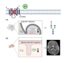
Clinical Description

SLC39A8-CDG is characterized by a severe, primarily neurologic phenotype with developmental delay, intellectual disability, muscular hypotonia, and variable additional neurologic symptoms including dyskinetic movements and spasticity. To date, 15 individuals have been identified with pathogenic variants in SLC39A8 [Boycott et al 2015, Park et al 2015, Riley et al 2017, Park et al 2020, Bonaventura et al 2021]. The following description of the phenotypic features associated with this condition is based on these reports.

Table 2.

SLC39A8-CDG: Frequency of Select Features

FeatureProportion of Persons w/FeatureComment
Developmental delay /
intellectual disability
15/15
Hypotonia15/15Truncal postural hypotonia
Feeding difficulties14/15
Movement disorder15/15Dyskinetic movements
Spasticity9/15Variable (mild to severe)
Epilepsy9/15
Growth deficiency3/15
Ophthalmologic manifestations14/15Strabismus, cortical blindness
Hearing impairment3/15

Developmental delay (DD) and intellectual disability (ID). All known individuals exhibit varying degrees of DD and subsequent ID. Milestones of motor development are typically reached with major delays, although some are never reached. Most older individuals have severe ID that does not allow for standardized testing. Communication is usually limited to single words and/or gestures.

Other neurodevelopmental features

  • Hypotonia. All described individuals presented with truncal muscular hypotonia, often more pronounced in early life. Head control is reduced but can be achieved in more mildly affected individuals.
  • Infant feeding difficulties. Feeding difficulties necessitating at least transient tube feeding are common in individuals with SLC39A8-CDG.
  • Movement disorders, especially marked dystonia, were reported in several individuals, at times overlapping with spasticity and thus resulting in spastic dystonia.
  • Spasticity to varying degrees has been observed in several affected individuals, correlating with a more severe disease course.
  • Reduced nerve conduction velocities, both motor and sensory, have been reported in several individuals with SLC39A8-CDG.

Epilepsy. The majority of reported individuals suffer from seizures, with onset in the first months of life. Some individuals have relatively mild well-controlled seizures. However, severely affected individuals often present with infantile epileptic spasms and hypsarrhythmia unresponsive to intense treatment including corticosteroids. Manganese treatment was associated with improved seizure control in singular case reports [Park et al 2018].

Growth. Poor weight gain with subsequent growth deficiency is a common feature of SLC39A8-CDG. While not all individuals have short stature at birth, many develop proportionate or disproportionate short stature with short limbs during childhood. Head size can be reduced, although this is not a consistent or common feature.

Ophthalmologic involvement. Strabismus has been described in the vast majority of affected individuals. Loss of vision has been identified, although it can be attributed to cortical blindness in some individuals. No specific ophthalmologic abnormalities have been described, but such manifestations cannot be ruled out based on the limited number of described individuals.

Hearing impairment. Sensorineural hearing impairment of variable severity has been observed in some individuals with SLC39A8-CDG and was typically detected on newborn hearing screen.

Liver disease. Elevated liver enzymes are routinely observed, although not usually clinically significant (e.g., resulting in liver failure). Treatment for liver disease is rarely required but might be indicated on an individual basis. Treatment response is poorly characterized.

Osteopenia. Osteopenia is frequently observed in affected individuals. Whether this is a primary manifestation of SLC39A8-CDG or secondary due to disuse is unclear.

Prognosis. The overall prognosis is highly variable. To date, three of the reported 15 individuals are deceased due to secondary complications of infections accompanied by exacerbations of seizures. In contrast, two mildly affected individuals diagnosed in adulthood have a stable phenotype without a progressive neurologic disorder [J Park, personal communication].

Genotype-Phenotype Correlations

No genotype-phenotype correlations have been identified.

Nomenclature

In 2009 the nomenclature for all types of CDG was changed to include the official gene (not italicized) followed by "-CDG" [Jaeken et al 2009].

Prevalence

The prevalence of SLC39A8-CDG is unknown. To date, 15 individuals have been described in the literature [Boycott et al 2015, Park et al 2015, Riley et al 2017, Park et al 2020, Bonaventura et al 2021]. Including additional unreported individuals, approximately 20 affected individuals are known.

Of note, the founder variant c.112G>C (p.Gly38Arg) has been identified in the North American Hutterite population [Boycott et al 2015, Park et al 2015].

Differential Diagnosis

Early infantile presentation (in those infants who have not yet had an MRI). Many metabolic and genetic disorders that present in infancy share at least some of the clinical features of SLC39A8-CDG. Metabolic disorders in the differential diagnosis of hypotonia, developmental delay, and growth deficiency are summarized in Table 3a.

Table 3a.

Metabolic Disorders to Consider in the Differential Diagnosis of SLC39A8-CDG in Infants Who Have Not Yet Had an MRI

GenesDisorderClinical CharacteristicsComment
169 genes 1Other CDG & CDDG (See CDG-N-Linked & Multiple Pathway Overview, PMM2-CDG, & NGLY1-CDDG.)
  • DD
  • Seizures
  • Liver disease
  • Abnormal transferrin glycosylation analysis
CDG & CDDG can be clinically indistinguishable. However, the combination of mitochondrial dysfunction & dysglycosylation w/↓ manganese levels is exclusive to SLC39A8-CDG.
>300 genes

Mitochondrial disorders

  • Commonly involve multiple organ systems
  • Movement disorder(s)
  • Unlike persons w/mitochondrial disorders, persons w/SLC39A8-CDG do not typically have episodes of metabolic decompensation or significantly ↑ acidemia (if not assoc w/hypoperfusion).
  • The disorders are further distinguished by the presence of abnormal transferrin glycoform analysis & deficient SLC39A8 activity in SLC39A8-CDG.
>20 genesPeroxisomal biogenesis defects (See Zellweger Spectrum Disorder.)
  • Multisystem involvement
  • DD/ID
  • Neurologic dysfunction
  • Liver disease
Unlike persons w/peroxisomal biogenesis defects, persons w/SLC39A8-CDG do not have abnormal VLCFAs.
>20 genesUrea cycle disorders / organic acidemias (See Propionic Acidemia, Glutaric Acidemia Type 1, Isolated Methylmalonic Acidemia, & Disorders of Intracellular Cobalamin Metabolism.)
  • Hypotonia
  • Growth deficiency
  • Feeding intolerance
  • DD/ID
  • Spasticity
  • Unlike persons w/urea cycle disorders / organic acidemias, persons w/SLC39A8-CDG do not typically have episodes of metabolic decompensation or hyperammonemia.
  • The disorders are further distinguished by the presence of abnormal transferrin glycoform analysis & ↓ levels of manganese in SLC39A8-CDG.

CDDG = congenital disorder of deglycosylation; CDG = congenital disorder of glycosylation; DD = developmental delay; ID = intellectual disability; VLCFAs = very long-chain fatty acids

1.

Disorders with overlapping MRI findings. See Table 3b.

Table 3b.

Metabolic Disorders with Overlapping MRI Findings in the Differential Diagnosis of SLC39A8-CDG

GeneDisorder 1Clinical Characteristics
MRI findingsClinical features
ABHD12 PHARC syndrome (OMIM 612674)Cerebellar atrophy
  • Ataxia
  • Cataracts
  • Hearing loss
  • Neurodegeneration
  • Retinitis pigmentosa
ALDH18A1 Delta-1-pyrroline-5-carboxylate synthetase deficiency 2Cerebellar atrophy
  • Cataracts
  • Cutis laxa
  • DD/ID
  • Faltering growth
  • Hypotonia
  • Joint hyperlaxity
  • Microcephaly
  • Short stature
ALDH5A1 Succinic semialdehyde dehydrogenase deficiency
  • Abnormalities of myelination
  • Cerebellar atrophy
  • Hyperintensity of T1-weighted signals in globus pallidus
  • Ataxia
  • DD
  • Epilepsy
  • Hyporeflexia
  • Hypotonia
FA2H Fatty acid hydroxylase-associated neurodegeneration
  • Confluent periventricular white matter abnormalities
  • Iron accumulation
  • Profound pontocerebellar atrophy
  • Cognitive decline
  • Optic atrophy
  • Progressive spastic paraparesis & dysmetria
  • Xeroderma
GCDH Glutaric acidemia type 1
  • Cerebellar atrophy
  • Cortical atrophy
  • Progressive disturbance of myelination
  • Signal changes &/or atrophy of basal ganglia
  • Striatal injury spreading in dorsoventral direction
  • Acute brain injury assoc w/infections
  • Choreoathetosis
  • Progressive dystonic cerebral palsy
L2HGDH L-2-hydroxyglutaric aciduria (OMIM 236792)
  • ↑ signal density of dentate nuclei & globi pallidi on T2-weighted images
  • Cerebellar atrophy
  • Leukoencephalopathy
  • Variety of neurologic malignancies
  • Progressive ataxia
  • DD/ID
  • Seizures
  • Spasticity
MVK Mevalonate kinase deficiency (OMIM 610377)Cerebellar atrophy
  • Ataxia
  • Autoinflammation
  • Dysmorphic features
  • Faltering growth
  • Hypotonia
  • ID
PLA2G6 Infantile neuroaxonal dystrophy, NBIA (See PLA2G6-Associated Neurodegeneration.)
  • Cerebellar atrophy
  • Hypointensities in globus pallidus & substantial nigra
  • Signal hyperintensity in cerebellar cortex
  • Cerebellar ataxia
  • Chronic denervation
  • Hypotonia
  • Progression to spastic tetraplegia
  • Progressive motor & cognitive deterioration
  • Visual disturbances
See footnote 3.Leigh syndrome (See Mitochondrial Disorders Overview & Nuclear Gene-Encoded Leigh Syndrome Spectrum Overview.)Symmetrical ↑ signal density of basal ganglia, esp putamina & globi pallidi, on T2-weighted imagesProgressive neurologic & motor decline

DD = developmental delay; ID = intellectual disability; NBIA = neurodegeneration with brain iron accumulation; PHARC = polyneuropathy, hearing loss, ataxia, retinitis pigmentosa, and cataract

1.

With the exception of delta-1-pyrroline-5-carboxylate synthetase deficiency (which can be inherited in either an autosomal dominant or autosomal recessive manner) and Leigh syndrome (which can be inherited in an autosomal recessive, autosomal dominant, X-linked, or maternal manner), the disorders in Table 3b are inherited in an autosomal recessive manner.

2.
3.

Many genes (nuclear and mitochondrial) are known to be associated with Leigh syndrome.

Management

No clinical practice guidelines for SLC39A8-CDG have been published.

Evaluations Following Initial Diagnosis

To establish the extent of disease and needs in an individual diagnosed with SLC39A8-CDG, the evaluations summarized in Table 4 (if not performed as part of the evaluation that led to the diagnosis) are recommended.

Table 4.

Recommended Evaluations Following Initial Diagnosis in Individuals with SLC39A8-CDG

System/ConcernEvaluationComment
Development Developmental assessment
  • To incl motor, adaptive, cognitive, speech-language eval
  • Eval for early intervention / special education
Neurologic Neurologic eval
  • To incl brain MRI
  • Consider EEG if seizures are a concern.
Feeding/Nutrition
  • Gastroenterology / nutrition / feeding team eval
  • Logotherapy eval to assess for swallowing abnormalities
  • To incl eval of aspiration risk & nutritional status
  • Consider eval for gastrostomy tube placement in persons w/dysphagia &/or aspiration risk.
Eyes Ophthalmologic evalTo assess for reduced vision, abnormal ocular movement, best corrected visual acuity, refractive errors, & strabismus
Hearing Audiologic evalAssess for hearing loss.
Musculoskeletal Orthopedics / physical medicine & rehab / PT & OT evalTo incl assessment of:
  • Gross motor & fine motor skills
  • Contractures, clubfoot, & kyphoscoliosis
  • Mobility, ADL, & need for adaptive devices
  • Need for PT (to improve gross motor skills) &/or OT (to improve fine motor skills)
Genetic counseling By genetics professionals 1To inform affected persons & their families re nature, MOI, & implications of SLC39A8-CDG to facilitate medical & personal decision making
Family support
& resources
Assess need for:

ADL = activities of daily living; MOI = mode of inheritance; OT = occupational therapy; PT = physical therapy

1.

Medical geneticist, certified genetic counselor, certified advanced genetic nurse

Treatment of Manifestations

Targeted Therapies

In GeneReviews, a targeted therapy is one that addresses the specific underlying mechanism of disease causation (regardless of whether the therapy is significantly efficacious for one or more manifestation of the genetic condition); would otherwise not be considered without knowledge of the underlying genetic cause of the condition; or could lead to a cure. —ED

Manganese supplementation is currently the most effective treatment for SLC39A8-CDG [Riley et al 2017, Park et al 2018]. This treatment is reported to lead to normalization of both transferrin glycosylation and other affected manganese-dependent enzymes and is associated with clinical improvement [Park et al 2018], especially improved seizure control in singular case reports.

  • Manganese sulfate is preferable to other manganese salts given its high solubility and widespread availability at pharmaceutical grade due to its use in nutritional applications.
  • Manganese sulfate doses of up to 20 mg/kg body weight per day have been used to treat SLC39A8-CDG.
  • Monitoring glycosylation using mass spectrometry-based methods is necessary to identify the appropriate manganese dose. Note: (1) Abnormal glycosylation persisted longer than other observed abnormal laboratory findings [Park et al 2018]. (2) Abnormal glycan structures were detected in the serum by matrix-assisted laser desorption/ionization time-of-flight mass spectrometry in individuals with SLC39A8-CDG with normal glycosylation profiles of transferrin [Park et al 2020].
  • Regular measurement of blood manganese levels is advised with the aim of adjusting the manganese dose to achieve manganese levels similar to those observed in healthy controls.
  • Brain MRI every one to two years is recommend in order to detect potential manganese deposits. Note: Long-term, high-dose manganese can be neurotoxic in healthy individuals [Erikson & Aschner 2019]. However, manganese toxicity has not been observed in individuals with SLC39A8-CDG treated with high doses of manganese sulfate.

Galactose supplementation. In addition to manganese supplementation, treatment with galactose was utilized based on the finding of hypogalactosylation of plasma glycoproteins in persons with SLC39A8-CDG [Park et al 2015]. Due to the manganese dependence of galactosyltransferases, reduction or even absence of manganese results in impaired glycosylation. Galactose supplementation increased intracellular pools of uridine diphosphate galactose [Tegtmeyer et al 2014], which in turn led to improved galactosylation by manganese-depleted galactosyltransferases.

  • Titration to identify adequate manganese sulfate dose should precede introduction of galactose.
    Note: Addition of galactose and subsequent normalization of glycosylation prevents the ability to identify the appropriate manganese dose based on the levels of dysglycosylation.
  • Galactose supplementation does not have any effect on unrelated enzyme dysfunctions caused by manganese depletion and should thus be considered only in combination with manganese supplementation.
  • Galactose doses of up to 3.75 g/kg body weight per day resulted in rapid normalization of glycosylation of the marker protein serum transferrin.

Supportive Care

Supportive care to improve quality of life, maximize function, and reduce complications is also recommended. This ideally involves multidisciplinary care by specialists in relevant fields (see Table 5).

Table 5.

Treatment of Manifestations in Individuals with SLC39A8-CDG

Manifestation/ConcernTreatmentConsiderations/Other
Developmental delay /
Intellectual disability
See Developmental Delay / Intellectual Disability Management Issues.
Spasticity Orthopedics / physical medicine & rehab / PT & OT incl stretching to help avoid contractures & fallsConsider need for positioning & mobility devices, disability parking placard.
Epilepsy
  • Supplementation w/manganese to ↓ seizure frequency & severity (See Targeted Therapies.)
  • Standardized treatment w/ASM by experienced neurologist
  • Many ASMs may be effective; none has been demonstrated effective specifically for this disorder.
  • Education of parents/caregivers 1
Feeding difficulties /
Poor weight gain
  • Feeding therapy
  • Gastrostomy tube placement may be required for persistent feeding issues.
Low threshold for clinical feeding eval &/or radiographic swallowing study when showing clinical signs or symptoms of dysphagia
Eyes Standard treatment for strabismus per ophthalmologist
Referral to low vision services as needed
  • Children: through early intervention programs &/or school district
  • Adults: low vision clinic &/or community vision services / OT / mobility services
Central visual impairment No specific treatmentEarly intervention program to stimulate visual development
Hearing Hearing aids may be helpful per otolaryngologist.Community hearing services through early intervention or school district
Musculoskeletal Referral to orthopedist when scoliosis becomes evident w/mgmt per orthopedist
Family/Community
  • Ensure appropriate social work involvement to connect families w/local resources, respite, & support.
  • Coordinate care to manage multiple subspecialty appointments, equipment, medications, & supplies.
  • Ongoing assessment of need for palliative care involvement &/or home nursing
  • Consider involvement in adaptive sports or Special Olympics.

ASM = anti-seizure medication; OT = occupational therapy; PT = physical therapy

1.

Education of parents/caregivers regarding common seizure presentations is appropriate. For information on non-medical interventions and coping strategies for children diagnosed with epilepsy, see Epilepsy Foundation Toolbox.

Developmental Delay / Intellectual Disability Management Issues

The following information represents typical management recommendations for individuals with developmental delay / intellectual disability in the US; standard recommendations may vary from country to country.

Ages 0-3 years. Referral to an early intervention program is recommended for access to occupational, physical, speech, and feeding therapy as well as infant mental health services, special educators, and sensory impairment specialists. In the US, early intervention is a federally funded program available in all states that provides in-home services to target individual therapy needs.

Ages 3-5 years. In the US, developmental preschool through the local public school district is recommended. Before placement, an evaluation is made to determine needed services and therapies and an individualized education plan (IEP) is developed for those who qualify based on established motor, language, social, or cognitive delay. The early intervention program typically assists with this transition. Developmental preschool is center based; for children too medically unstable to attend, home-based services are provided.

All ages. Consultation with a developmental pediatrician is recommended to ensure the involvement of appropriate community, state, and educational agencies (US) and to support parents in maximizing quality of life. Some issues to consider:

  • IEP services:
    • An IEP provides specially designed instruction and related services to children who qualify.
    • IEP services will be reviewed annually to determine whether any changes are needed.
    • Special education law requires that children participating in an IEP be in the least restrictive environment feasible at school and included in general education as much as possible, when and where appropriate.
    • Vision and hearing consultants should be a part of the child's IEP team to support access to academic material.
    • PT, OT, and speech services will be provided in the IEP to the extent that the need affects the child's access to academic material. Beyond that, private supportive therapies based on the affected individual's needs may be considered. Specific recommendations regarding type of therapy can be made by a developmental pediatrician.
    • As a child enters the teen years, a transition plan should be discussed and incorporated in the IEP. For those receiving IEP services, the public school district is required to provide services until age 21.
  • A 504 plan (Section 504: a US federal statute that prohibits discrimination based on disability) can be considered for those who require accommodations or modifications such as front-of-class seating, assistive technology devices, classroom scribes, extra time between classes, modified assignments, and enlarged text.
  • Developmental Disabilities Administration (DDA) enrollment is recommended. DDA is a US public agency that provides services and support to qualified individuals. Eligibility differs by state but is typically determined by diagnosis and/or associated cognitive/adaptive disabilities.
  • Families with limited income and resources may also qualify for supplemental security income (SSI) for their child with a disability.
Motor Dysfunction

Gross motor dysfunction

  • Physical therapy is recommended to maximize mobility and to reduce the risk for later-onset orthopedic complications (e.g., contractures, scoliosis, hip dislocation).
  • Consider use of durable medical equipment and positioning devices as needed (e.g., wheelchairs, walkers, bath chairs, orthotics, adaptive strollers).
  • For muscle tone abnormalities including hypertonia or dystonia, consider involving appropriate specialists to aid in management of baclofen, tizanidine, Botox®, anti-parkinsonian medications, or orthopedic procedures.

Fine motor dysfunction. Occupational therapy is recommended for difficulty with fine motor skills that affect adaptive function such as feeding, grooming, dressing, and writing.

Oral motor dysfunction should be assessed at each visit and clinical feeding evaluations and/or radiographic swallowing studies should be obtained for choking/gagging during feeds, poor weight gain, frequent respiratory illnesses, or feeding refusal that is not otherwise explained. Assuming that the child is safe to eat by mouth, feeding therapy (typically from an occupational or speech therapist) is recommended to help improve coordination or sensory-related feeding issues. Feeds can be thickened or chilled for safety. When feeding dysfunction is severe, an NG-tube or G-tube may be necessary.

Communication issues. Consider evaluation for alternative means of communication (e.g., augmentative and alternative communication [AAC]) for individuals who have expressive language difficulties. An AAC evaluation can be completed by a speech-language pathologist who has expertise in the area. The evaluation will consider cognitive abilities and sensory impairments to determine the most appropriate form of communication. AAC devices can range from low-tech, such as picture exchange communication, to high-tech, such as voice-generating devices. Contrary to popular belief, AAC devices do not hinder verbal development of speech, but rather support optimal speech and language development.

Surveillance

To monitor existing manifestations, the individual's response to supportive care, and the emergence of new manifestations, the evaluations summarized in Table 6 are recommended.

Table 6.

Recommended Surveillance for Individuals with SLC39A8-CDG

System/ConcernEvaluationFrequency
Development Monitor developmental progress & educational needs.At each visit
Neurologic
  • Assess for abnormal glycosylation using mass spectrometry.
  • Measure blood manganese levels.
As needed to titrate manganese dose
Brain MRIEvery 1-2 yrs
Monitor those w/seizures as clinically indicated.At each visit
EEGFrequency per neurologist
Assess for new manifestations such as seizures, changes in tone, & movement disorders.At each visit
Feeding
  • Measurement of growth parameters
  • Eval of nutritional status & safety of oral intake
  • Monitor for evidence of aspiration (e.g., respiratory insufficiency).
At each visit
Ophthalmologic involvement
  • Assessment of visual acuity
  • Assessment for strabismus
Per treating ophthalmologist(s)
Liver AST, ALT, albuminAnnually in those w/evidence of liver disease; further follow up as needed
Musculoskeletal
  • Physical medicine, OT/PT assessment of mobility, self-help skills
  • Clinical exam for scoliosis
At each visit
Assess for clinical manifestations of osteopenia/osteoporosis.Every 1-2 yrs or as needed
DXA scanEvery 3-5 yrs starting in adolescence
Family/Community Assess family need for social work support (e.g., palliative/respite care, home nursing, other local resources), care coordination, or follow-up genetic counseling if new questions arise (e.g., family planning).At each visit

ALT = alanine transaminase; AST = aspartate transaminase; DXA = dual-energy x-ray absorptiometry; OT = occupational therapy; PT = physical therapy

Agents/Circumstances to Avoid

Fever. It is well established that increases in body temperature are associated with further impairment of the residual glycosylation in congenital disorders of glycosylation. Therefore, antipyretic treatment to reduce and/or prevent fever is recommended.

Hepatotoxic drugs. In those with evidence of liver dysfunction, hepatotoxic agents should be avoided or used with extreme caution.

Drugs contraindicated in mitochondriopathies. Due to secondary impairment of mitochondrial function, agents contraindicated in mitochondriopathies (e.g., valproate) should be avoided in individuals with SLC39A8-CDG.

Evaluation of Relatives at Risk

All at-risk sibs of any age should have molecular testing for the familial SLC39A8 pathogenic variants in order to identify as early as possible those who would benefit from prompt initiation of manganese and galactose supplementation. This is especially recommended if additional – seemingly non-related – neuropsychiatric symptoms are present.

For at-risk newborn sibs, analysis of blood and urine manganese levels as well as protein N-glycosylation should be performed simultaneously with molecular genetic testing, as results from these tests are potentially available earlier than results of genetic testing. Altered glycosylation and severely reduced blood manganese levels are indicative of SLC39A8-CDG.

See Genetic Counseling for issues related to testing of at-risk relatives for genetic counseling purposes.

Therapies Under Investigation

Search ClinicalTrials.gov in the US and EU Clinical Trials Register in Europe for access to information on clinical studies for a wide range of diseases and conditions. Note: There may not be clinical trials for this disorder.

Genetic Counseling

Genetic counseling is the process of providing individuals and families with information on the nature, mode(s) of inheritance, and implications of genetic disorders to help them make informed medical and personal decisions. The following section deals with genetic risk assessment and the use of family history and genetic testing to clarify genetic status for family members; it is not meant to address all personal, cultural, or ethical issues that may arise or to substitute for consultation with a genetics professional. —ED.

Mode of Inheritance

SLC39A8-CDG is inherited in an autosomal recessive manner.

Risk to Family Members

Parents of a proband

  • The parents of an affected child are presumed to be heterozygous for an SLC39A8 pathogenic variant.
  • Molecular genetic testing is recommended for the parents of a proband to confirm that both parents are heterozygous for an SLC39A8 pathogenic variant and to allow reliable recurrence risk assessment.
  • If a pathogenic variant is detected in only one parent and parental identity testing has confirmed biological maternity and paternity, it is possible that one of the pathogenic variants identified in the proband occurred as a de novo event in the proband or as a postzygotic de novo event in a mosaic parent [Jónsson et al 2017]. If the proband appears to have homozygous pathogenic variants (i.e., the same two pathogenic variants), additional possibilities to consider include:
  • Heterozygotes (carriers) are asymptomatic and are not at risk of developing the disorder.

Sibs of a proband

  • If both parents are known to be heterozygous for an SLC39A8 pathogenic variant, each sib of an affected individual has at conception a 25% chance of being affected, a 50% chance of being an asymptomatic carrier, and a 25% chance of being unaffected and not a carrier.
  • Heterozygotes (carriers) are asymptomatic and are not at risk of developing the disorder.

Offspring of a proband. To date, individuals with SLC39A8-CDG are not known to reproduce.

Other family members. Each sib of the proband's parents is at a 50% risk of being a carrier of an SLC39A8 pathogenic variant.

Carrier Detection

Carrier testing for at-risk relatives requires prior identification of the SLC39A8 pathogenic variants in the family.

Related Genetic Counseling Issues

See Management, Evaluation of Relatives at Risk for information on evaluating at-risk relatives for the purpose of early diagnosis and treatment.

Family planning

  • The optimal time for determination of genetic risk and discussion of the availability of prenatal/preimplantation genetic testing is before pregnancy.
  • It is appropriate to offer genetic counseling (including discussion of potential risks to offspring and reproductive options) to young adults who are carriers or are at risk of being carriers.
  • Carrier testing for reproductive partners of known carriers should be considered, particularly if both partners are of the same ethnic background. An SLC39A8 founder variant has been identified in individuals of Hutterite descent (see Table 7).

Prenatal Testing and Preimplantation Genetic Testing

Once the SLC39A8 pathogenic variants have been identified in an affected family member, prenatal and preimplantation genetic testing are possible.

Differences in perspective may exist among medical professionals and within families regarding the use of prenatal testing. While most centers would consider use of prenatal testing to be a personal decision, discussion of these issues may be helpful.

Resources

GeneReviews staff has selected the following disease-specific and/or umbrella support organizations and/or registries for the benefit of individuals with this disorder and their families. GeneReviews is not responsible for the information provided by other organizations. For information on selection criteria, click here.

Molecular Genetics

Information in the Molecular Genetics and OMIM tables may differ from that elsewhere in the GeneReview: tables may contain more recent information. —ED.

Table A.

SLC39A8-CDG: Genes and Databases

GeneChromosome LocusProteinHGMDClinVar
SLC39A8 4q24 Metal cation symporter ZIP8 SLC39A8 SLC39A8

Data are compiled from the following standard references: gene from HGNC; chromosome locus from OMIM; protein from UniProt. For a description of databases (Locus Specific, HGMD, ClinVar) to which links are provided, click here.

Table B.

OMIM Entries for SLC39A8-CDG (View All in OMIM)

608732SOLUTE CARRIER FAMILY 39 (ZINC TRANSPORTER), MEMBER 8; SLC39A8
616721CONGENITAL DISORDER OF GLYCOSYLATION, TYPE IIn; CDG2N

Molecular Pathogenesis

SLC39A8 encodes the metal cation symporter ZIP8, a major manganese uptake channel [Fujishiro & Kambe 2022] (see Figure 1). SLC39A8 pathogenic variants disrupting the function of the protein result in decreased blood levels of manganese and subsequent intracellular manganese depletion [Choi et al 2018].

Figure 1. . Molecular pathogenesis of SLC39A8-CDG.

Figure 1.

Molecular pathogenesis of SLC39A8-CDG. Dysfunction of the manganese transporter SLC39A8 causes intracellular manganese depletion. (A) A reduced Mn2+ concentration in the Golgi apparatus impairs the function of manganese-dependent glycosylation enzymes. (more...)

Due to its relevance as a cofactor for a plethora of enzymes [Chen et al 2018], manganese depletion in SLC39A8-CDG causes a varied and multisystem phenotype. Importantly, the manganese dependence of both galactosyl transferases and the mitochondrial superoxide dismutase results in a combined mitochondrial and glycosylation disorder phenotype.

Mechanism of disease causation. Loss of function, with impaired manganese transport across the channel encoded by SLC39A8 [Choi et al 2018]

Table 7.

Notable SLC39A8 Pathogenic Variants

Reference SequencesDNA Nucleotide ChangePredicted Protein ChangeComment [Reference]
NM_022154​.5
NP_071437​.3
c.112G>Cp.Gly38ArgFounder variant in the Hutterite population [Park et al 2015].

Variants listed in the table have been provided by the authors. GeneReviews staff have not independently verified the classification of variants.

GeneReviews follows the standard naming conventions of the Human Genome Variation Society (varnomen​.hgvs.org). See Quick Reference for an explanation of nomenclature.

Chapter Notes

Author Notes

Julien H Park (ed.retsneumku@krap.neiluj) and Thorsten Marquardt (ed.retsneum-inu@tauqram) are actively involved in clinical and preclinical research regarding individuals with SLC39A8-CDG. They would be happy to communicate with persons who have any questions regarding diagnosis and treatment of SLC39A8-CDG or other considerations.

They are also interested in hearing from clinicians treating families affected by SLC39A8-CDG in whom no causative variant has been identified through molecular genetic testing.

See the Congenital Metabolic Diseases working group at Universitätsklinikum Münster.

Acknowledgments

The authors acknowledge the commitment of the patients and families supporting the ongoing research in the field of SLC39A8-CDG.

Revision History

  • 6 April 2023 (sw) Review posted live
  • 22 December 2022 (jp) Original submission

References

Literature Cited

  • Bonaventura E, Barone R, Sturiale L, Pasquariello R, Alessandrì MG, Pinto AM, Renieri A, Panteghini C, Garavaglia B, Cioni G, Battini R. Clinical, molecular and glycophenotype insights in SLC39A8-CDG. Orphanet J Rare Dis. 2021;16:307. [PMC free article: PMC8272319] [PubMed: 34246313]
  • Boycott KM, Beaulieu CL, Kernohan KD, Gebril OH, Mhanni A, Chudley AE, Redl D, Qin W, Hampson S, Küry S, Tetreault M, Puffenberger EG, Scott JN, Bezieau S, Reis A, Uebe S, Schumacher J, Hegele RA, McLeod DR, Gálvez-Peralta M, Majewski J, Ramaekers VT, Nebert DW, Innes AM, Parboosingh JS, Abou Jamra R, et al. Autosomal-recessive intellectual disability with cerebellar atrophy syndrome caused by mutation of the manganese and zinc transporter gene SLC39A8. Am J Hum Genet. 2015;97:886-93. [PMC free article: PMC4678439] [PubMed: 26637978]
  • Chen P, Bornhorst J, Aschner M. Manganese metabolism in humans. Front Biosci (Landmark Ed). 2018;23:1655-79. [PubMed: 29293455]
  • Choi EK, Nguyen TT, Gupta N, Iwase S, Seo YA. Functional analysis of SLC39A8 mutations and their implications for manganese deficiency and mitochondrial disorders. Sci Rep. 2018;8:3163. [PMC free article: PMC5816659] [PubMed: 29453449]
  • Erikson KM, Aschner M. Manganese: its role in disease and health. Met Ions Life Sci. 2019;19. [PubMed: 30855111]
  • Fujishiro H, Kambe T. Manganese transport in mammals by zinc transporter family proteins, ZNT and ZIP. J Pharmacol Sci. 2022;148:125-33. [PubMed: 34924116]
  • Jaeken J, Hennet T, Matthijs G, Freeze HH. CDG nomenclature: time for a change! Biochim Biophys Acta. 2009;1792:825-6. [PMC free article: PMC3917312] [PubMed: 19765534]
  • Jónsson H, Sulem P, Kehr B, Kristmundsdottir S, Zink F, Hjartarson E, Hardarson MT, Hjorleifsson KE, Eggertsson HP, Gudjonsson SA, Ward LD, Arnadottir GA, Helgason EA, Helgason H, Gylfason A, Jonasdottir A, Jonasdottir A, Rafnar T, Frigge M, Stacey SN, Th Magnusson O, Thorsteinsdottir U, Masson G, Kong A, Halldorsson BV, Helgason A, Gudbjartsson DF, Stefansson K. Parental influence on human germline de novo mutations in 1,548 trios from Iceland. Nature. 2017;549:519-22. [PubMed: 28959963]
  • Keen CL, Ensunsa JL, Watson MH, Baly DL, Donovan SM, Monaco MH, Clegg MS. Nutritional aspects of manganese from experimental studies. Neurotoxicology. 1999;20:213-23. [PubMed: 10385885]
  • Krajewski WW, Collins R, Holmberg-Schiavone L, Jones TA, Karlberg T, Mowbray SL. Crystal structures of mammalian glutamine synthetases illustrate substrate-induced conformational changes and provide opportunities for drug and herbicide design. J Mol Biol. 2008;375:217-28. [PubMed: 18005987]
  • Kuhn NJ, Ward S, Piponski M, Young TW. Purification of human hepatic arginase and its manganese (II)-dependent and pH-dependent interconversion between active and inactive forms: a possible pH-sensing function of the enzyme on the ornithine cycle. Arch Biochem Biophys. 1995;320:24-34. [PubMed: 7793981]
  • Marco-Marín C, Escamilla-Honrubia JM, Llácer JL, Seri M, Panza E, Rubio V. Δ1-pyrroline-5-carboxylate synthetase deficiency: an emergent multifaceted urea cycle-related disorder. J Inherit Metab Dis. 2020;43:657-70. [PubMed: 32017139]
  • Narimatsu Y, Joshi HJ, Nason R, Van Coillie J, Karlsson R, Sun L, Ye Z, Chen YH, Schjoldager KT, Steentoft C, Furukawa S, Bensing BA, Sullam PM, Thompson AJ, Paulson JC, Büll C, Adema GJ, Mandel U, Hansen L, Bennett EP, Varki A, Vakhrushev SY, Yang Z, Clausen H. An atlas of human glycosylation pathways enables display of the human glycome by gene engineered cells. Mol Cell. 2019;75:394-407.e5. [PMC free article: PMC6660356] [PubMed: 31227230]
  • Park JH, Hogrebe M, Fobker M, Brackmann R, Fiedler B, Reunert J, Rust S, Tsiakas K, Santer R, Grüneberg M, Marquardt T. SLC39A8 deficiency: biochemical correction and major clinical improvement by manganese therapy. Genet Med. 2018;20:259-68. [PubMed: 28749473]
  • Park JH, Hogrebe M, Grüneberg M, DuChesne I, von der Heiden AL, Reunert J, Schlingmann KP, Boycott KM, Beaulieu CL, Mhanni AA, Innes AM, Hörtnagel K, Biskup S, Gleixner EM, Kurlemann G, Fiedler B, Omran H, Rutsch F, Wada Y, Tsiakas K, Santer R, Nebert DW, Rust S, Marquardt T. SLC39A8 deficiency: a disorder of manganese transport and glycosylation. Am J Hum Genet. 2015;97:894-903. [PMC free article: PMC4678430] [PubMed: 26637979]
  • Park JH, Mealer RG, Elias AF, Hoffmann S, Grüneberg M, Biskup S, Fobker M, Haven J, Mangels U, Reunert J, Rust S, Schoof J, Schwanke C, Smoller JW, Cummings RD, Marquardt T. N-glycome analysis detects dysglycosylation missed by conventional methods in SLC39A8 deficiency. J Inherit Metab Dis. 2020;43:1370-81. [PMC free article: PMC8086894] [PubMed: 32852845]
  • Richards S, Aziz N, Bale S, Bick D, Das S, Gastier-Foster J, Grody WW, Hegde M, Lyon E, Spector E, Voelkerding K, Rehm HL, et al. Standards and guidelines for the interpretation of sequence variants: a joint consensus recommendation of the American College of Medical Genetics and Genomics and the Association for Molecular Pathology. Genet Med. 2015;17:405-24. [PMC free article: PMC4544753] [PubMed: 25741868]
  • Riley LG, Cowley MJ, Gayevskiy V, Roscioli T, Thorburn DR, Prelog K, Bahlo M, Sue CM, Balasubramaniam S, Christodoulou J. A SLC39A8 variant causes manganese deficiency, and glycosylation and mitochondrial disorders. J Inherit Metab Dis. 2017;40:261-9. [PubMed: 27995398]
  • Stenson PD, Mort M, Ball EV, Chapman M, Evans K, Azevedo L, Hayden M, Heywood S, Millar DS, Phillips AD, Cooper DN. The Human Gene Mutation Database (HGMD®): optimizing its use in a clinical diagnostic or research setting. Hum Genet. 2020;139:1197-207. [PMC free article: PMC7497289] [PubMed: 32596782]
  • Tegtmeyer LC, Rust S, van Scherpenzeel M, Ng BG, Losfeld ME, Timal S, Raymond K, He P, Ichikawa M, Veltman J, Huijben K, Shin YS, Sharma V, Adamowicz M, Lammens M, Reunert J, Witten A, Schrapers E, Matthijs G, Jaeken J, Rymen D, Stojkovic T, Laforêt P, Petit F, Aumaître O, Czarnowska E, Piraud M, Podskarbi T, Stanley CA, Matalon R, Burda P, Seyyedi S, Debus V, Socha P, Sykut-Cegielska J, van Spronsen F, de Meirleir L, Vajro P, DeClue T, Ficicioglu C, Wada Y, Wevers RA, Vanderschaeghe D, Callewaert N, Fingerhut R, van Schaftingen E, Freeze HH, Morava E, Lefeber DJ, Marquardt T. Multiple phenotypes in phosphoglucomutase 1 deficiency. N Engl J Med. 2014;370:533-42. [PMC free article: PMC4373661] [PubMed: 24499211]
Copyright © 1993-2024, University of Washington, Seattle. GeneReviews is a registered trademark of the University of Washington, Seattle. All rights reserved.

GeneReviews® chapters are owned by the University of Washington. Permission is hereby granted to reproduce, distribute, and translate copies of content materials for noncommercial research purposes only, provided that (i) credit for source (http://www.genereviews.org/) and copyright (© 1993-2024 University of Washington) are included with each copy; (ii) a link to the original material is provided whenever the material is published elsewhere on the Web; and (iii) reproducers, distributors, and/or translators comply with the GeneReviews® Copyright Notice and Usage Disclaimer. No further modifications are allowed. For clarity, excerpts of GeneReviews chapters for use in lab reports and clinic notes are a permitted use.

For more information, see the GeneReviews® Copyright Notice and Usage Disclaimer.

For questions regarding permissions or whether a specified use is allowed, contact: ude.wu@tssamda.

Bookshelf ID: NBK590152PMID: 37023243

Views

Key Sections in this GeneReview

Tests in GTR by Gene

Related information

  • OMIM
    Related OMIM records
  • PMC
    PubMed Central citations
  • PubMed
    Links to PubMed
  • Gene
    Locus Links

Similar articles in PubMed

See reviews...See all...

Recent Activity

Your browsing activity is empty.

Activity recording is turned off.

Turn recording back on

See more...