U.S. flag

An official website of the United States government

NCBI Bookshelf. A service of the National Library of Medicine, National Institutes of Health.

Adam MP, Feldman J, Mirzaa GM, et al., editors. GeneReviews® [Internet]. Seattle (WA): University of Washington, Seattle; 1993-2023.

Cover of GeneReviews®

GeneReviews® [Internet].

Show details

Classic Ehlers-Danlos Syndrome

Synonyms: Ehlers-Danlos Syndrome, Classic Type; EDS, Classic Type

, MD, PhD, , MD, and , MD, PhD.

Author Information and Affiliations

Initial Posting: ; Last Update: July 26, 2018.

Estimated reading time: 24 minutes

Summary

Clinical characteristics.

Classic Ehlers-Danlos syndrome (cEDS) is a connective tissue disorder characterized by skin hyperextensibility, atrophic scarring, and generalized joint hypermobility (GJH). The skin is soft and doughy to the touch, and hyperextensible, extending easily and snapping back after release (unlike lax, redundant skin, as in cutis laxa). The skin is fragile, as manifested by splitting of the dermis following relatively minor trauma, especially over pressure points (knees, elbows) and areas prone to trauma (shins, forehead, chin). Wound healing is poor, and stretching of scars after apparently successful primary wound healing is characteristic. Complications of joint hypermobility, such as dislocations of the shoulder, patella, digits, hip, radius, and clavicle, usually resolve spontaneously or are easily managed by the affected individual. Other features include hypotonia with delayed motor development, fatigue and muscle cramps, and easy bruising. Mitral valve prolapse can occur infrequently, but tends to be of little clinical consequence. Aortic root dilatation has been reported, appears to be more common in young individuals, and rarely progresses.

Diagnosis/testing.

The diagnosis of cEDS is established in a proband with the minimal clinical diagnostic criteria (skin hyperextensibility and atrophic scarring and either GJH or ≥3 minor clinical criteria) and identification on molecular genetic testing of a heterozygous pathogenic variant in COL5A1, COL5A2, or (less commonly) COL1A1.

Management.

Treatment of manifestations: Children with hypotonia and delayed motor development benefit from physiotherapy. Non-weight-bearing exercise promotes muscle strength and coordination. Anti-inflammatory drugs may alleviate joint pain. Those with hypotonia, joint instability, and chronic pain may need to adapt lifestyles accordingly. Dermal wounds are closed without tension, preferably in two layers. For other wounds, deep stitches are applied generously; cutaneous stitches are left in place twice as long as usual; and the borders of adjacent skin are carefully taped to prevent stretching of the scar. DDAVP® (deamino-delta-D-arginine vasopressin) may be useful to normalize bleeding time. Cardiovascular problems are treated in a standard manner.

Prevention of primary manifestations: Young children with skin fragility can wear pads or bandages over the forehead, knees, and shins to avoid skin tears. Older children can wear soccer pads or ski stockings with shin padding during activities. Ascorbic acid (vitamin C) may reduce bruising.

Surveillance: Yearly echocardiogram when aortic dilatation and/or mitral valve prolapse are present.

Agents/circumstances to avoid: Sports with heavy joint strain; acetylsalicylate (aspirin).

Genetic counseling.

Classic EDS is inherited in an autosomal dominant manner. It is estimated that approximately 50% of affected individuals have an affected parent, and approximately 50% of affected individuals have the disorder as the result of a de novo pathogenic variant. Each child of an affected individual has a 50% chance of inheriting the pathogenic variant. Once the pathogenic variant has been identified in an affected family member, prenatal and preimplantation genetic testing are possible.

Diagnosis

Suggestive Findings

The diagnosis of classic Ehlers-Danlos syndrome (cEDS) can be suspected based on clinical examination and family history. Diagnostic criteria were developed by a medical advisory group in a conference at Villefranche in 1997 [Beighton et al 1998] and recently revised by the International EDS Consortium [Malfait et al 2017].

  • A major criterion has high diagnostic specificity because it is present in the vast majority of affected individuals and/or it is characteristic for the disorder and allows differentiation from other EDS subtypes and/or other heritable connective tissue disorders.
  • A minor criterion is a sign of lesser diagnostic specificity, but its presence supports the diagnosis.

Major Diagnostic Criteria for cEDS

Skin hyperextensibility. See Figure 1.

Figure 1.

Figure 1.

Skin hyperextensibility

  • Skin hyperextensibility should be measured by pinching and lifting the cutaneous and subcutaneous layers of the skin on the volar surface at the middle of the nondominant forearm as described by Remvig et al [2009].
  • Skin is hyperextensible if it can be stretched over a standardized cutoff in three of the following areas: 1.5 cm for the distal part of the forearms and the dorsum of the hands; 3 cm for neck, elbows, and knees.

Atrophic scarring. See Figure 2.

Figure 2.

Figure 2.

Widened atrophic scars

Generalized joint hypermobility (GJH). Joint hypermobility (see Figure 3) depends on an individual's age, sex, and family and ethnic background.

Figure 3.

Figure 3.

Passive flexion of thumbs to the forearm: manifestation of joint hypermobility

  • Joint hypermobility in cEDS is general, affecting both large and small joints, and is usually noted when a child starts to walk.
  • It should be assessed using the Beighton scale, the most widely accepted grading system for the objective semi-quantification of joint hypermobility (see Table 1).
  • Since laxity decreases with age, individuals with a Beighton score of <5 may be considered positive based on historical observations (see Five-point questionnaire).

Table 1.

Beighton Criteria for Joint Hypermobility

Joint/FindingNegativeUnilateralBilateral
Passive dorsiflexion of the 5th finger >90°012
Passive flexion of thumbs to the forearm012
Hyperextension of the elbows beyond 10°012
Hyperextension of the knees beyond 10°012
Forward flexion of the trunk with knees fully extended and palms resting on the floor01

A total score of ≥5 defines hypermobility.

Five-point questionnaire (adapted from Hakim & Grahame [2003])

1.

Can you now (or could you ever) place your hands flat on the floor without bending your knees?

2.

Can you now (or could you ever) bend your thumb to touch your forearm?

3.

As a child, did you amuse your friends by contorting your body into strange shapes or could you do the splits?

4.

As a child or teenager, did your shoulder or kneecap dislocate on more than one occasion?

5.

Do you consider yourself "double-jointed?"

Note: A "yes" answer to ≥2 questions suggests joint hypermobility with 80%-85% sensitivity and 80%-90% specificity.

Minor Diagnostic Criteria for cEDS

  • Easy bruising
  • Soft, doughy skin
  • Skin fragility (or traumatic splitting)
  • Molluscoid pseudotumors: fleshy, heaped-up lesions associated with scars over pressure points such as the elbows and knees
  • Subcutaneous spheroids: small spherical hard bodies, frequently mobile, and palpable on the forearms and shins. Spheroids may be calcified and detectable radiologically.
  • Hernia (or history thereof)
  • Epicanthal folds
  • Complications of joint hypermobility (e.g., sprains, dislocations/subluxations, pain, flexible flat foot)
  • Family history of a first-degree relative who meets clinical criteria

Classic Ehlers-Danlos syndrome (cEDS) should be suspected in individuals with both of the following:

Establishing the Diagnosis

The diagnosis of classic EDS is established in a proband with the minimal clinical diagnostic criteria and a heterozygous pathogenic (or likely pathogenic) variant in one of the genes listed in Table 2 identified by molecular genetic testing.

Note: (1) Per ACMG/AMP variant interpretation guidelines, the terms "pathogenic variants" and "likely pathogenic variants" are synonymous in a clinical setting, meaning that both are considered diagnostic and both can be used for clinical decision making [Richards et al 2015]. Reference to "pathogenic variants" in this section is understood to include any likely pathogenic variants. (2) Identification of a heterozygous variant of uncertain significance in one of the genes listed in Table 2 does not establish or rule out the diagnosis.

Molecular genetic testing approaches can include concurrent (or serial) single-gene testing, use of a multigene panel, and more comprehensive genomic testing:

  • Concurrent single-gene testing. Sequence analysis of COL5A1, COL5A2, and COL1A1 is performed first, followed by gene-targeted deletion/duplication analysis if no pathogenic variant is found.
    Note: If serial gene analysis is to be performed, sequence analysis of COL5A1 is performed first, followed by sequence analysis of COL5A2 and then gene-targeted deletion/duplication analysis if no pathogenic variant is found. If the causative variant is not identified, COL1A1 sequencing should be considered.
  • A multigene panel that includes COL5A1, COL5A2, COL1A1, and other genes of interest (see Differential Diagnosis) may also be considered. Note: (1) The genes included in the panel and the diagnostic sensitivity of the testing used for each gene vary by laboratory and are likely to change over time. (2) Some multigene panels may include genes not associated with the condition discussed in this GeneReview; thus, clinicians need to determine which multigene panel is most likely to identify the genetic cause of the condition while limiting identification of variants of uncertain significance and pathogenic variants in genes that do not explain the underlying phenotype. (3) In some laboratories, panel options may include a custom laboratory-designed panel and/or custom phenotype-focused exome analysis that includes genes specified by the clinician. (4) Methods used in a panel may include sequence analysis, deletion/duplication analysis, and/or other non-sequencing-based tests.
    For an introduction to multigene panels click here. More detailed information for clinicians ordering genetic tests can be found here.
  • More comprehensive genomic testing (when available) including exome sequencing and genome sequencing may be considered. Such testing may provide or suggest a diagnosis not previously considered (e.g., mutation of a different gene or genes that results in a similar clinical presentation).
    For an introduction to comprehensive genomic testing click here. More detailed information for clinicians ordering genomic testing can be found here.
    Note: When a pathogenic variant cannot be found, a type V collagen abnormality can sometimes be demonstrated by the COL5A1 null allele test. The individual being tested must be heterozygous for a polymorphic marker in COL5A1; cDNA from a skin biopsy is then tested to look for the presence of one or both markers. If one marker is not expressed, that allele is assumed to be nonfunctional (i.e., "null"). This type of testing is not widely available.

Table 2.

Molecular Genetic Testing Used in Classic Ehlers-Danlos Syndrome (cEDS)

Gene 1, 2Proportion of cEDS Attributed to Pathogenic Variants in GeneProportion of Pathogenic Variants 3 Detectable by Method
Sequence analysis 4Gene-targeted deletion/duplication analysis 5
COL1A1 <1% 6100% 6None reported 6
COL5A1 75%-78% 799% 71% 7
COL5A2 14% 7100% 7Unknown 8
Unknown<10% 7NA
1.

Genes are listed in alphabetic order.

2.
3.

See Molecular Genetics for information on variants detected in this gene.

4.

Sequence analysis detects variants that are benign, likely benign, of uncertain significance, likely pathogenic, or pathogenic. Variants may include small intragenic deletions/insertions and missense, nonsense, and splice site variants; typically, exon or whole-gene deletions/duplications are not detected. For issues to consider in interpretation of sequence analysis results, click here.

5.

Gene-targeted deletion/duplication analysis detects intragenic deletions or duplications. Methods used may include a range of techniques such as quantitative PCR, long-range PCR, multiplex ligation-dependent probe amplification (MLPA), and a gene-targeted microarray designed to detect single-exon deletions or duplications.

6.
7.
8.

No data on detection rate of gene-targeted deletion/duplication analysis are available.

Note: If genetic testing is not available, transmission electron microscopy (TEM) findings of collagen flowers on skin biopsy can support the clinical diagnosis, although not confirm it.

Clinical Characteristics

Clinical Description

Classic Ehlers-Danlos syndrome (cEDS) is a connective tissue disorder characterized by skin hyperextensibility, abnormal wound healing, and generalized joint hypermobility.
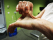
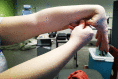
 Previously, two subtypes, EDS type I and EDS type II, differing only in phenotypic severity, were recognized; it is now apparent that they form a continuum of clinical findings.

Skin

Cutaneous hyperextensibility is one of the cardinal features of EDS in general and of cEDS in particular. Skin extends easily and snaps back after release (unlike lax, redundant skin, as in cutis laxa).

The skin is soft and doughy to the touch.

The skin is fragile, as manifested by splitting of the dermis following relatively minor trauma, especially over pressure points (knees, elbows) and areas prone to trauma (shins, forehead, chin). Skin fragility may cause dehiscence of sutured incisions in skin or mucosa.
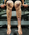
Wound healing is poor, and stretching of scars after apparently successful primary wound healing is characteristic. Scars become wide, with a "cigarette-paper"-like (papyraceous) appearance.

Other dermatologic features in cEDS:

  • Molluscoid pseudotumors
  • Subcutaneous spheroids
  • Piezogenic papules: small, painful, reversible herniations of underlying adipose tissue globules through the fascia into the dermis, such as on medial and lateral aspects of the feet upon standing
  • Elastosis perforans serpiginosa: a rare skin condition of unknown etiology characterized by skin-colored to erythematous keratotic papules, some enlarging outwards in serpiginous or arcuate configurations, leaving slightly atrophic centers
  • Acrocyanosis: a painless disorder caused by constriction or narrowing of the small blood vessels in the skin (affecting mainly the hands) in which the affected areas turn blue and become cold and sweaty; localized swelling may also occur
  • Chilblains: cold injuries, characterized by a red swollen skin that is tender and hot to the touch, and may itch; can develop in less than two hours in skin exposed to cold

Tissue Fragility

Manifestations of generalized tissue extensibility and fragility are observed in multiple organs:

  • Cervical insufficiency during pregnancy
  • Inguinal and umbilical hernia
  • Hiatal and incisional hernia
  • Recurrent rectal prolapse in early childhood

Joints

Complications of joint hypermobility including dislocations of the shoulder, patella, digits, hip, radius, and clavicle may occur and usually resolve spontaneously or are easily managed by the affected individual. Some individuals with cEDS may experience chronic joint and limb pain, despite normal skeletal radiographs.

Other problems related to the joint hypermobility are joint instability, foot deformities such as congenital clubfoot or pes planus, temporomandibular joint dysfunction, joint effusions, and osteoarthritis [Hagberg et al 2004, De Coster et al 2005a, De Coster et al 2005b].

Neurologic Features

Primary muscular hypotonia may occur and may cause delayed motor development, problems with ambulation, and mild motor disturbance in individuals with cEDS. Fatigue and muscle cramps are relatively frequent. Rarely, CSF leak has been reported to cause postural hypotension and headache [Schievink et al 2004].

Easy Bruising

Easy bruising is a common finding and manifests as spontaneous ecchymoses, frequently recurring in the same areas and causing a characteristic brownish discoloration of the skin, especially in exposed areas such as shins and knees. There is a tendency toward prolonged bleeding (e.g., following brushing of the teeth) in spite of a normal coagulation status.

Cardiovascular

Structural cardiac malformations are uncommon in cEDS.

Mitral valve prolapse and (less frequently) tricuspid valve prolapse may occur. Stringent criteria should be used for the diagnosis of mitral valve prolapse. When it does occur, mitral valve prolapse tends to be of little clinical consequence [Atzinger et al 2011].

Aortic root dilatation has been reported in cEDS [Wenstrup et al 2002, McDonnell et al 2006, Atzinger et al 2011]. It appears to be more common in young individuals and rarely progresses [Atzinger et al 2011].

Spontaneous rupture of large arteries, along with intracranial aneurysms and arteriovenous fistulae, may occur in the rare individual with a severe form of cEDS.

Pregnancy

Pregnancy in a woman with cEDS places both the newborn and the mother at risk for complications (see Pregnancy Management). As a whole, the complications are more frequent than in the normal population, although it is difficult to quantitate the incidence of each complication in affected individuals because no good studies exist.

  • Preterm rupture of the membranes and prematurity can occur when the mother is affected, and also when the fetus is affected, especially in the most severe forms.
  • Because of hypotonia, breech presentation is more frequent if the baby is affected and may lead to dislocation of the hips or shoulder of the newborn.

Genotype-Phenotype Correlations

No genotype/phenotype correlations have emerged to date.

Although numbers are still limited, pathogenic variants in COL5A2 are thought to result in a phenotype at the more severe end of the classic EDS spectrum.

Penetrance

It is unknown whether penetrance is 100% or reduced. It is presumed to be the same for males and females.

Nomenclature

As a result of the 1997 Villefranche conference on EDS [Beighton et al 1998], the former EDS type I and type II were reclassified as EDS, classic type. In 2017, the International EDS Consortium proposed a revised EDS classification system. The new nomenclature for EDS, classic type is classic EDS, or cEDS [Malfait et al 2017].

Prevalence

The prevalence of cEDS has been estimated at 1:20,000 [Byers 2001]. However, it is likely that some individuals with milder manifestations of the disease, previously classified as EDS type II, do not come to medical attention and thus go undetected.

Differential Diagnosis

Other forms of Ehlers-Danlos syndrome (EDS) should be considered in individuals with easy bruising, joint hypermobility, and/or chronic joint dislocation. Clinical overlap is seen with all other forms of EDS, in particular with the following subtypes.

Table 3.

Other EDS disorders to Consider in the Differential Diagnosis of Classic EDS

DiffDx DisorderGene(s)MOIClinical Features of DiffDx Disorder
Overlapping w/cEDSDistinguishing from cEDS
Classic-like EDS TNXB AR
  • Skin hyperextensibility
  • Velvety skin
  • GJH
  • Easy bruising
  • AR inheritance
  • Absence of atrophic scarring
Cardiac valvular EDS (OMIM 225320) COL1A2 AR
  • Skin hyperextensibility
  • Atrophic scarring
  • Easy bruising
  • (Generalized) joint hypermobility
  • AR inheritance
  • Severe progressive cardiac valvular problems
Hypermobile EDS UnknownAD
  • GJH
  • Soft, velvety skin
  • Mild skin hyperextensibility
  • Mild atrophic scarring
Absence of truly papyraceous &/or hemosiderotic scars
Arthrochalasia EDS (OMIM 130060, 617821) COL1A1
COL1A2
AD
  • GJH
  • Skin hyperextensibility
  • Atrophic scarring
  • Easy bruising
Bilateral congenital hip dislocation
Dermatosparaxis EDS (OMIM 225410) ADAMTS2 AR
  • Soft, doughy skin texture
  • Atrophic scarring
  • Skin hyperextensibility
  • GJH
  • Extreme skin fragility (usually > than in cEDS)
  • Redundant, almost lax, skin
  • Unusual craniofacial features
  • Postnatal growth restriction
  • AR inheritance
PLOD1-related kyphoscoliotic EDS PLOD1 AR
  • GJH
  • Skin hyperextensibility
  • Easy bruising
  • Atrophic scarring
Congenital muscle hypotonia
FKBP14-related kyphoscoliotic EDS FKBP14 AR
  • GJH
  • Skin hyperextensibility
  • Easy bruising
  • Congenital muscle hypotonia
  • Muscle atrophy
  • Congenital hearing impairment

AD = autosomal dominant; AR = autosomal recessive; DiffDx = differential diagnosis; GJH = generalized joint hypermobility; MOI = mode of inheritance

Management

For a detailed review of complications and management, see Bowen et al [2017].

Evaluations Following Initial Diagnosis

To establish the extent of disease in an individual diagnosed with classic Ehlers-Danlos syndrome (cEDS), the following evaluations are recommended if they have not already been completed:

  • Clinical examination of the skin with assessment of skin hyperextensibility, atrophic scars and bruises, and other manifestations of cEDS
  • Evaluation of joint mobility with use of the Beighton score
  • Evaluation for hypotonia and motor development in infants and children
  • A baseline echocardiogram with aortic diameter measurement at diagnosis
  • Evaluation of clotting factors if severe easy bruising is present
  • Consultation with a clinical geneticist and/or genetic counselor

Treatment of Manifestations

In children with hypotonia and delayed motor development, a physiotherapeutic program is important.

Non-weight-bearing muscular exercise, such as swimming, is useful to promote muscular development and coordination.

Individuals with muscle hypotonia and joint instability with chronic pain may have to adjust lifestyle and professional choices accordingly. Emotional support and behavioral and psychological therapy may help in developing acceptance and coping skills.

Dermal wounds should be closed without tension, preferably in two layers. Deep stitches should be applied generously. Cutaneous stitches should be left in place twice as long as usual and additional fixation of adjacent skin with adhesive tape can help prevent stretching of the scar.

DDAVP® (deamino-delta-D-arginine vasopressin) may be useful to normalize bleeding time. It may be beneficial in case of bruising or epistaxis, or before procedures such as dental extractions.

For recommendations on treatment of joint laxity and dislocations, see Hypermobile EDS. (Note: Surgical stabilization of joints may lead to disappointing, or only temporary, improvement.)

Anti-inflammatory drugs may help with joint pain.

Long-term chronic pain may result in the need for mental health services.

Cardiovascular problems should be treated in a standard manner.

Prevention of Primary Manifestations

Very young children with pronounced skin fragility can wear protective pads or bandages over the forehead, knees, and shins in order to avoid skin tears. Older children who are active can wear soccer pads or ski stockings with shin padding during activities. Contact sports should be avoided.

For recommendations on prevention of primary manifestations of joint laxity and dislocations, see Hypermobile EDS, Management: Prevention of Primary Manifestations.

Ascorbic acid (vitamin C) may reduce easy bruising but has no effect on the primary findings of skin hyperextensibility, atrophic scarring, and joint hypermobility. In general, a dose of two grams per day is recommended for adults, with proportionally reduced doses for children; however, there is no limitation.

Prevention of Secondary Complications

For recommendations on prevention of secondary manifestations of joint laxity and dislocations, see Hypermobile EDS, Management: Prevention of Secondary Complications.

Surveillance

If no abnormalities are found on echocardiogram in an adult, a follow-up echocardiogram is not necessary. (Because longitudinal data on progression of aortic dilatation are not available, specific recommendations for follow up in individuals with a normal aortic diameter are not available.)

If no abnormalities are found on echocardiogram in a child, follow up directed by the pediatric cardiologist is recommended.

Yearly echocardiogram is warranted if an abnormality such as aortic dilatation or mitral valve prolapse is present.

Agents/Circumstances to Avoid

The following should be avoided:

  • Sports with heavy joint strain (contact sports, fighting sports, football, running)
  • Acetylsalicylate (aspirin)

Evaluation of Relatives at Risk

It is appropriate to clarify the genetic status of apparently asymptomatic older and younger at-risk relatives of an affected individual by molecular genetic testing of the pathogenic variant in the family in order to identify as early as possible those who would benefit from prompt initiation of treatment and preventive measures.

See Genetic Counseling for issues related to testing of at-risk relatives for genetic counseling purposes.

Pregnancy Management

Because of the increased risk for skin lacerations, postpartum hemorrhages, and prolapse of the uterus and/or bladder, monitoring of women throughout pregnancy and in the postpartum period is recommended.

Ascorbic acid (vitamin C) may reduce easy bruising (see Prevention of Primary Manifestations); in general, 2 g/day is recommended for adults; however, no strict guidelines exist regarding recommended dose during the third trimester of pregnancy.

Monitoring of pregnant women for preterm labor is warranted during the third trimester when the risk for premature rupture of the membranes is increased.

Therapies Under Investigation

Search ClinicalTrials.gov in the US and EU Clinical Trials Register in Europe for information on clinical studies for a wide range of diseases and conditions. Note: There may not be clinical trials for this disorder.

Genetic Counseling

Genetic counseling is the process of providing individuals and families with information on the nature, mode(s) of inheritance, and implications of genetic disorders to help them make informed medical and personal decisions. The following section deals with genetic risk assessment and the use of family history and genetic testing to clarify genetic status for family members; it is not meant to address all personal, cultural, or ethical issues that may arise or to substitute for consultation with a genetics professional. —ED.

Mode of Inheritance

Classic Ehlers-Danlos syndrome (cEDS) is inherited in an autosomal dominant manner.

Risk to Family Members

Parents of a proband

  • It is estimated that 50% of individuals diagnosed with cEDS have an affected parent.
  • It is estimated that 50% of individuals diagnosed with cEDS have the disorder as the result of a de novo pathogenic variant.
  • The parents of a proband with an apparent de novo pathogenic variant should be evaluated by physical examination of the skin with special attention to delayed wound healing, easy bruising, joint hypermobility or recurrent dislocations, and chronic articular pain.
  • Molecular genetic testing is recommended for the parents of a proband with an apparent de novo pathogenic variant.
  • If the pathogenic variant found in the proband cannot be detected in the leukocyte DNA of either parent, possible explanations include a de novo pathogenic variant in the proband or germline mosaicism in a parent. Though theoretically possible, no instances of germline mosaicism have been reported.
  • The family history of some individuals diagnosed with cEDS may appear to be negative because of failure to recognize the disorder in family members or, theoretically, reduced penetrance (it is unknown whether penetrance is 100% or reduced). Therefore, an apparently negative family history cannot be confirmed unless appropriate clinical evaluation and/or molecular genetic testing has been performed on the parents of the proband.

Sibs of a proband. The risk to the sibs of the proband depends on the clinical/genetic status of the proband's parents:

Offspring of a proband. Each child of an individual with classic EDS has a 50% chance of inheriting the pathogenic variant. Intrafamilial phenotypic variability is observed.

Other family members The risk to other family members depends on the status of the proband's parents: if a parent has the pathogenic variant, the parent's family members may be at risk.

Related Genetic Counseling Issues

See Management, Evaluation of Relatives at Risk for information on evaluating at-risk relatives for the purpose of early diagnosis and treatment.

At-risk individuals. The severity, specific symptoms, and progression of the disorder are variable and cannot be predicted based on family history or results of molecular genetic testing.

Considerations in families with an apparent de novo pathogenic variant. When neither parent of a proband with an autosomal dominant condition has the pathogenic variant identified in the proband or clinical evidence of the disorder, the pathogenic variant is likely de novo. However, non-medical explanations including alternate paternity or maternity (e.g., with assisted reproduction) and undisclosed adoption could also be explored.

Family planning

  • The optimal time for determination of genetic risk and discussion of the availability of prenatal/preimplantation genetic testing is before pregnancy.
  • It is appropriate to offer genetic counseling (including discussion of potential risks to offspring and reproductive options) to young adults who are affected or at risk.

DNA banking. Because it is likely that testing methodology and our understanding of genes, pathogenic mechanisms, and diseases will improve in the future, consideration should be given to banking DNA of probands in whom a molecular diagnosis has not been confirmed (i.e., the causative pathogenic mechanism is unknown). For more information, see Huang et al [2022].

Prenatal Testing and Preimplantation Genetic Testing

Once the pathogenic variant has been identified in an affected family member, prenatal and preimplantation genetic testing are possible.

Differences in perspective may exist among medical professionals and within families regarding the use of prenatal testing. While most centers would consider use of prenatal testing to be a personal decision, discussion of these issues may be helpful.

Resources

GeneReviews staff has selected the following disease-specific and/or umbrella support organizations and/or registries for the benefit of individuals with this disorder and their families. GeneReviews is not responsible for the information provided by other organizations. For information on selection criteria, click here.

Molecular Genetics

Information in the Molecular Genetics and OMIM tables may differ from that elsewhere in the GeneReview: tables may contain more recent information. —ED.

Table A.

Classic Ehlers-Danlos Syndrome: Genes and Databases

Data are compiled from the following standard references: gene from HGNC; chromosome locus from OMIM; protein from UniProt. For a description of databases (Locus Specific, HGMD, ClinVar) to which links are provided, click here.

Table B.

OMIM Entries for Classic Ehlers-Danlos Syndrome (View All in OMIM)

120150COLLAGEN, TYPE I, ALPHA-1; COL1A1
120190COLLAGEN, TYPE V, ALPHA-2; COL5A2
120215COLLAGEN, TYPE V, ALPHA-1; COL5A1
130000EHLERS-DANLOS SYNDROME, CLASSIC TYPE, 1; EDSCL1

COL1A1

Gene structure. COL1A1 is 18 kilobases in size and is composed of 52 exons (NM_000088.3).

Pathogenic variants. Although most pathogenic variants in COL1A1 cause osteogenesis imperfecta, specific missense and splice site variants have been associated with specific EDS subtypes. In particular, Nuytinck et al [2000] reported two children with classic EDS with a c.934C>T (p.Arg312Cys) pathogenic variant. Malfait et al [2007] identified the same variant in an adult with signs of cEDS, who suffered from a rupture of medium-sized arteries. This pathogenic variant was subsequently found in two other adults with EDS and complications of vascular fragility [Ritelli et al 2013, Gaines et al 2015].

Table 4.

COL1A1 Pathogenic Variants Discussed in This GeneReview

DNA Nucleotide ChangePredicted Protein ChangeReference Sequences
c.934C>Tp.Arg312Cys NM_000088​.4
NP_000079​.2

Variants listed in the table have been provided by the authors. GeneReviews staff have not independently verified the classification of variants.

GeneReviews follows the standard naming conventions of the Human Genome Variation Society (varnomen​.hgvs.org). See Quick Reference for an explanation of nomenclature.

Normal gene product. COL1A1 encodes the 1,464-amino acid pro α1 chains of type I collagen, whose triple helix comprises two α1 chains and one α2 chain. Type I is a fibril-forming collagen found in most connective tissues and is abundant in bone, cornea, dermis, and tendon.

Abnormal gene product. The pathogenetic basis for the phenotype resulting from this COL1A1 Arg-to-Cys substitution is currently not well understood. Ultrastructural studies of dermal collagen fibrils have shown fibrils with variable diameters, slightly irregular contour, and, in the case of p.Arg312Cys, flower-like abnormalities [Malfait et al 2007].

Several mechanisms have been proposed as possibly involved in the pathogenesis including [Malfait et al 2007]:

  • Local destabilization of the triple helix due to loss of the stabilizing arginine residue;
  • Introduction of a cysteine residue, which can lead to disulfide bonding with other collagenous or noncollagenous proteins, either intracellularly or in the extracellular matrix (ECM), thereby disturbing normal physiologic interactions;
  • Interference with pericellular processing of the amino-propeptide of procollagen type I; and/or
  • Local unwinding of the region surrounding the mutations, thereby disturbing specific interactions with type I collagen ligands.

COL5A1

Gene structure. The COL5A1 cDNA comprises 66 exons distributed over more than 150 kb of genomic DNA. For a detailed summary of gene and protein information, see Table A, Gene.

Pathogenic variants. Several types of pathogenic variants have been identified in COL5A1:

  • The most common types of molecular defect lead to haploinsufficiency for COL5A1 mRNA. In approximately 40%-50% of individuals with cEDS, nonsense or frameshift variants are responsible for a nonfunctional COL5A1 allele [Schwarze et al 2000, Wenstrup et al 2000, Schwarze et al 2001, Malfait et al 2005, Symoens et al 2012, Ritelli et al 2013]. The predicted consequence of haploinsufficiency is synthesis of approximately half the amount of normal type V collagen.
  • In a small proportion of individuals, a COL5A1 variant affects the structural integrity of type V collagen, resulting in the production of a functionally defective type V collagen protein (dominant-negative variant). These structural variants are most commonly splice site variants that result in exon skipping and a few single-nucleotide variants that result in the substitution for glycine in the triple-helical region of the collagen molecule.
  • A unique single-nucleotide variant in a highly conserved cysteine residue, p.Cys1639Ser, in the C-terminal propeptide of the α1(V) collagen chain has also been identified.
  • In contrast to other collagen disorders, few pathogenic variants in glycine residues have been found. A p.Gly530Ser substitution in the amino-terminal propeptide of the α1(V) chain may be disease modifying when present in the heterozygous state and disease causing in the homozygous state [Giunta & Steinmann 2000, Giunta et al 2002].

Table 5.

COL5A1 Pathogenic Variants Discussed in This GeneReview

DNA Nucleotide ChangePredicted Protein ChangeReference Sequences
c.1588G>Ap.Gly530Ser NM_000093​.3
NP_000084​.3
c.4916G>Cp.Cys1639Ser

Variants listed in the table have been provided by the authors. GeneReviews staff have not independently verified the classification of variants.

GeneReviews follows the standard naming conventions of the Human Genome Variation Society (varnomen​.hgvs.org). See Quick Reference for an explanation of nomenclature.

Normal gene product. Collagen α1 (V) chain (type V collagen chains). Type V collagen is a quantitatively minor fibrillar collagen that is widely distributed in a variety of tissues. It is present mainly as [α1(V)]2 α2(V) heterotrimers in skin, bone, and tendon. It forms heterotypic fibrils with type I collagen and regulates the diameter of those fibrils, presumably through its very large amino-terminal propeptide. Data indicate that type V collagen controls collagen fibril assembly in several tissues [Wenstrup et al 2004].

Abnormal gene product. Missense variants in the triple helical domain of the α1(V) or α2(V) chains are likely to have dominant-negative activity; that is, the abnormal form can interfere with protein from the normal allele. Diminished amounts, caused by premature termination of codons in COL5A1 or mRNA product, may alter normal collagen fibrillogenesis.

COL5A2

Gene structure. The COL5A2 cDNA comprises 51 exons distributed over 67 kb. For a detailed summary of gene and protein information, see Table A, Gene.

Pathogenic variants. Structural pathogenic variants in COL5A2 have been demonstrated in few individuals with classic EDS. These structural variants are most commonly splice site variants that result in exon skipping [Michalickova et al 1998, Malfait et al 2005, Symoens at al 2012] and a few nucleotide variants that result in substitution for glycine in the triple-helical region of the collagen molecule [Richards et al 1998, Symoens et al 2012, Ritelli et al 2013].

Normal gene product. Collagen α2(V) chains (type V collagen chains). Type V collagen is a quantitatively minor fibrillar collagen that is widely distributed in a variety of tissues. It is present mainly as [α1(V)]2, α2(V) heterotrimers in skin, bone, and tendon. It forms heterotypic fibrils with type I collagen and regulates the diameter of those fibrils, presumably through its very large amino-terminal propeptide.

Abnormal gene product. Missense variants in the triple helical domain of the α1(V) or α2(V) chains are likely to have dominant-negative activity; that is, the abnormal forms can interfere with protein derived from the normal allele.

Chapter Notes

Revision History

  • 26 July 2018 (ha) Comprehensive update posted live
  • 18 August 2011 (me) Comprehensive update posted live
  • 11 May 2010 (cd) Revision: changes in testing
  • 24 July 2008 (me) Comprehensive update posted live
  • 29 May 2007 (cd) Revision: sequence analysis of entire coding region available for COL5A1 and COL5A2 and prenatal testing available
  • 10 April 2006 (me) Comprehensive update posted live
  • 29 October 2003 (ca) Review posted live
  • 20 June 2003 (rw, ad) Original submission

References

Published Guidelines / Consensus Statements

  • Bowen JM, Sobey GJ, Burrows NP, Colombi M, Lavallee ME, Malfait F, Francomano CA. Ehlers-Danlos syndrome, classical type. Am J Med Genet C Semin Med Genet. 2017;175:27-39. [PubMed]
  • Malfait F, Francomano C, Byers P, Belmont J, Berglund B, Black J, Bloom L, Bowen JM, Brady AF, Burrows NP, Castori M, Cohen H, Colombi M, Demirdas S, De Backer J, De Paepe A, Fournel-Gigleux S, Frank M, Ghali N, Giunta C, Grahame R, Hakim A, Jeunemaitre X, Johnson D, Juul-Kristensen B, Kapferer-Seebacher I, Kazkaz H, Kosho T, Lavallee ME, Levy H, Mendoza-Londono R, Pepin M, Pope FM, Reinstein E, Robert L, Rohrbach M, Sanders L, Sobey GJ, Van Damme T, Vandersteen A, van Mourik C, Voermans N, Wheeldon N, Zschocke J, Tinkle B. The 2017 international classification of the Ehlers–Danlos syndromes. Am J Med Genet C Semin Med Genet. 2017;175:8-26. [PubMed]

Literature Cited

  • Atzinger CL, Meyer RA, Khoury PR, Gao Z, Tinkle BT. Cross-sectional and longitudinal assessment of aortic root dilation and valvular anomalies in hypermobile and classic Ehlers-Danlos syndrome. J Pediatr. 2011;158:826–830.e1. [PubMed: 21193204]
  • Beighton P, De Paepe A, Steinmann B, Tsipouras P, Wenstrup RJ. Ehlers-Danlos syndromes: revised nosology, Villefranche, 1997. Ehlers- Danlos National Foundation (USA) and Ehlers-Danlos Support Group (UK). Am J Med Genet. 1998;77:31–7. [PubMed: 9557891]
  • Brady AF, Demirdas S, Fournel-Gigleux S, Ghali N, Giunta C, Kapferer-Seebacher I, Kosho T, Mendoza-Londono R, Pope MF, Rohrbach M, Van Damme T, Vandersteen A, van Mourik C, Voermans N, Zschocke J, Malfait F. The Ehlers-Danlos syndromes, rare types. Am J Med Genet C Semin Med Genet. 2017;175:70–115. [PubMed: 28306225]
  • Bowen JM, Sobey GJ, Burrows NP, Colombi M, Lavallee ME, Malfait F, Francomano CA. Ehlers-Danlos syndrome, classical type. Am J Med Genet C Semin Med Genet. 2017;175:27–39. [PubMed: 28192633]
  • Byers PH. Disorders of collagen biosynthesis and structure. In: Scriver CR, Beaudet AL, Sly WS, Valle D, eds. The Metabolic and Molecular Bases of Inherited Disease. 2 ed. Edinburgh, UK: Churchill Livingstone; 2001:1065-81.
  • Cabral WA, Makareeva E, Colige A, Letocha AD, Ty JM, Yeowell HN, Pals G, Leikin S, Marini JC. Mutations near amino end of alpha-1(I) collagen cause combined osteogenesis imperfecta/Ehlers-Danlos syndrome by interference with N-propeptide processing. J Biol Chem. 2005;280:19259–69. [PubMed: 15728585]
  • De Coster PJ, Martens LC, De Paepe A. Oral health in prevalent types of Ehlers-Danlos syndromes. J Oral Pathol Med. 2005a;34:298–307. [PubMed: 15817074]
  • De Coster PJ, Van den Berghe LI, Martens LC. Generalized joint hypermobility and temporomandibular disorders: inherited connective tissue disease as a model with maximum expression. J Orofac Pain. 2005b;19:47–57. [PubMed: 15779539]
  • Gaines R, Tinkle BT, Halandras PM, Al-Nouri O, Crisostomo P, Cho JS. Spontaneous ruptured dissection of the right common iliac artery in a patient with classic Ehlers-Danlos syndrome phenotype. Ann Vasc Surg. 2015;29:595.e11–4. [PubMed: 25597651]
  • Giunta C, Nuytinck L, Raghunath M, Hausser I, De Paepe A, Steinmann B. Homozygous Gly530Ser substitution in COL5A1 causes mild classical Ehlers-Danlos syndrome. Am J Med Genet. 2002;109:284–90. [PubMed: 11992482]
  • Giunta C, Steinmann B. Compound heterozygosity for a disease-causing G1489E [correction of G1489D] and disease-modifying G530S substitution in COL5A1 of a patient with the classical type of Ehlers-Danlos syndrome: an explanation of intrafamilial variability? Am J Med Genet. 2000;90:72–9. [PubMed: 10602121]
  • Hagberg C, Berglund B, Korpe L, Andersson-Norinder J. Ehlers-Danlos Syndrome (EDS) focusing on oral symptoms: a questionnaire study. Orthod Craniofac Res. 2004;7:178–85. [PubMed: 15359504]
  • Hakim AJ, Grahame R. A simple questionnaire to detect hypermobility: an adjunct to the assessment of patients with diffuse musculoskeletal pain. Int J Clin Pract. 2003;57:163–6. [PubMed: 12723715]
  • Huang SJ, Amendola LM, Sternen DL. Variation among DNA banking consent forms: points for clinicians to bank on. J Community Genet. 2022;13:389–97. [PMC free article: PMC9314484] [PubMed: 35834113]
  • Malfait F, Coucke P, Symoens S, Loeys B, Nuytinck L, De Paepe A. The molecular basis of classic Ehlers-Danlos syndrome: a comprehensive study of biochemical and molecular findings in 48 unrelated patients. Hum Mutat. 2005;25:28–37. [PubMed: 15580559]
  • Malfait F, Francomano C, Byers P, Belmont J, Berglund B, Black J, Bloom L, Bowen JM, Brady AF, Burrows NP, Castori M, Cohen H, Colombi M, Demirdas S, De Backer J, De Paepe A, Fournel-Gigleux S, Frank M, Ghali N, Giunta C, Grahame R, Hakim A, Jeunemaitre X, Johnson D, Juul-Kristensen B, Kapferer-Seebacher I, Kazkaz H, Kosho T, Lavallee ME, Levy H, Mendoza-Londono R, Pepin M, Pope FM, Reinstein E, Robert L, Rohrbach M, Sanders L, Sobey GJ, Van Damme T, Vandersteen A, van Mourik C, Voermans N, Wheeldon N, Zschocke J, Tinkle B. The 2017 international classification of the Ehlers–Danlos syndromes. Am J Med Genet C Semin Med Genet. 2017;175:8–26. [PubMed: 28306229]
  • Malfait F, Symoens S, De Backer J, Hermanns-Lê T, Sakalihasan N, Lapière CM, Coucke P, De Paepe A. Three arginine to cysteine substitutions in the pro-alpha (I)-collagen chain cause Ehlers-Danlos syndrome with a propensity to arterial rupture in early adulthood. Hum Mutat. 2007;28:387–95. [PubMed: 17211858]
  • McDonnell NB, Gorman BL, Mandel KW, Schurman SH, Assanah-Carroll A, Mayer SA, Najjar SS, Francomano CA. Echocardiographic findings in classical and hypermobile Ehlers-Danlos syndromes. Am J Med Genet A. 2006;140:129–36. [PubMed: 16353246]
  • Michalickova K, Susic M, Willing MC, Wenstrup RJ, Cole WG. Mutations of the alpha2(V) chain of type V collagen impair matrix assembly and produce Ehlers-Danlos syndrome type I. Hum Mol Genet. 1998;7:249–55. [PubMed: 9425231]
  • Nuytinck L, Freund M, Lagae L, Pierard GE, Hermanns-Le T, De Paepe A. Classical Ehlers-Danlos syndrome caused by a mutation in type I collagen. Am J Hum Genet. 2000;66:1398–402. [PMC free article: PMC1288203] [PubMed: 10739762]
  • Rahbari R, Wuster A, Lindsay SJ, Hardwick RJ, Alexandrov LB, Turki SA, Dominiczak A, Morris A, Porteous D, Smith B, Stratton MR, Hurles ME, et al. Timing, rates and spectra of human germline mutation. Nat Genet. 2016;48:126–33. [PMC free article: PMC4731925] [PubMed: 26656846]
  • Remvig L, Duhn PH, Ullman S, Kobayasi T, Hansen B, Juul-Kristensen B, Jurvelin JS, Arokoski J. Skin extensibility and consistency in patients with Ehlers-Danlos syndrome and benign joint hypermobility syndrome. Scand J Rheumatol. 2009;38:227–30. [PubMed: 19169910]
  • Richards AJ, Martin S, Nicholls AC, Harrison JB, Pope FM, Burrows NP. A single base mutation in COL5A2 causes Ehlers-Danlos syndrome type II. J Med Genet. 1998;35:846–8. [PMC free article: PMC1051462] [PubMed: 9783710]
  • Richards S, Aziz N, Bale S, Bick D, Das S, Gastier-Foster J, Grody WW, Hegde M, Lyon E, Spector E, Voelkerding K, Rehm HL, et al. Standards and guidelines for the interpretation of sequence variants: a joint consensus recommendation of the American College of Medical Genetics and Genomics and the Association for Molecular Pathology. Genet Med. 2015;17:405–24. [PMC free article: PMC4544753] [PubMed: 25741868]
  • Ritelli M, Dordoni C, Venturini M, Chiarelli N, Quinzani S, Traversa M, Zoppi N, Vascellaro A, Wischmeijer A, Manfredini E, Garavelli L, Calzavara-Pinton P, Colombi M. Clinical and molecular characterization of 40 patients with classic Ehlers-Danlos syndrome: identification of 18 COL5A1 and 2 COL5A2 novel mutations. Orphanet J Rare Dis. 2013;8:58. [PMC free article: PMC3653713] [PubMed: 23587214]
  • Schievink WI, Gordon OK, Tourje J. Connective tissue disorders with spontaneous spinal cerebrospinal fluid leaks and intracranial hypotension: a prospective study. Neurosurgery. 2004;54:65–70. [PubMed: 14683542]
  • Schwarze U, Atkinson M, Hoffman GG, Greenspan DS, Byers PH. Null alleles of the COL5A1 gene of type V collagen are a cause of the classical forms of Ehlers-Danlos syndrome (types I and II). Am J Hum Genet. 2000;66:1757–65. [PMC free article: PMC1378060] [PubMed: 10796876]
  • Schwarze U, Schievink WI, Petty E, Jaff MR, Babovic-Vuksanovic D, Cherry KJ, Pepin M, Byers PH. Haploinsufficiency for one COL3A1 allele of type III procollagen results in a phenotype similar to the vascular form of Ehlers-Danlos syndrome, Ehlers-Danlos syndrome type IV. Am J Hum Genet. 2001;69:989–1001. [PMC free article: PMC1274375] [PubMed: 11577371]
  • Symoens S, Syx D, Malfait F, Callewaert B, De Backer J, Vanakker O, Coucke P, De Paepe A. Comprehensive molecular analysis demonstrates type V collagen mutations in over 90% of patients with classic EDS and allows to refine diagnostic criteria. Hum Mutat. 2012;33:1485–93. [PubMed: 22696272]
  • Wenstrup RJ, Florer JB, Brunskill EW, Bell SM, Chervoneva I, Birk DE. Type V collagen controls the initiation of collagen fibril assembly. J Biol Chem. 2004;279:53331–7. [PubMed: 15383546]
  • Wenstrup RJ, Florer JB, Willing MC, Giunta C, Steinmann B, Young F, Susic M, Cole WG. COL5A1 haploinsufficiency is a common molecular mechanism underlying the classical form of EDS. Am J Hum Genet. 2000;66:1766–76. [PMC free article: PMC1378044] [PubMed: 10777716]
  • Wenstrup RJ, Meyer RA, Lyle JS, Hoechstetter L, Rose PS, Levy HP, Francomano CA. Prevalence of aortic root dilation in the Ehlers-Danlos syndrome. Genet Med. 2002;4:112–7. [PubMed: 12180144]
Copyright © 1993-2023, University of Washington, Seattle. GeneReviews is a registered trademark of the University of Washington, Seattle. All rights reserved.

GeneReviews® chapters are owned by the University of Washington. Permission is hereby granted to reproduce, distribute, and translate copies of content materials for noncommercial research purposes only, provided that (i) credit for source (http://www.genereviews.org/) and copyright (© 1993-2023 University of Washington) are included with each copy; (ii) a link to the original material is provided whenever the material is published elsewhere on the Web; and (iii) reproducers, distributors, and/or translators comply with the GeneReviews® Copyright Notice and Usage Disclaimer. No further modifications are allowed. For clarity, excerpts of GeneReviews chapters for use in lab reports and clinic notes are a permitted use.

For more information, see the GeneReviews® Copyright Notice and Usage Disclaimer.

For questions regarding permissions or whether a specified use is allowed, contact: ude.wu@tssamda.

Bookshelf ID: NBK1244PMID: 20301422

Views

Tests in GTR by Gene

Related information

  • MedGen
    Related information in MedGen
  • OMIM
    Related OMIM records
  • PMC
    PubMed Central citations
  • PubMed
    Links to PubMed
  • Gene
    Locus Links

Similar articles in PubMed

See reviews...See all...

Recent Activity

Your browsing activity is empty.

Activity recording is turned off.

Turn recording back on

See more...