The ultimate test of pathogenic potential involves induction of disease in vivo, and animal inoculation experiments will always play an essential part in studies examining the mechanisms of disease induction. However, the ability to grow and quantitate retroviruses in vitro is critical to understanding these agents. In vitro approaches can also provide insight into basic pathogenic mechanisms. These techniques facilitate study of the complex mixtures of viruses usually found in naturally infected hosts and allow biological cloning of the agents, a process that is often critical for sorting out the role of individual viruses in disease induction. Changes in the morphology and growth patterns of infected cell cultures provide simple detection systems for many viruses and form the basis for quantitative assays that monitor infectivity. In vitro cultures also simplify the derivation and characterization of viral mutants and allow biochemical and molecular analyses of the host-virus relationship at the cellular level. These latter experiments have been particularly illuminating for many of the retroviruses that induce tumors. Indeed, they set the stage for the explosive progress that has been made in understanding signal transduction in normal and malignant cells over the past decade.
Although many types of cells can be used to grow and assay retroviruses in vitro, the single most commonly used cell type is fibroblastoid cells derived from embryos. Ironically, fibroblastoid cells are usually not directly involved in disease induction. However, they can be grown easily and they support high-titer replication of many viruses, making them an important research tool. These cells can be used as primary cultures, such as the chick embryo fibroblasts used to study avian retroviruses, or as established, immortalized rodent cell lines such as 3T3 cells, which have been particularly useful for studies of murine retroviruses. Similar cell systems have been used to study retroviruses that infect other types of animals. Cultures of normal and neoplastic hematopoietic cells have also been widely used to grow and assay viruses that affect these types of cells.
Cellular Transformation
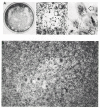
Many retroviruses carry oncogenes and almost all of these induce the changes in cellular growth and morphology of fibroblastoid cells that are collectively termed transformation. Most transformed cells grow in a disorganized and multilayered pattern (Fig. 1) reflecting changes in the mechanisms that allow normal cells to sense the microenvironment. Cytoskeletal reorganization, a feature related to the morphologic changes that characterize the transformed cell, also occurs. Other common changes include the ability of cells to grow in semisolid media, such as agar, or to grow in reduced levels of serum or growth factors, changes in nutrient transport and glucose metabolism, and alterations in expression and modification of surface proteins. All of these properties are reminiscent of the growth properties of tumor cells, and in many cases, cells transformed in vitro form tumors when injected into syngeneic or immunocompromised animals. Thus, transformation has been viewed as the in vitro equivalent of tumorigenesis.
Most retroviruses that transform cells in vitro are replication-defective because some or all of the genes required for viral replication were deleted when the oncogene was captured (see Chapter 4. Stocks of these viruses contain both the transforming virus and a replication-competent retrovirus that provides the gene products required for replication and packaging of the transforming viral genome. Cultures infected with concentrated viral stocks become transformed and release infectious, transforming virus. However, when individual cells are infected with highly diluted stocks, two types of cells can be obtained. One of these retains normal morphology and releases virus that does not induce transformation; the other type is transformed but does not release infectious virus. These latter cells are termed nonproducer cells. Transforming virus can be recovered from nonproducer cells by infecting them with an appropriate replication-competent virus. In this setting, the replication-competent virus is referred to as a helper virus. Nonproducer cells provide an important tool for analyzing the genome and protein products of replication-defective oncogene-containing viruses and were the first way in which these agents were biologically cloned.
The ease and speed with which transformation can be detected in vitro form the basis for assays to measure the infectivity and transforming ability of many viruses that contain oncogenes (Temin and Rubin 1958; Hartley and Rowe 1966). When susceptible cells are plated at appropriate concentrations and infected with diluted viral stocks, individual areas of transformed cells called foci can be visualized. The number of foci is a quantitative measure of the infectivity of the stock and is represented as focus-forming units (ffu). After the initial infection event, a focus can grow to a visible size either by spread of virus to neighboring cells or by division of the initially infected cell. A focus of transformed chick embryo fibroblasts or immortalized 3T3 cells can be induced by a single infectious unit of transforming virus (Temin and Rubin 1958; Aaronson et al. 1970).
In vitro transformation assays with hematopoietic cells have been used to study many oncogene-containing viruses that induce hematopoietic tumors (for review, see Graf and Beug 1978; Pierce 1989). Unlike most fibroblast transformation assays, these assays are usually not used to titer the viruses. They are used to study basic mechanisms of host-virus interaction and to identify the types of cells that can be transformed by different viruses. These assays involve infection of heterogeneous cell populations obtained from bone marrow, fetal liver, yolk sac, or other tissues rich in hematopoietic progenitor cells. The cells can be used immediately after harvest from the animal or fractionated using physical or surface-marker parameters to enrich for particular precursors. After infection, the cells are plated under conditions that do not allow expansion of normal cells. The use of semisolid medium and conditions under which a colony is generated from a single transformed cell provide clonally derived transformed cells for studies assessing parameters such as the growth, differentiation, and tumorigenicity of the transformants. Some hematopoietic transformation assays are based on the ability of the virus to confer factor-independent growth on the cells. Although most of the viruses that can be used in such assays contain oncogenes, human T-cell leukemia virus (HTLV), a complex retrovirus that lacks an oncogene (see section below, Oncogenesis: HTLV and BLV), can immortalize T cells in vitro (Chosa et al. 1982; Yamamoto et al. 1982; Chen et al. 1983; Miyoshi et al. 1983; Popovic et al. 1983).
Assay of Nontransforming Retroviruses
Only a subset of retroviruses that lack oncogenes induce visible changes in the growth of the cells used to propagate them. Infection can be detected by examining cultures for expression of viral proteins, by using assays that monitor release of viral particles such as reverse transcriptase assays, or by monitoring infectivity in vivo or in vitro (for review, see Weiss et al. 1982). These assays and polymerase chain reaction (PCR)-based approaches that detect integrated proviruses or viral messenger RNAs have proven useful for studies of viruses that are difficult to propagate in vitro or that remain cell-associated such as HTLV. In addition, these strategies, coupled with assays based on detection of antiviral antibodies, provide important tools for diagnosis of HTLV infection in human populations and for the detection of many retroviral infections that can threaten the health of pets and farm animals (for review, see Hardy 1993; Montelaro et al. 1993; Pedersen 1993; Cann and Chen 1996).
Under special conditions, a variety of simple retroviruses induce cytopathology, providing simple methods of detection and assay. Avian leukosis viruses (ALVs) with a subgroup-B or subgroup-D envelope can be assayed by plaque formation under appropriate conditions (Kawai and Hanafusa 1972; Wyke and Linial 1973; Weller and Temin 1981; Dorner and Coffin 1986) as can other viruses including bovine leukemia virus (BLV) and Mason-Pfizer monkey virus (M-PMV) (Ahmed et al. 1975; Diglio and Ferrer 1976). Mink cell focus-inducing (MCF) viruses and endogenous-virus-derived murine leukemia viruses (MLVs) induce cytopathology in mink lung cells and can be titered on these cells (Hartley et al. 1977). Many lentiviruses, including visna/maedi virus (VMV) and feline immunodeficiency virus (FIV), induce cytopathic effects in certain cell types (Sigurdsson et al. 1960; Haase et al. 1982; Pedersen et al. 1987; Yamamoto et al. 1989); HTLV can also induce syncytia (Fig. 2A) (Hoshino et al. 1983a; Nagy et al. 1983; Ho et al. 1984; Hoxie et al. 1984).
Many MLVs and some other viruses can be assayed using XC cells (Rowe et al. 1970), a rat cell line derived from a Rous sarcoma virus (RSV)-induced tumor (Svoboda et al. 1963). XC cells form syncytia when they come in contact with MLV budding from other cells (Fig. 2B). Dilutions of virus are used to infect monolayers of 3T3 or other susceptible cells which are then irradiated and overlaid with XC cells. The number of syncytia formed is a measure of viral titer. S+L– cells, a mouse cell line infected with Moloney murine sarcoma virus (Mo-MSV) which does not display a transformed phenotype, can also be used to assay MLV (Bassin et al. 1971). These cells round up and retract when infected with MLVs, leaving a plaque.
Publication Details
Copyright
Publisher
Cold Spring Harbor Laboratory Press, Cold Spring Harbor (NY)
NLM Citation
Coffin JM, Hughes SH, Varmus HE, editors. Retroviruses. Cold Spring Harbor (NY): Cold Spring Harbor Laboratory Press; 1997. Growth and Assay of Pathogenic Retroviruses.


