NCBI Bookshelf. A service of the National Library of Medicine, National Institutes of Health.
Coffin JM, Hughes SH, Varmus HE, editors. Retroviruses. Cold Spring Harbor (NY): Cold Spring Harbor Laboratory Press; 1997.
A variety of retroviruses induce diseases of the CNS, all of which are characterized by progressive loss of neuronal function (Table 5). Two of these viruses, HTLV-1 and HIV (see Chapter 11, can induce devastating CNS disease in humans, so understanding the pathogenesis of these infections has obvious medical significance. The animal retroviruses are also very important. These induce pathological changes in the same areas of the CNS as multiple sclerosis, amyotrophic lateral sclerosis, Creutzfeld-Jakob disease, and other poorly understood human CNS diseases. These viruses provide simple genetic tools to identify products that induce neuronal damage, a first step toward understanding the mechanisms that underlie neuronal loss. The excellent animal models available for the neurotropic MLVs allow analysis of the factors that facilitate viral spread and infection of the CNS. These studies could reveal insights into the mechanisms by which other, less readily studied infectious agents seed the CNS. In addition, development of many CNS diseases is influenced by the genetic background of the host. The murine models provide excellent tools for identifying such genes. Even though some of these will almost certainly control viral replication and spread, others that affect the immune response and additional modulating factors may have important roles in many types of CNS disease.
Table 5
Retroviruses Involved in CNS Disease.
Understanding the pathogenesis of retrovirus-induced CNS infections is especially challenging. Some aspects of CNS physiology are still mysterious. For example, the complex interactions that occur between neurons and the supporting glial cells (astrocytes, oligodendrocytes, and microglia) that provide the necessary microenvironment within the CNS are not well understood. These interactions are particularly important in retrovirus-induced CNS diseases because neuronal loss stems from changes in this environment. Indeed, the nondividing neurons are not even infected in most of these disorders. Also complicating these analyses is the lack of in vitro models in which normal and infected cells can be manipulated. Although excellent in vivo models exist for the MLVs, most of the other viruses in this group are associated with naturally occurring infections in large animals. Some of these are difficult to study in laboratory settings with cloned viral stocks. In addition, the latent period required for disease induction is very long and only a fraction of infected animals develop CNS disease (see Table 5).
Although considerable diversity can be found in the specifics of the CNS diseases induced by neurotropic retroviruses (Table 6), the diseases can be grouped into two major types: spongiform encephalopathy, a disease in which neuronal degeneration occurs in the absence of an inflammatory response; and CNS diseases in which an inflammatory response is the hallmark of the pathogenic process (Andrews and Andrews 1976). These processes can be illustrated by considering the pathogenesis of two viruses: Cas-Br-E MLV, a neurotropic MLV, and HTLV-1. More detailed information comparing these infections with those induced by other neurotropic retroviruses is presented in Table 6 and in recent reviews (Haase 1986; Cheevers and McGuire 1988; McGuire et al. 1990; Portis et al. 1990; Wong 1990; Gessain and Gout 1992; Montelaro et al. 1993; Pedersen 1993).
Table 6
Characteristics of CNS Infections.
Spongiform Encephalopathy Induced by Cas-Br-E MLV
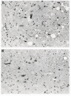
The first clues that murine retroviruses could cause neurologic disorders came from studies on wild mice from the Lake Casitas region of California (Gardner et al. 1973; Officer et al. 1973). A significant number of these animals develop hind limb paralysis associated with high levels of replication-competent, ecotropic MLVs. These viruses, typified by the well-studied Cas-Br-E MLV isolate, induce a similar disease in laboratory mice (Gardner 1978). Histologic examination of diseased mice reveals neuronal loss, as well as proliferation and hypertrophy of glial cells in the absence of an inflammatory response (Fig. 17) (Andrews and Gardner 1974; Andrews and Andrews 1976; Oldstone et al. 1977; Swarz et al. 1981; Zachary et al. 1986). Both neurons and glial cells accumulate vacuoles, giving the tissue a characteristic spongy appearance, and the myelin sheath that covers and insulates axons degenerates. Lesions are particularly prominent in the anterior horn cells of the lumbar spinal cord. These motor neurons control the lower extremities, and their loss is reflected in the hind limb paralysis often seen in infected mice.
Infection of the CNS and Induction of Lesions
Entry of viruses into the CNS is restricted by the blood-brain barrier, formed by tight endothelial cell junctions within the blood vessels that line the CNS. Cas-Br-E MLV-infected mice contain high titers of circulating virus, facilitating infection of vascular endothelial cells early in the disease (Swarz et al. 1981; Bilello et al. 1986; Pitts et al. 1987; Kay et al. 1991; Lynch et al. 1991; Nagra et al. 1992). Some of the virus produced by these cells is released directly into the CNS where it infects cells within the brain. Cas-Br-E MLV targets endothelial cells in particular regions of the brain, and the pattern of endothelial cell infection correlates closely with the pattern of infection found within the brain (Pitts et al. 1987; Baszler and Zachery 1990; Kay et al. 1991). Unfortunately, very little is known about the mechanisms that control this important aspect of pathogenesis. Interestingly, susceptibility is developmentally regulated; only very young mice that have not developed an immune response are susceptible to neurologic disease (Hoffman et al. 1984; Czub et al. 1991, 1992; Lynch and Portis 1993; Park et al. 1994a; Lynch et al. 1995).
Cas-Br-E MLV that has penetrated the blood-brain barrier targets microglial cells (Lynch et al. 1991; Gravel et al. 1993). The infected cells are morphologically similar to activated microglia, a feature that could reflect proliferation or hypertrophy of the infected cells. These cells frequently clump and form characteristic micronodules that are found in regions of neuropathology (Gravel et al. 1993). Similar histologic changes are found in neurodegenerative lesions induced by HIV (Koenig et al. 1986), SIV (Desrosiers 1990), and FIV (Dow et al. 1990, 1992), strengthening the possible role of microglia in the disease process. In addition, the most severe lesions are usually associated with areas of heaviest microglial cell infection (Kay et al. 1991; Lynch et al. 1991). Despite this strong correlation, many regions with large numbers of virus-infected cells remain disease-free (Morey and Wiley 1990; Kay et al. 1991; Lynch et al. 1991). Clarification of the mechanisms by which some regions resist the detrimental effects of the virus could shed light on the mechanisms by which damage is induced and might provide clues to fruitful therapeutic approaches for HIV-associated myelopathy and possibly other human neurologic diseases.
Viral Determinants of CNS Disease
Analyses of chimeric viruses containing sequences from Cas-Br-E MLV and from closely related, nonneurovirulent strains have revealed that sequences within the SU protein are important for neuropathogenesis (DesGroseillers et al. 1984a; Paquette et al. 1989; Portis et al. 1990). Similar experiments with five other neurotropic MLVs (see Table 5) reveal that SU is also important for neuropathogenesis in these strains (Szurek et al. 1988; Matsuda et al. 1993; Park et al. 1994c; Portis et al. 1995). One of these, the ts1 variant of Mo-MLV, carries a single point mutation that distinguishes it from strains of Mo-MLV that do not induce CNS disease (Szurek et al. 1988). Consistent with the important role of SU in neuropathogenesis, transgenic mice expressing only the Cas-Br-E MLV-encoded SU molecule develop neurodegenerative lesions (Kay et al. 1993). However, the lesions found in these animals are not as severe as those present in virus-infected animals, perhaps because levels of SU expression are lower in the transgenic mice. In addition, it is not known if transgenic mice expressing SU from a nonneurotropic strain would develop neurological lesions. This is an important control because sequences within the LTR, the gag-pol, and R-U5-leader region also affect neuropathogenesis, probably by influencing the levels and patterns of viral replication within the CNS (DesGroseillers et al. 1985; Paquette et al. 1990; Portis et al. 1990, 1991).
Mechanism of Disease Induction
The way in which Cas-Br-E MLV (or indeed any of the neurotropic MLVs) induces neuronal loss remains a matter of speculation. Because mutations affecting SU protein are correlated with neuropathogenesis and neurons in diseased areas are not infected (Paquette et al. 1990; Baszler and Zachary 1991; Kay et al. 1991; Lynch et al. 1991; Gravel et al. 1993), most models postulate that these cells are damaged by indirect effects of SU expression. One model suggests that SU, produced by infected microglial cells, binds to an unknown receptor on the surface of neurons, triggering cell damage (Jolicoeur 1990; Jolicoeur et al. 1991c). Such a receptor might normally interact with a factor that is required for survival of the neuron; binding of SU would block this interaction. This model is reminiscent of the pathogenic interactions between Env proteins and the Epo receptor in the FV system (Li et al. 1990; see above Oncogenesis, Tumor Induction by Simple C-type Retroviruses That Lack Oncogenes). Since multiple receptors may be involved in maintaining neuronal health, testing of this highly specific model is difficult. Other models suggest that production of the SU protein could stimulate the release of toxic substances that damage neurons or diminish the supportive and scavenger functions normally provided by the glial cells. Either of these effects could indirectly affect the health of the neurons.
Inflammatory CNS Disease Induced by HTLV-1
A low frequency of HTLV-1-infected individuals develops a chronic CNS disorder affecting the spinal cord called tropical spastic paraparesis or HTLV-1-associated myelopathy (TSP/HAM). This name reflects the history surrounding the discovery of HTLV-associated neurologic disease. TSP is one of several tropical myelopathies that were originally grouped together on the basis of geographic clustering (Cruickshank 1956). HTLV-1 was implicated in a subset of these disorders when a high frequency of anti-HTLV-1-antibodies was found in patients with similar pathologic findings (Gessain et al. 1985; Rodgers-Johnson et al. 1985). About the same time, a similar set of neurologic abnormalities, referred to as HAM, was associated with HTLV-1 infection in Japan (Osame et al. 1986b). There is now a general consensus that these are the same disease (Roman and Osame 1988). Neurologic disease has not been found in association with HTLV-2 infection.
The pathological hallmark of HTLV-1-induced CNS disease is a severe demyelination associated with a vigorous inflammatory response involving T cells, macrophages, and other hematopoietic cells (Akizuki et al. 1988; Piccardo et al. 1988; Rodgers-Johnson et al. 1990). This response can damage the CNS by a direct immunologic attack against virus-infected cells or other cells. In TSP/HAM, large numbers of CD8+ cytotoxic T cells are particularly prominent in inflamed areas; many of these react with the HTLV Tax protein, suggesting that a specific response to infected cells is important in causing the disease (Bhigjee et al. 1991; Hara et al. 1994). The release of cytokines and chemokines by the infiltrating cells and the swelling that results from inflammation can damage both neurons and supporting glial cells.
Infection of the CNS
The mechanism by which HTLV-1 enters the CNS is poorly understood, and the types of cells that are in fected by the virus are not known. Indeed, detecting expression of the virus or even the virus itself has been difficult. HTLV-1 sequences and antibodies against HTLV-1 have been detected in cerebrospinal fluid, indicating that the virus is present in the CNS (Hirose et al. 1986; Bhagavati et al. 1988; Ceroni et al. 1988; Jacobson et al. 1988b; McKhann et al. 1989). PCR and electron microscopy approaches have found evidence of HTLV-1 within the CNS in some cases (Akizuki et al. 1988; Liberski et al. 1988; Bhigjee et al. 1991; Iannone et al. 1992). Cytotoxic T cells that are found in the lesions are one likely source of virus (Bhigjee et al. 1991; Hara et al. 1994). Astrocytes may also be infected (Lehky et al. 1995). HTLV-1 can replicate in human monocytes and microglial cells in vitro (Hoffman et al. 1992b) but whether this mimics the situation in vivo is not known. Certainly it seems likely that some cells are latently infected, a feature that has been suggested to characterize lentiviral CNS infections (Haase et al. 1990; Staskus et al. 1991a).
Viral Factors Controlling CNS Disease
Very little is known about the viral determinants that affect induction of TSP/HAM. Gross molecular characterization of HTLV-1 strains isolated from patients with TSP/HAM has not revealed unique features that could influence development of CNS disease (Imamura et al. 1988; Gessain et al. 1989; Daenke et al. 1990; Evangelista et al. 1990; Kinoshita et al. 1991). In cases where differences have been found, they seem to reflect the types of virus present in a particular geographic area rather than the presence of variants in CNS lesions (Komurian et al. 1991; Ratner et al. 1991; Schulz et al. 1991; Gessain et al. 1993; Mahieux et al. 1995; Renjifo et al. 1995). However, a point mutation is sufficient for neuropathogenesis in ts1 Mo-MLV (Szurek et al. 1988; see above). Thus, a careful genetic analysis of neuropathogenic strains and an experimental system to study their properties are required before firm conclusions can be reached.
Mechanisms of Neuropathogenesis
Given the difficulties inherent in studying HTLV-1-induced CNS disease, it is not surprising that the mechanisms involved in pathogenesis are poorly understood. Unlike ATL, TSP/HAM can develop within several years of HTLV-1 infection (Osame et al. 1986a, 1987) and patients usually have a polyclonal HTLV-1 infection characterized by high levels of viral DNA (Greenberg et al. 1989; Yoshida et al. 1989; Gessain et al. 1990; Kira et al. 1991). The inflammatory response is the hallmark of this disease, but the factors that initiate and sustain it are not known (Jacobson 1992; Hollsberg and Hafler 1993, 1995). An autoimmune response mediated by infiltrating T cells that is directed against CNS products or a cytotoxic response against infected CNS cells could be involved. Late in the disease, patients with TSP/HAM contain high levels of cytotoxic T cells, many of which react with Tax (Jacobson et al. 1990, 1992; Elovaara et al. 1993). These cells are also found in CNS lesions (Bhigjee et al. 1991; Hara et al. 1994), suggesting that they may have a role in pathogenesis. Viral products are present at relatively low levels within the CNS, but these levels may be sufficient to induce disease. As mentioned earlier, low levels of Cas-Br-E MLV SU expressed by transgenic mice are sufficient to induce CNS lesions (Kay et al. 1993). The extended course of the disease could reflect the time required for inflammation to reach a critical level and neuronal damage to exceed a tolerable threshold.
If the pathogenesis of these viruses is to be understood, an experimental model must be developed in which viral replication, spread, and infection of the CNS can be studied. The only animal model available involves rats inoculated with HTLV-1-infected cells. Although these animals develop CNS lesions, the disease does not duplicate many of the important features of TSP/HAM (Ishiguro et al. 1992; Kushida et al. 1994). If a better animal system could be found, the recently isolated infectious HTLV-1 clones (Kimata et al. 1994; Derse et al. 1995) could be exploited to test the role of different gene products in pathogenesis. Although none of the available HTLV-1 transgenic mice develop parenchymal CNS disease (Hinrichs et al. 1987; Nerenberg et al. 1987; Iwakura et al. 1991; Grossman et al. 1995), this approach might also be fruitful if the relevant HTLV-1 products could be identified and targeted to the appropriate cell types. Such models would not reveal features of the disease that are caused by viral replication, but questions relating to the targets of the inflammatory response might be approachable.
- Neurological Diseases - RetrovirusesNeurological Diseases - Retroviruses
- Bacillus subtilis strain MMS15 16S ribosomal RNA gene, partial sequenceBacillus subtilis strain MMS15 16S ribosomal RNA gene, partial sequencegi|1007377228|gb|KT933118.1|Nucleotide
Your browsing activity is empty.
Activity recording is turned off.
See more...

